. 2024 Aug;20(8):1053-1065.
doi: 10.1038/s41589-024-01568-7. Epub 2024 Feb 29.

In situ analysis of osmolyte mechanisms of proteome thermal stabilization

Affiliations

In situ analysis of osmolyte mechanisms of proteome thermal stabilization

Monika Pepelnjak et al. Nat Chem Biol. 2024 Aug.

Abstract

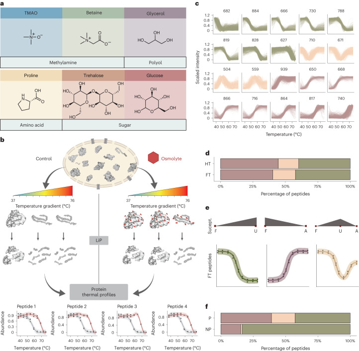
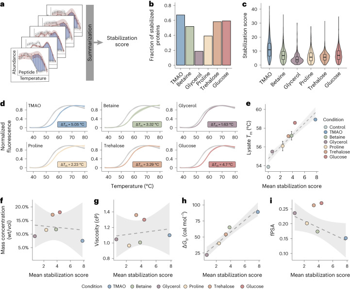
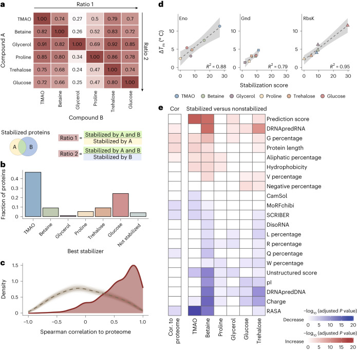
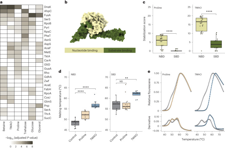
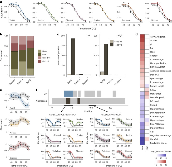
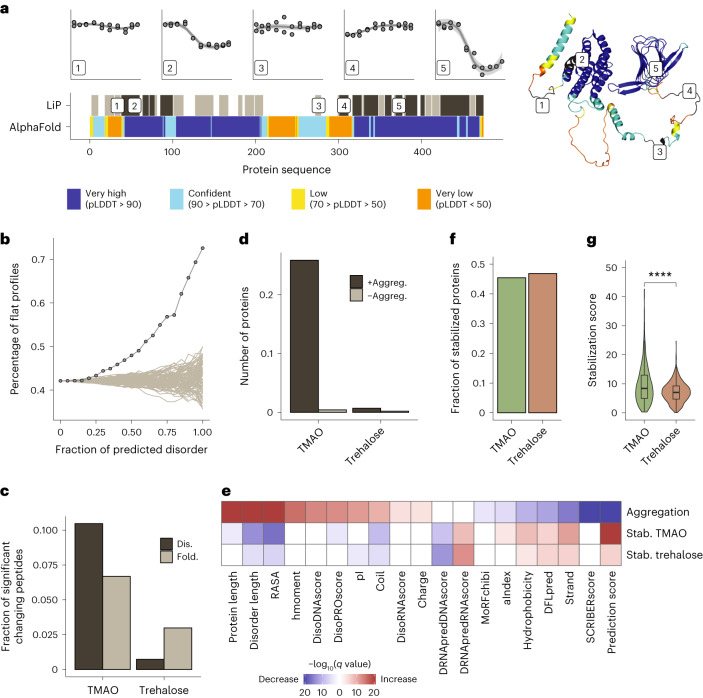
Organisms use organic molecules called osmolytes to adapt to environmental conditions. In vitro studies indicate that osmolytes thermally stabilize proteins, but mechanisms are controversial, and systematic studies within the cellular milieu are lacking. We analyzed Escherichia coli and human protein thermal stabilization by osmolytes in situ and across the proteome. Using structural proteomics, we probed osmolyte effects on protein thermal stability, structure and aggregation, revealing common mechanisms but also osmolyte- and protein-specific effects. All tested osmolytes (trimethylamine N-oxide, betaine, glycerol, proline, trehalose and glucose) stabilized many proteins, predominantly via a preferential exclusion mechanism, and caused an upward shift in temperatures at which most proteins aggregated. Thermal profiling of the human proteome provided evidence for intrinsic disorder in situ but also identified potential structure in predicted disordered regions. Our analysis provides mechanistic insight into osmolyte function within a complex biological matrix and sheds light on the in situ prevalence of intrinsically disordered regions.


Conflict of interest statement

P.P. is an inventor of a patent licensed by Biognosys AG that covers the LiP–MS method used in this manuscript (patent number WO-A-2014082733). P.P. is also a scientific advisor of Biognosys AG. The remaining authors declare no competing interests.

Figures

Fig. 1
Fig. 1. LiP–MS thermal profiling to study osmolyte effects.
a, Chemical structures of osmolytes used in the study. All osmolytes were used at 1 M except for trehalose (0.5 M). b, Overview of the experimental procedure. Aliquots of an E. coli lysate were subjected to a thermal gradient (ten temperatures; 37–76 °C) in the presence or absence of an osmolyte. Subsequent proteolysis with PK under native conditions yielded proteolytic fragments that are informative about the folded state of proteins, with PK accessibility increasing after protein unfolding. Trypsin digestion under denaturing conditions generated peptides that can be measured with MS. Scaled relative abundance of individual peptides across the temperature gradient allows profiling of protein thermal unfolding, and osmolyte effects can be studied by comparing profiles between control (gray curves) and osmolyte (red curves) conditions. Figure created with BioRender.com. c, Peptide thermal profile clusters in the absence of osmolytes. Both fully tryptic (FT) and half-tryptic (HT) peptides are shown. The numbers of peptides in each cluster are indicated. Colors indicate profiles with decreasing intensity (group 1, green), profiles with increasing intensity (group 2, purple) and nonmonotonous profiles (group 3, orange). d, Percentage of cluster groups from c shown separately for FT and HT peptides. e, Interpretation of FT peptide behavior in the three cluster groups from c. We interpret changing FT peptide intensity as a function of increased or decreased proteolytic susceptibility (Suscept.) as indicating protein in a folded (F), unfolded (U) or aggregated (A) state. For HT peptides, the opposite effect (that is, a flipped thermal profile) is expected (Supplementary Fig. 1f). f, Percentage of cluster groups from c separated by proteins defined as nonprecipitators (NP) and precipitators (P) in TPP; only FT peptides are shown (see Supplementary Fig. 1g for HT peptides).
Fig. 2
Fig. 2. Osmolytes have a global effect on protein stability.
a, Overview of the analytical procedure. A peptide-level score was calculated by summing the distances (blue) between thermal profiles in the absence (gray) and presence (red) of osmolyte at temperature values where confidence intervals do not overlap. Peptide-level scores were then combined after correcting for peptide length and overlap into a stabilization score for each protein (Supplementary Fig. 2c and Methods). b, Fraction of stabilized proteins out of all detected proteins for each osmolyte. c, Distribution of stabilization scores for proteins significantly stabilized by each osmolyte. Horizontal lines define the median, and boxes define the 25th and 75th percentiles; whiskers represent the maximum and minimum values. Each box plot represents the stabilization scores of stabilized proteins (205 proteins for glycerol, 791 for glucose, 885 for trehalose, 702 for betaine, 423 for proline and 733 for TMAO) calculated based on two LiP–MS replicates per temperature. d, Melting curves for E. coli cell lysate measured by DSF under control conditions (gray) and after the addition of osmolytes. Error bars show mean ± s.d. (n = 5 replicates per condition). e, Linear regression between DSF-measured lysate melting temperatures and mean stabilization scores for all detected proteins. Error bars show mean ± s.d. (n = 5 replicates). The shaded area represents the confidence interval of the linear fit (dashed line). fi, Linear regression between mean stabilization score and osmolyte mass concentration (wt/vol; f), osmolyte viscosity at 25 °C (g), transfer free energy (Gtr) for backbone model substrate transferred from water to 1 M osmolyte (h) and fraction polar surface area (fPSA; i). Transfer free energy for glucose was not available in the literature. In all cases, the shaded area represents the confidence interval of the linear fit (dashed line); cP, centipoise.
Fig. 3
Fig. 3. Biophysical features of osmolyte-stabilized proteins.
a, The heat map shows pairwise overlap of osmolyte-stabilized proteins. Ratio 1 and ratio 2 are calculated as indicated. b, Fraction of proteins stabilized most strongly by each osmolyte out of all proteins detected in all six datasets. c, Density estimate distributions of the Spearman correlation coefficient between the stability score per protein and the mean stability score across the lysate (red line) or a randomized mean stability (dashed gray line) obtained from 1,000 repeated calculations (light gray lines; appears as a gray band). d, Linear regression between DSF-calculated ∆Tm and protein stabilization score for the indicated purified proteins. The shaded area represents the confidence interval of the linear fit (dashed line). Error bars (smaller than point size) show the standard deviation (n = 4 replicates). e, Heat map showing the most significantly different features (adjusted P value of <0.0001 in at least one comparison) between proteins with high correlation and low correlation of the stabilization score with that of the global proteome (first column, Cor) or between proteins significantly stabilized and those not stabilized by the indicated individual osmolytes. The color indicates whether the feature is higher (red) or lower (blue) in proteins with high correlation (first column) and in stabilized proteins (other columns) than in the rest. Feature significance was determined by two-sided t-test followed by correction by multiple hypothesis testing (Benjamini–Hochberg). Other tested features are shown in Supplementary Fig. 3. The term ‘X percentage’ refers to the fraction of the indicated amino acid. See Methods for the calculation or prediction of features.
Fig. 4
Fig. 4. Stabilization of multidomain proteins.
a, Heat map showing multidomain proteins with domains differentially stabilized by the indicated osmolytes (adjusted P value of <0.05; as determined by two-sided t-test and Benjamini–Hochberg correction). b, Structure of the chaperone DnaK (PDB ID: 4JNE) with the nucleotide binding domain (light green) and substrate binding domain (dark green). c, Distribution of the indicated osmolyte stabilization scores for amino acids mapping to the nucleotide-binding domain (NBD) and substrate-binding domain (SBD) of DnaK. Horizontal lines define the median, and boxes define the 25th and 75th percentiles; whiskers represent the maximum and minimum values. Significance was determined using two-sided Wilcoxon tests; ****P < 0.0001. Each dot represents an amino acid level stabilization score calculated based on two LiP–MS replicates per temperature. d, Distribution of melting temperatures derived from peptides mapping to the indicated domains of DnaK in the presence and absence of the indicated osmolytes. Each point represents one peptide curve measured in duplicate (n ≥ 7 peptides per condition). Box plots are as in c; **P < 0.01; ****P < 0.0001; NS, not significant. e, Protein melting curves measured with DSF and the first derivative of the melting curve for purified DnaK are plotted for control (gray), proline (yellow) and TMAO (blue).
Fig. 5
Fig. 5. Osmolyte effects on protein aggregation.
a, TPP-determined insolubility profile of E. coli lysate in the absence (gray) and presence (colors) of osmolytes. Plots show protein concentration in the soluble fraction scaled to the value at 37 °C. Shaded areas indicate the confidence interval of the fit. b, Comparison of protein stabilization analysis by LiP–MS and TPP. The plot shows the fraction of proteins reported stabilized by both methods (both), neither method (none) and one method (only LiP and only TPP). Only proteins defined as precipitators in TPP and detected in both datasets are included. c, Number of proteins significantly changed in abundance in the soluble fraction at low (37 °C and 42 °C) and high (68.2 °C, 72.5 °C and 76 °C) temperatures in TPP in the presence and absence of the indicated osmolytes. Significance was calculated separately for proteins with increased aggregation (+Aggreg.; fold change of <−1; dark gray) and decreased aggregation (–Aggreg.; fold change of >1; light gray) in the presence of osmolyte; adjusted P value of <0.05 (data were analyzed by two-sided t-test with a Benjamini–Hochberg correction). d, Heat map showing the most significantly different features (adjusted P value of <0.01; data were analyzed by t-test with a Benjamini–Hochberg correction) between proteins with increased aggregation in TMAO at high or low temperature (as in c) and all other detected proteins. Color indicates higher (red) or lower (blue) features in the increased aggregation group. Rg, radius of gyration; aIndex, aliphatic index. e, TPP profile for Frr in the absence (gray) and presence of the indicated osmolytes. The shaded area indicates the confidence interval of the fit. f, LiP–MS thermal profiles for two representative peptides from Frr for the indicated osmolytes. The top plot shows peptide positions along the Frr protein sequence. Top track, all detected peptides; blue, peptides with an increased aggregation profile under at least one condition; gray, peptides with no aggregation profile under any condition. Bottom track, predicted aggregation-prone regions (dark gray). The shaded area indicates the confidence interval of the fit.
Fig. 6
Fig. 6. Osmolyte effects on the human proteome.
a, Analysis of predicted disorder in regions of CAP-1 with different thermal melting behavior. Plots (top) show thermal profiles of five example peptides mapping to indicated regions along the protein sequence. The top barcode (LiP) shows all peptides (dark gray) with a measurable thermal melting profile out of all detected peptides (light gray) along the protein sequence. The lower barcode shows the AlphaFold pLDDT prediction score; very low scores typically correspond to disordered protein regions. The AlphaFold-predicted structure is shown (right), with peptides annotated. b, Percentage of peptides with flat profiles (lines with dots) for proteins with increasing fractions of predicted disorder. All detected peptides/proteins are plotted. Gray lines show tests in which protein disorder was randomized. c, Fraction of peptides that show a structural change following osmolyte treatment relative to control out of all detected peptides calculated separately for peptides predicted to be folded (Fold) or disordered (Dis). d, Number of proteins with increased (+Aggreg.) or decreased (–Aggreg.) aggregation after the addition of osmolytes to HEK293T cell lysates at 37 °C. e, Biophysical features of human proteins affected by osmolytes. The heat map shows significantly enriched or depleted features for proteins that precipitate in the presence of TMAO versus those that do not precipitate (row 1, aggregation) or that are stabilized (Stab.) in the presence of the indicated osmolytes versus nonstabilized proteins (rows 2 and 3). P values were determined by two-sided t-tests followed by Benjamini–Hochberg multiple testing correction. hmoment, hydrophobic moment. f, Fraction of proteins stabilized in HEK293T cell lysates in the presence of TMAO and trehalose. g, Distribution of stabilization scores for proteins significantly stabilized by TMAO (899 proteins) and trehalose (948 proteins) based on two LiP–MS replicates per temperature. Horizontal lines indicate the median, boxes indicate the 25th and 75th percentiles, and whiskers indicate the maximum and minimum values. Significance was determined by two-sided Wilcoxon tests; ****P < 0.0001.

References

    1. Ye, Y. et al. Global metabolomic responses of Escherichia coli to heat stress. J. Proteome Res.11, 2559–2566 (2012). - PubMed
    1. Kim, S. et al. Heat-responsive and time-resolved transcriptome and metabolome analyses of Escherichia coli uncover thermo-tolerant mechanisms. Sci. Rep.10, 17715 (2020). - PMC - PubMed
    1. Jozefczuk, S. et al. Metabolomic and transcriptomic stress response of Escherichia coli. Mol. Syst. Biol.6, 364 (2010). - PMC - PubMed
    1. Sévin, D. C., Stählin, J. N., Pollak, G. R., Kuehne, A. & Sauer, U. Global metabolic responses to salt stress in fifteen species. PLoS ONE11, e0148888 (2016). - PMC - PubMed
    1. Hincha, D. K. & Hagemann, M. Stabilization of model membranes during drying by compatible solutes involved in the stress tolerance of plants and microorganisms. Biochem. J.383, 277–283 (2004). - PMC - PubMed

Publication types

MeSH terms

LinkOut - more resources