. 2024 Feb 29;14(1):5048.
doi: 10.1038/s41598-024-55632-2.

SDF-1 involvement in orthodontic tooth movement after tooth extraction

Affiliations

SDF-1 involvement in orthodontic tooth movement after tooth extraction

Duangtawan Rintanalert et al. Sci Rep. .

Erratum in

Abstract

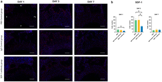
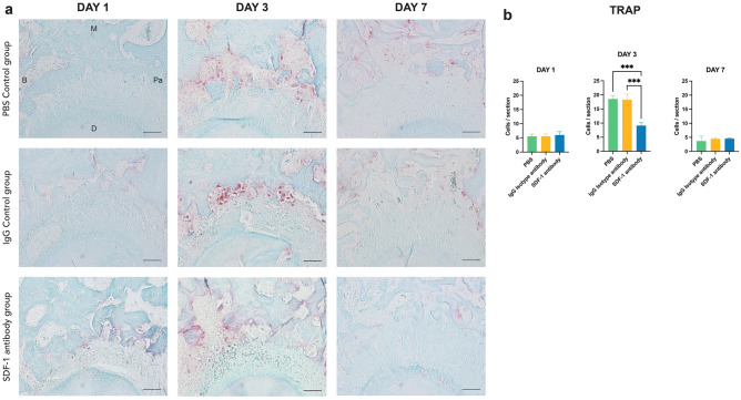
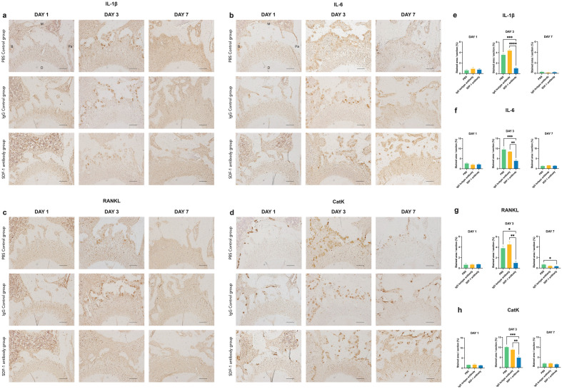
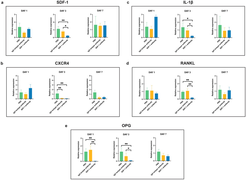
The stromal cell-derived factor 1 (SDF-1)/chemokine receptor type 4 (CXCR4) axis plays a key role in alveolar bone metabolism during orthodontic tooth movement (OTM). Herein, the effects of the SDF-1/CXCR4 axis on the regional acceleratory phenomenon (RAP) in OTM velocity and on changes in the surrounding periodontium after adjacent tooth extraction in rats were investigated. Six-week-old male Wistar/ST rats underwent left maxillary first molar (M1) extraction and mesial OTM of the left maxillary second molar (M2) with a 10-g force closed-coil spring. Phosphate-buffered saline, immunoglobulin G (IgG) isotype control antibody, or anti-SDF-1 neutralizing monoclonal antibody were injected at the M1 and M2 interproximal areas (10 μg/0.1 mL) for the first three days. Analyses were performed after 1, 3, and 7 days (n = 7). The results demonstrated a significant increase in SDF-1 expression from day 1, which was effectively blocked via anti-SDF-1 neutralizing monoclonal antibody injection. On day 3, the M2 OTM distance and the number of positively stained osteoclasts significantly reduced alongside a reduction in inflammatory markers in the experimental group. Our results demonstrated that serial local injection of the anti-SDF-1 neutralizing monoclonal antibody reduces M2 OTM, osteoclast accumulation, and localized inflammatory responses in an OTM model with tooth extraction-induced RAP.

Keywords: Neutralizing antibody; Orthodontic tooth movement (OTM); Regional acceleratory phenomenon (RAP); Stromal cell-derived factor 1 (SDF-1); Tooth extraction.


Conflict of interest statement

The authors declare no competing interests.

Figures

Figure 1
Figure 1
Immunofluorescent evidence of increased stromal cell-derived factor 1 (SDF-1) expression on the experimental side of the left maxillary second molar (M2) mesial root on days 1,3, and 7 (a). SDF-1 immunofluorescence (IF) staining of M2 mesial root at the compression side on day 3 of the experimental side at × 10 and × 20 magnification (b). Positively stained cells with Alexa Fluor® 594 are red, and the nuclei with DAPI are blue. Scale bars = 50 μm. Abbreviations: B, buccal; M, mesial; Pa, palatal; D, distal.
Figure 2
Figure 2
Qualitative reverse transcription polymerase chain reaction comparing the untreated control side and the experimental side of the control phosphate-buffered saline (PBS) group to verify the expression of our targeted chemokine (a) the stromal cell-derived factor 1 (SDF-1) and its receptor, (b) the chemokine receptor type 4 (CXCR4). A significant increase in the SDF-1 relative expression was detectable on days 1, 3, and 7 on the experimental side, whereas CXCR4 expression significantly increased only on day 7 in the experimental side. Values are presented as means ± standard deviation (n = 4). * p < 0.05, ** p < 0.01.
Figure 3
Figure 3
The maxillary second molar (M2) tooth movement distance after mechanical loading. Measured using the direct distance from M2 distal surface to maxillary third molar (M3) mesial surface, no significant differences were found in all groups at all time points (a). A measurement with the reference line created from the opposite M3 of the untreated side revealed a significant difference on day 3 of the experimental stromal cell-derived factor 1 (SDF-1) antibody group where less distance could be observed in comparison to the two control groups (b). Measurement of M3 tooth movement distance using the reference line created from the opposite M3 of the right maxillary untreated side. A significant difference was detectable on day 3 in the SDF-1 antibody group where less M3 tooth movement was measured in comparison to the two control groups (c). Values are presented as means ± standard deviation (n = 7). * p < 0.05, ** p < 0.01.
Figure 4
Figure 4
Immunofluorescence (IF) staining of stromal cell-derived factor 1 (SDF-1) using Alexa Fluor® 594 (red) and DAPI (blue) for nuclear staining at the compression side of maxillary second molar (M2) mesial root on days 1,3, and 7. Positively stained cells were detected abundantly on day 3, whereas less were observed on days 1 and 7. Following neutralization via anti-SDF-1 neutralizing monoclonal antibody injection, reduced positive signals could be detected on day 3 of the SDF-1 antibody group compared to the two control groups (a). Number of positively stained cells at the compression side of M2 mesial root on days 1, 3, and 7. Statistically significant differences can be seen on day 1 between the SDF-1 antibody group and the PBS control group, and on day 3 between the SDF-1 antibody group and the two control groups (b). Scale bars = 50 μm. Abbreviations: B; buccal, M; mesial, Pa; palatal, D; distal. Values are presented as means ± standard deviation (n = 3). * p < 0.05.
Figure 5
Figure 5
Representative image of tartrate-resistant acid phosphatase (TRAP) staining at the compression side of M2 mesial roots on day 1, 3, and 7. TRAP-positive multinucleated cells were observed abundantly on day 3 in both control groups, whereas less were observed on days 1 and 7 in all groups. Focusing on the compression side of the mesio-palatal root on day 3 of all three groups, the stromal cell-derived factor 1 (SDF-1) antibody group demonstrated less TRAP-positive multinuclear osteoclasts in the PDL compared to the two control groups (a). Number of TRAP-stained cells at the compression side of M2 mesial root on days 1, 3, and 7. Statistically significant differences can be seen on day 3 between the SDF-1 antibody group and the two control groups (b). Scale bars = 50 μm. Abbreviations: B; buccal, M; mesial, Pa; palatal, D; distal. Values are presented as mean ± standard deviation (n = 3). *** p < 0.001.
Figure 6
Figure 6
Immunohistochemistry-stained images of the M2 compression side, and quantitative analysis of positively stained areas of interleukin (IL)-1β) (a,e), IL-6 (b,f), receptor activator of nuclear factor-kappa B ligand (RANKL) (c, g), and Cathepsin K (CatK) (d,h). In all tested antibodies, significant reductions in positively stained areas per slide of the M2 compression side were observed on day 3 in the SDF-1 experimental group. * p < 0.05, ** p < 0.01, ** p < 0.001.
Figure 7
Figure 7
Qualitative reverse transcription polymerase chain reaction analyzing key genes involved in the inflammation, bone remodeling and stromal cell-derived factor 1 (SDF-1)/ chemokine receptor type 4 (CXCR4) axis related to the OTM. Relative gene expressions of SDF-1 (a), CXCR4 (b), interleukin-1 beta (IL-1β) (c), receptor activator of nuclear factor-kappa B ligand (RANKL) (d), and osteoprotegerin (OPG) (e) were analyzed using GAPDH as an internal reference. Expressions of all tested key genes were significantly reduced on day 3 in the experimental group, compared to the two controls. Additionally, a significant reduction in OPG was also found on day 1 in the experimental group. Values are presented as means ± standard deviation (n = 4). * p < 0.05, ** p < 0.01.

Similar articles

References

    1. Häsler R, Schmid G, Ingervall B, Gebauer U. A clinical comparison of the rate of maxillary canine retraction into healed and recent extraction sites—a pilot study. Eur. J. Orthod. 1997;19:711–719. doi: 10.1093/ejo/19.6.711. - DOI - PubMed
    1. Abu Alhaija ES, Al Shayeb RA, Al-Khateeb S, Daher HO, Daher SO. A comparative assessment of the amount and rate of orthodontic space closure toward a healed vs recent lower premolar extraction site: A split-mouth randomized clinical trial. Angle Orthod. 2022;92(4):463–470. doi: 10.2319/102921-797.1. - DOI - PMC - PubMed
    1. Frost HM. The regional acceleratory phenomenon: A review. Henry Ford Hosp. Med. J. 1983;31:3–9. - PubMed
    1. Fu T, Liu S, Zhao H, Cao M, Zhang R. Effectiveness and safety of minimally invasive orthodontic tooth movement acceleration: A systematic review and meta-analysis. J. Dent. Res. 2019;98:1469–1479. doi: 10.1177/0022034519878412. - DOI - PubMed
    1. Verna C, Cattaneo PM, Dalstra M. Corticotomy affects both the modus and magnitude of orthodontic tooth movement. Eur. J. Orthod. 2018;40:107–112. doi: 10.1093/ejo/cjx041. - DOI - PubMed

LinkOut - more resources