. 2024 Feb 29;43(1):63.
doi: 10.1186/s13046-024-02989-x.

IL6-STAT3-C/EBPβ-IL6 positive feedback loop in tumor-associated macrophages promotes the EMT and metastasis of lung adenocarcinoma

Affiliations

IL6-STAT3-C/EBPβ-IL6 positive feedback loop in tumor-associated macrophages promotes the EMT and metastasis of lung adenocarcinoma

Zhengyang Hu et al. J Exp Clin Cancer Res. .

Abstract

Background: Lung cancer is one of the most common tumors in the world, and metastasis is one of the major causes of tumor-related death in lung cancer patients. Tumor-associated macrophages (TAMs) are a major component of the tumor microenvironment (TME) and are frequently associated with tumor metastasis in human cancers. However, the regulatory mechanisms of TAMs in lung cancer metastasis remain unclear.

Methods: Single-cell sequencing analysis of lung cancer and normal tissues from public databases and from 14 patients who underwent surgery at Zhongshan Hospital was performed. In vitro co-culture experiments were performed to evaluate the effects of TAMs on lung cancer migration and invasion. Changes in the expression of IL-6, STAT3, C/EBPΒ, and EMT pathway were verified using RT-qPCR, western blotting, and immunofluorescence. Dual luciferase reporter assays and ChIP were used to reveal potential regulatory sites on the transcription factor sets. In addition, the effects of TAMs on lung cancer progression and metastasis were confirmed by in vivo models.

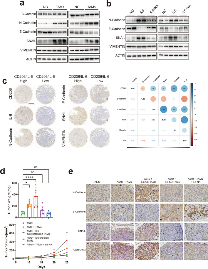
Results: TAM infiltration is associated with tumor progression and poor prognosis. IL-6 secreted by TAMs can activate the JAK2/STAT3 pathway through autocrine secretion, and STAT3 acts as a transcription factor to activate the expression of C/EBPβ, which further promotes the transcription and expression of IL-6, forming positive feedback loops for IL6-STAT3-C/EBPβ-IL6 in TAMs. IL-6 secreted by TAMs promotes lung cancer progression and metastasis in vivo and in vitro by activating the EMT pathway, which can be attenuated by the use of JAK2/STAT3 pathway inhibitors or IL-6 monoclonal antibodies.

Conclusions: Our data suggest that TAMs promote IL-6 expression by forming an IL6-STAT3-C/EBPβ-IL6 positive feedback loop. Released IL-6 can induce the EMT pathway in lung cancer to enhance migration, invasion, and metastasis. The use of IL-6-neutralizing antibody can partially counteract the promotion of LUAD by TAMs. A novel mechanism of macrophage-promoted tumor progression was revealed, and the IL6-STAT3-C/EBPβ-IL6 signaling cascade may be a potential therapeutic target against lung cancer.

Keywords: Epithelial-mesenchymal transition; Interleukin-6; Lung adenocarcinoma; Metastasis; Tumor-associated macrophages.


Conflict of interest statement

The authors declare that they have no competing interests.

Figures

Fig. 1
Fig. 1
Annotation and function of myeloid cells and macrophages by scRNA-seq in tumor and normal tissue. a UMAP plot colored by different clusters. b Dot plot of mean expression of myeloid cells and macrophages’ characteristic genes in different clusters. c UMAP plot colored by different cell types. d Complex heatmap of selected marker genes in each cell cluster. Up: Tissue preference of each cluster. Down: Relative expression of marker genes associated with each cell subset. e Relative contribution of each cell type in normal vs. tumor tissue and early vs. advanced LUAD. f Kaplan–Meier curves of survival analysis in TCGA LUAD patients based on the TAMs infiltration. g Left: Difference genes between normal tissue, early LUAD, and advanced LUAD, with a threshold of |log2FC| > 0.5 & p_val_adj < 0.05, and take the intersection of the differential genes of the three groups. Right: Heatmap of 26 genes that differ among the three groups. h Immunofluorescence shows the expression of TAMs marker and IL-6 (Red CD68, Pink CD163, Green IL-6, Blue Dapi), scale bar represents 50 μm
Fig. 2
Fig. 2
TAMs promote LUAD cell proliferation, migration and invasion by secreting IL-6. a Cell migration and invasion ability of LUAD cells (A549 and H358) alone, stimulated with IL-6 or stimulated with IL-6 followed by addition of IL-6 neutralizing antibody was determined by the transwell assay. b Cell proliferation ability of LUAD cells (A549 and H358) alone, stimulated with IL-6 or stimulated with IL-6 followed by addition of IL-6 neutralizing antibody was determined by the CCK8 assay. c The tumor size and tumor weight in the A549 alone, A549 + IL-6 stimulation and A549 + IL-6 stimulation + IL-6-NA groups. d RT-qPCR analyzed the expression of the most often released cytokines by macrophages after IL-6 stimulation. e Elisa analysis of IL-6 showed changes in macrophage IL-6 expression over time after IL-6 stimulation. f & g Cell migration, invasion and proliferation ability of LUAD cells (A549 and H358) alone, co-culture with M2-like macrophages or co-culture with M2-like macrophages and stimulated with IL-6 was determined by the transwell assay and CCK8 assay
Fig. 3
Fig. 3
IL-6 promotes LUAD progression by activating the JAK2/STAT3 pathway in M2-like macrophages. a GSVA analysis of differential pathways between AMs, TAMs in early and advanced LUAd. b Effects of IL-6 stimulation on common pathway expression in TAMs detected by western blot analysis. c Expression of the JAK2/STAT3 pathway with IL-6-stimulated, IL-6-stimulated with the addition of IL-6-NA in M2-like macrophages were analyzed by western blot. d Effect of AZD1480 and WP1066 on JAK2/STAT3 pathway expression in THP1 derived M2-like macrophages after IL-6 stimulation were analyzed by western blot. e Cell migration and invasion ability of LUAD cells alone, co-culture with M2-like macrophages or co-culture with M2-like macrophages followed by addition of AZD1480 and WP1066 was determined by the transwell assay. f RT-qPCR and Elisa analysis showed IL-6 expression in M2-like macrophages after IL-6 stimulation or IL-6 stimulation with the addition of AZD1480 and WP1066 (Left: Elisa results. Right: RT-qPCR results). g IL-6 expression after STAT3 overexpression or knockdown was analyzed by western blot. h RT-qPCR and Elisa analysis showed IL-6 expression in M2-like macrophages after STAT3 overexpression or knockdown
Fig. 4
Fig. 4
pSTAT3 promotes tumor progression by regulating C/EBPβ expression in TAMs. a Cell migration and invasion ability of LUAD cells, C/EBPβ overexpression or C/EBPβ knockdown was determined by the transwell assay. b Effects of IL-6 stimulation on C/EBPβ expression in TAMs. c Expression of C/EBPβ after STAT3 overexpression or knockdown. d RT-qPCR and Elisa analysis showed IL-6 expression in M2-like macrophages after C/EBPβ overexpression or knockdown. e Expression of pSTAT3 and C/EBPβ in the nucleus after IL-6 stimulation. f The localization of pSTAT3 and C/EBPβ were analyzed by immunofluorescence staining. The nuclei were stained with 4′,6-diamidino-2-phenylindole (DAPI; blue). The line charts represent fluorescence intensity (MFI), presenting the distance from α to β, scale bar represents 100 μm
Fig. 5
Fig. 5
Regulatory role of STAT3-C/EBPβ-IL6. a C/EBPβ and IL-6 expression after STAT3 knockdown or overexpression. b STAT3 and IL-6 expression after C/EBPβ knockdown or overexpression. c IL-6 expression after STAT3 knockdown and C/EBPβ overexpression or STAT3 overexpression and C/EBPβ knockdown. d IL-6 expression after STAT3 knockdown and rescued by IL-6 stimulation. e IL-6 expression after C/EBPβ knockdown and rescued by IL-6 stimulation
Fig. 6
Fig. 6
Mechanisms of STAT3 regulation of C/EBPβ and C/EBPβ regulation of IL-6. a ChIP assay demonstrated the binding of STAT3 to the C/EBPβ promoter in THP1-derived M2-like macrophages. b RT-qPCR of the ChIP products confirmed the binding capacity of STAT3 to the C/EBPβ promoter. c Selective mutation analyses identified STAT3-responsive regions in the C/EBPβ promoter. d Luciferase reporter plasmids with wild and mutated C/EBPβ promoters were transfected into 293 T cells. Relative luciferase activity was determined using IL-6 stimulation or STAT3 overexpression. e ChIP assay demonstrated the binding of C/EBPβ to the IL-6 promoter in. f RT-qPCR of the ChIP products confirmed the binding capacity of C/EBPβ to the IL-6 promoter. g Selective mutation analyses identified C/EBPβ-responsive regions in the IL-6 promoter. h Luciferase reporter plasmids with wild and mutated IL-6 promoters were transfected into 293 T cells. Relative luciferase activity was determined using IL-6 stimulation, C/EBPβ or STAT3 overexpression
Fig. 7
Fig. 7
IL-6 promotes LUAD migration and invasion through activation of EMT pathway in vivo and in vitro. a Expression of EMT-related genes after co-culture with M2-like macrophages. b Expression of EMT-related genes using IL-6 stimulation or IL-6-neutralising antibodies. c IHC analyzed the expression of CD206, IL-6, E-cadherin, N-cadherin, Vimentin and Snail, and their relationships of tumors from LUAD patients. d The tumor size and tumor weight in the A549 alone, A549 + control TAMs, A549 + IL-6 overexpression TAMs, A549 + IL-6 knockout TAMs, and A549 + TAMs + IL-6 neutralizing antibodies groups. e IHC analyzed the expression of E-cadherin, N-cadherin, Snail and Vimentin protein of tumors from the subgroups above. Scale bar represents 10 μm

References

    1. Sung H, Ferlay J, Siegel RL, et al. Global Cancer statistics 2020: GLOBOCAN estimates of incidence and mortality worldwide for 36 cancers in 185 countries. CA Cancer J Clin. 2021;71(3):209–249. doi: 10.3322/caac.21660. - DOI - PubMed
    1. Hu Z, Li M, Chen Z, et al. Advances in clinical trials of targeted therapy and immunotherapy of lung cancer in 2018. Transl Lung Cancer Res. 2019;8(6):1091–1106. doi: 10.21037/tlcr.2019.10.17. - DOI - PMC - PubMed
    1. Nadal E, Massuti B, Domine M, et al. Immunotherapy with checkpoint inhibitors in non-small cell lung cancer: insights from long-term survivors. Cancer Immunol Immunother. 2019;68(3):341–352. doi: 10.1007/s00262-019-02310-2. - DOI - PMC - PubMed
    1. Hirsch FR, Scagliotti GV, Mulshine JL, et al. Lung cancer: current therapies and new targeted treatments. Lancet. 2017;389(10066):299–311. doi: 10.1016/S0140-6736(16)30958-8. - DOI - PubMed
    1. Gettinger S, Horn L, Jackman D, et al. Five-year Follow-up of Nivolumab in previously treated advanced non-small-cell lung Cancer: results from the CA209-003 study. J Clin Oncol. 2018;36(17):1675–1684. doi: 10.1200/JCO.2017.77.0412. - DOI - PubMed

MeSH terms