. 2024 Feb 29;22(1):216.
doi: 10.1186/s12967-023-04700-8.

Ubiquitylation of RUNX3 by RNA-binding ubiquitin ligase MEX3C promotes tumorigenesis in lung adenocarcinoma

Affiliations

Ubiquitylation of RUNX3 by RNA-binding ubiquitin ligase MEX3C promotes tumorigenesis in lung adenocarcinoma

Zelai He et al. J Transl Med. .

Abstract

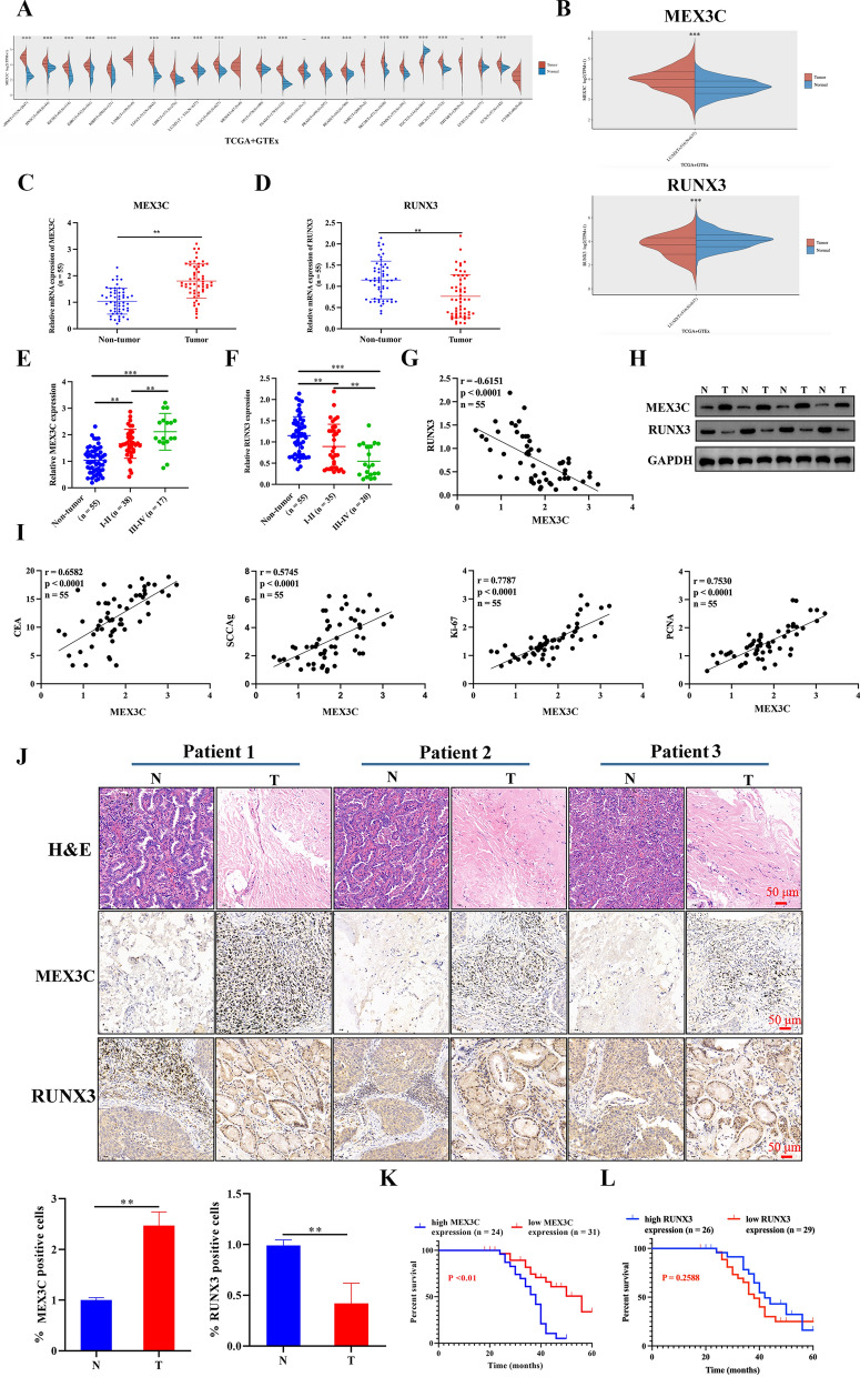
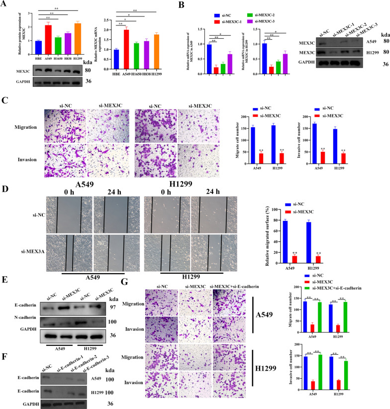
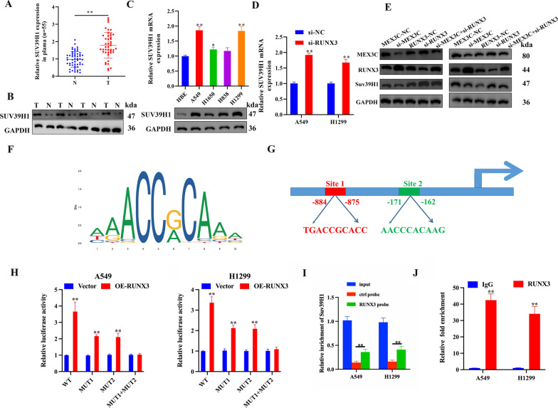
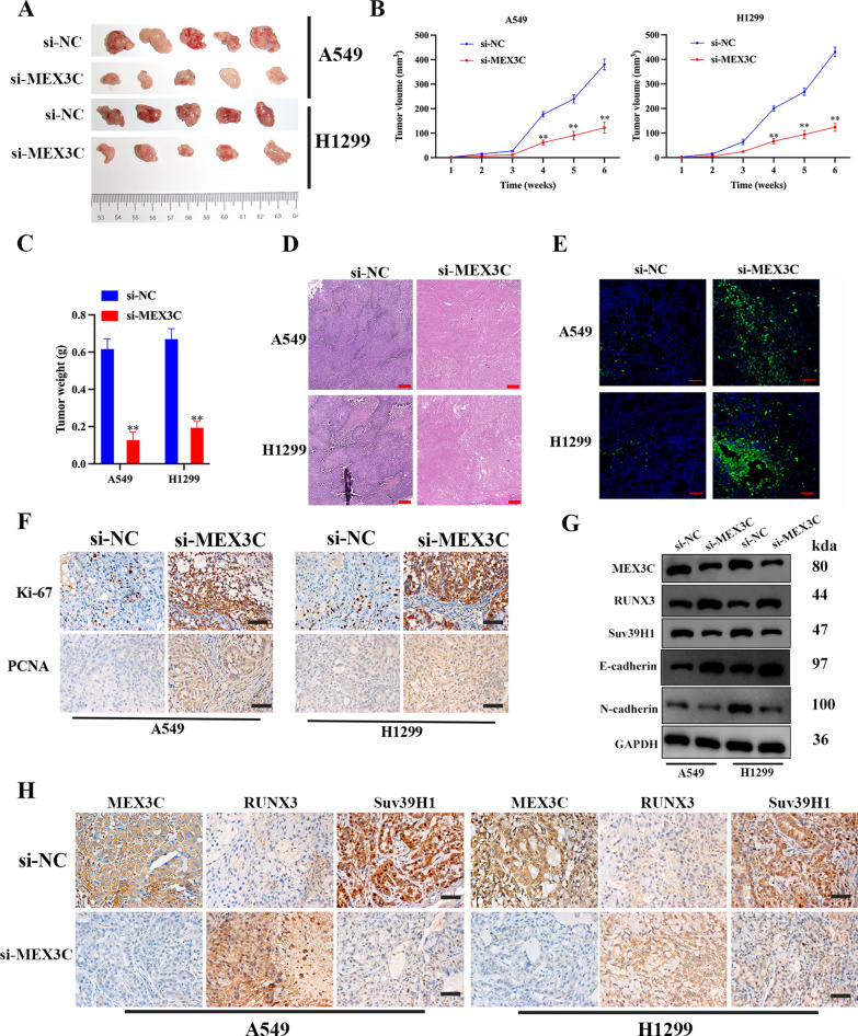
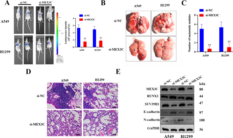
Lung adenocarcinoma (LUAD) is the most common pathological type of lung cancer, but the early diagnosis rate is low. The RNA-binding ubiquitin ligase MEX3C promotes tumorigenesis in several cancers but its mechanism of action in LUAD is unclear. In this study, the biological activity of MEX3C was assessed in LUAD. MEX3C and RUNX3 mRNA levels in the tissues of LUAD patients were determined using reverse transcription‑quantitative PCR. The involvement of MEX3C in the growth and metastasis of LUAD cells was measured by EdU assay, CCK-8, colony formation, Transwell assay, TUNEL, and flow cytometry. Expression of apoptosis and epithelial-mesenchymal transition related proteins were determined using western blotting analysis. LUAD cells transfected with si-MEX3C were administered to mice subcutaneously to monitor tumor progression and metastasis. We found that MEX3C is strongly upregulated in LUAD tissue sections, and involved in proliferation and migration. A549 and H1299 cells had significantly higher levels of MEX3C expression compared to control HBE cells. Knockdown of MEX3C dramatically decreased cell proliferation, migration, and invasion, and accelerated apoptosis. Mechanistically, we demonstrate MEX3C induces ubiquitylation and degradation of tumor suppressor RUNX3. Moreover, RUNX3 transcriptionally represses Suv39H1, as revealed by RNA pull-down and chromatin immunoprecipitation assays. The in vivo mice model demonstrated that knockdown of MEX3C reduced LUAD growth and metastasis significantly. Collectively, we reveal a novel MEX3C-RUNX3-Suv39H1 signaling axis driving LUAD pathogenesis. Targeting MEX3C may represent a promising therapeutic strategy against LUAD.

Keywords: LUAD; MEX3C; Metastasis; RUNX3; Suv39H1; Tumor growth.


Conflict of interest statement

None to declare.

Figures

Fig. 1
Fig. 1
Upregulation of MEX3C and downregulation of RUNX3 in LUAD. A The expression distribution of MEX3C gene in 24 type of tumor tissues and normal tissues. The abscissa represents different tumor tissues, and the ordinate represents the expression distribution of gene. B MEX3C and RUNX3 mRNA expression of tumor and normal tissues in LUAD datasets were analyzed using the TCGA + GTEx databases. C, D qRT-PCR was used to detected the MEX3C and RUNX3 mRNA levels in LUAD tissues and paired non-tumor tissues, n = 55. E, F MEX3C and RUNX3 mRNA expression in LUAD tissues at different stages and healthy subjects were detected using qRT-PCR. G Relative MEX3C mRNA levels were negatively correlated with relative RUNX3 mRNA levels in the LUAD tissues (r = − 0.6151, p < 0.0001). H Protein levels of MEX3C and RUNX3 in 3 pairs of clinical lung cancer specimens detected by western blotting. T and N indicate lung cancer tissue and paired adjacent normal tissue, respectively, n = 4. I Correlation analysis of MEX3C with CEA, SCCAg, Ki-67, and PCNA mRNA levels in the LUAD tissues via Pearson analysis. J H&E and IHC staining of MEX3C and RUNX3 in 3 pairs of NSCLC and adjacent samples, n = 3. Quantification of IHC staining. K, L Prognostic analysis of MEX3C and RUNX3 for LUAD. Bars represent mean ± SD. *P < 0.05; **P < 0.01
Fig. 2
Fig. 2
Knockdown of MEX3C in LUAD cells suppresses migration and invasion in vitro. A MEX3C mRNA and protein expression in human LUAD cell lines (A549, H1650, H838, and H1299) and a normal human bronchial epithelial cell-line HBE were analyzed by qRT-PCR and Western blotting, respectively. B The transcription of MEX3C in A549 and H1299 cells transfected with si-NC lentivirus or si-MEX3C lentivirus for 48 h was detected by qRT-PCR and western blotting analysis. C Cell migration and invasion of A549 and H1299 cells treated with or without knockdown of MEX3C were evaluated by Transwell assay. D A wound healing assay was used to assess the cell migration ability of A549 and H1299 cells treated with si-NC or si-MEX3C for 24 h. E EMT-related proteins E-cadherin and N-cadherin expression were detected by western blotting analysis. F Knockdown E-cadherin in A549 and H1299 cells using siRNA, E-cadherin expression was detected by western blotting. G The knockdown of E-cadherin was able to reverse the effect of MEX3C suppression on the migration and invasion of LUAD cell lines. Bars represent mean ± SD from 3 independent experiments. *P < 0.05, **P < 0.01
Fig. 3
Fig. 3
Knockdown of MEX3C promotes apoptosis and inhibits proliferation in LUAD cells. A Colony formation was used to evaluated proliferation for A549 and H1299 cells with or without MEX3C knockdown. B Proliferation of A549 and H1299 cells was detected using an Edu assay, scale bar, 50 μm. C TUNEL staining was used to determine the potential impact of MEX3C knockdown on apoptosis in A549 and H1299 cells, scale bar, 50 μm. D Flow cytometry analysis was performed to measure the percentage of apoptotic cells in A549 and H1299 cells after indicated treatment. E The expression levels of apoptosis-related proteins (Bcl-2, Bax, and Cleaved-Caspase-3) in MEX3C-silencing A549 and H1299 cells were detected by western blot. F Expression of Cleaved-Caspase-3 in A549/H1299 cells as analyzed by immunofluorescence assay, scale bar, 20 μm. Bars represent mean ± SD from 3 independent experiments. **P < 0.01
Fig. 4
Fig. 4
MEX3C mediated RUNX3 ubiquitination and degradation. A, B Transduction of A549 and H1299 cells with overexpressed MEX3C (OE-MEX3C) or si-MEX3C. Overexpression efficiency of MEX3C in A549 and H1299 cells using qRT-PCR (A) and western blotting (B). C, D By using qRT-PCR and western blotting, the mRNA (C) and protein (D) levels of RUNX3 were determined in MEX3C knockdown or overexpression A549 and H1299 cells. E A549 and H1299 cells were transduced with OE-MEX3C and subsequently grown with MG132 at a concentration of 10 μmol/L for 24 h. The presence of RUNX3 protein was determined using western blotting. F, G The interaction between MEX3C and RUNX3 was detected by Co-IP assay. H In vivo ubiquitination assay of HEK293 cells transfected with plasmids expressing Flag-tagged RUNX3, Myc-tagged MEX3C, and HA-tagged ubiquitin. I A549 and H1299 cells were transduced with OE-MEX3C. The ubiquitination level of RUNX3 was determined by ubiquitination assays. Bars represent mean ± SD from 3 independent experiments. **P < 0.01. ns, no significance
Fig. 5
Fig. 5
The effects of MEX3C suppression on LUAD cells could be reversed by inhibiting the expression of RUNX3. A, B Colony formation was evaluated for A549 and H1299 cells treated with si-MEX3C and/or si-RUNX3. C–E Representative images showing the results of Transwell cell migration and invasion assays of A549 and H1299 cells treated with si-MEX3C and/or si-RUNX3, scale bar, 50 μm. F Detection of proliferation in A549 and H1299 cells using an EdU assay, scale bar, 50 μm. G Flow cytometry assay to detect the cell apoptosis of A549 and H1299 cells treated with si-MEX3C and/or si-RUNX3. H Apoptosis and EMT-related protein expression were detected by western blot analysis. I Expression of Cleaved-Caspase-3 in A549/H1299 cells as analyzed by immunofluorescence assay, scale bar, 20 μm. Bars represent mean ± SD from 3 independent experiments. **P < 0.01
Fig. 6
Fig. 6
RUNX3 transcriptionally downregulates Suv39H1 expression. A Suv39H1 mRNA levels in 55 LUAD tissues and paired noncancerous lung tissues were detected by qRT-PCR. B Protein expression of Suv39H1 in 4 pairs of clinical LUAD specimens. N and T mean adjacent normal tissue and paired lung cancer tissue, respectively. C qRT-PCR and western blot analysis of Suv39H1 expression in HBE and LUAD cell lines. D qRT-PCR analysis of Suv39H1 expression in A549 and H1299 cells after MEX3C knockdown. E Western blot analysis of MEX3C, RUNX3, and Suv39H1 expression in A549 and H1299 cells transfected with si-MEX3C and/or si-RUNX3. F, G The binding motif of RUNX3 was obtained from JASPAR dataset. Two binding sites for RUNX3 in the Suv39H1 promoter were also predicted by using JASPAR. H Assessment of the Suv39H1 promoter RUNX3 binding site via dual luciferase reporter assay. I RNA pull-down assay was performed to determine whether RUNX3 targets Suv39H1. J ChIP assessment of RUNX3 binding to the Suv39H1 promoter. Immunoprecipitation from A549 and H1299 cells using RUNX3 antibody or mouse immunoglobulin G (IgG). Bars represent mean ± SD from 3 independent experiments. *P < 0.05, **P < 0.01
Fig. 7
Fig. 7
MEX3C promotes tumor growth in vivo. A Animal vivo images of MEX3C silencing A549 and H1299 xenograft nude mice (GFP fluorescent images) show that MEX3C silencing reduces the growth of xenografts (n = 5). A Images of A549 and H1299 xenograft tumors. B, C The histograms of tumor volumes and weight revealed that A549 and H1299 cells with MEX3C inhibited generated smaller xenografts when compared to the control cells. D The morphology of tumor was determined by H&E staining (scale bar, 200 μm) (E). Apoptosis ratio was determined by TUNEL assay (scale bar, 200 μm). F Immunohistochemistry was used to determine and compare the expression levels of Ki-67 and PCNA between the si-NC and si-MEX3C groups. Scale bar, 100 µm. G, H Western blot analysis and Immunohistochemistry were used to determine the protein levels of MEX3C, RUNX3 and Suv39H1 as well as EMT-related proteins in tumors of mice models of si-NC and si-MEX3C groups. Data were expressed as mean ± SD, n = 5, **P < 0.01
Fig. 8
Fig. 8
MEX3C accelerates lung cancer metastasis in vivo. A Nude mice were injected subcutaneously with A549 and H1299 cells expressing either si-NC or si-MEX3C. The tumor metastasis was detected by using IVIS® bioluminescence imaging system 8 weeks after injection. Quantitative data of tumor metastases detected by IVIS system. B, C The lungs were removed and imaged 8 weeks after injection via the tail vein. The number of metastatic nodules in the lungs was counted. D The H&E staining of lung tissues from different group of mice, scale bar, 50 μm. E Western blot analysis was performed to detect the protein expression of MEX3C, RUNX3, and Suv39H1, E-cadherin and N-cadherin. Data were expressed as mean ± SD, n = 5, **P < 0.01

Similar articles

Cited by

References

    1. Ferlay J, Colombet M, Soerjomataram I, Parkin DM, Piñeros M, Znaor A, et al. Cancer statistics for the year 2020: an overview. Int J Cancer. 2021 doi: 10.1002/ijc.33588. - DOI - PubMed
    1. Šutić M, Vukić A, Baranašić J, Försti A, Džubur F, Samaržija M, et al. Diagnostic, predictive, and prognostic biomarkers in non-small cell lung cancer (NSCLC) management. J Pers Med. 2021 doi: 10.3390/jpm11111102. - DOI - PMC - PubMed
    1. Ahern E, Solomon BJ, Hui R, Pavlakis N, O'Byrne K, Hughes BGM. Neoadjuvant immunotherapy for non-small cell lung cancer: right drugs, right patient, right time? J Immunother Cancer. 2021 doi: 10.1136/jitc-2020-002248. - DOI - PMC - PubMed
    1. Majeed U, Manochakian R, Zhao Y, Lou Y. Targeted therapy in advanced non-small cell lung cancer: current advances and future trends. J Hematol Oncol. 2021;14:108. doi: 10.1186/s13045-021-01121-2. - DOI - PMC - PubMed
    1. Schegoleva AA, Khozyainova AA, Fedorov AA, Gerashchenko TS, Rodionov EO, Topolnitsky EB, et al. Prognosis of different types of non-small cell lung cancer progression: current state and perspectives. Cell Physiol Biochem. 2021;55:29–48. doi: 10.33594/000000340. - DOI - PubMed

Publication types

MeSH terms