

Anaplastic Lymphoma Kinase (ALK)-Negative Anaplastic Large Cell Non-Hodgkin Lymphoma as a Rare Differential Diagnosis of Lung Cancer: A Case Report
- PMID: 38425329
- PMCID: PMC10904285
- DOI: 10.7759/cureus.55258
Anaplastic Lymphoma Kinase (ALK)-Negative Anaplastic Large Cell Non-Hodgkin Lymphoma as a Rare Differential Diagnosis of Lung Cancer: A Case Report
Abstract
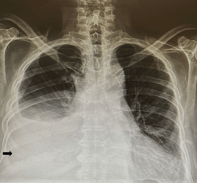
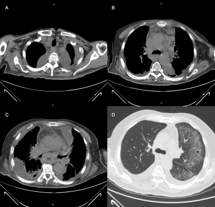
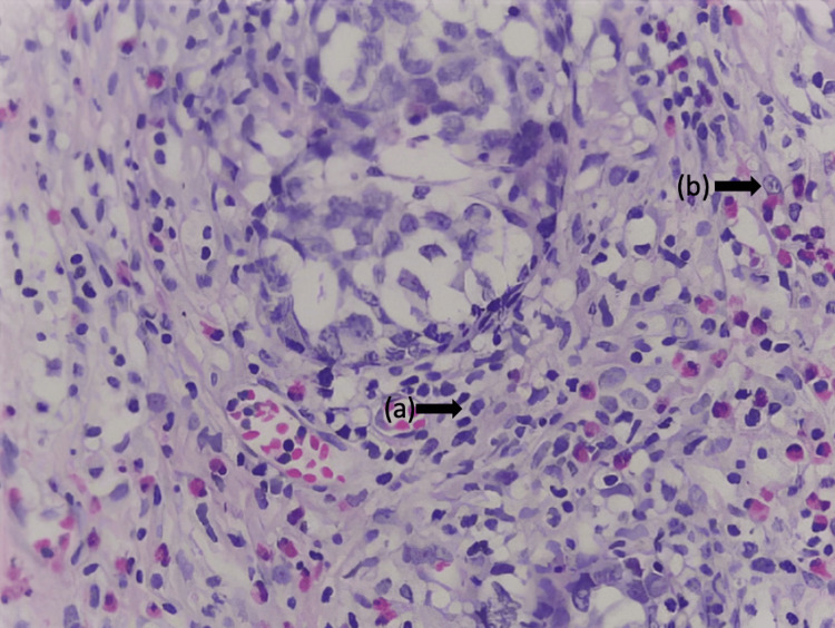
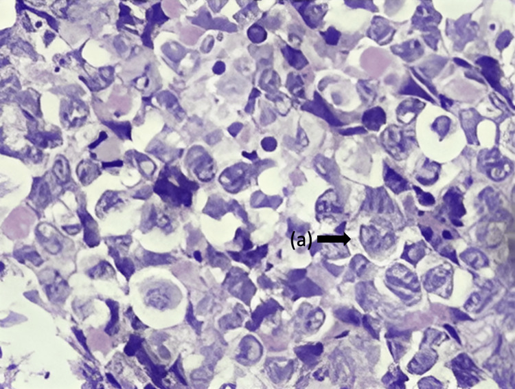
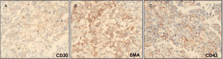
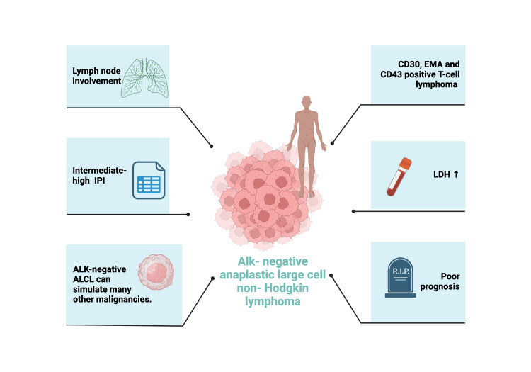
Anaplastic large cell lymphomas (ALCL) are a group of sporadic malignancies that generally have an aggressive clinical course, especially the subtype of anaplastic lymphoma kinase (ALK)-negative ALCL. The appropriate diagnostic study modalities must be chosen to make an accurate diagnosis and promptly initiate specific treatment. We present the clinical case of a 72-year-old male patient with dyspnea on small efforts accompanied by diaphoresis and a weight loss of 10 kg in two months. Physical examination revealed adenopathy in the cervical region and bilateral pleural effusion. The pleural and lung biopsies revealed poorly differentiated metastatic adenocarcinomas. A multidisciplinary analysis was carried out; the typical clinical-radiographic presentation of adenocarcinoma was ruled out with immunohistochemistry, thus determining a diagnosis of ALK-negative anaplastic large cell non-Hodgkin's lymphoma. This case represented a diagnostic and therapeutic challenge since it is a rare entity with a poor prognosis, and there are only a few studies about the choice of appropriate chemotherapy in these patients.
Keywords: anaplastic large cell lymphoma (alcl); anaplastic lymphoma kinase (alk); cd30; mature t-cell lymphoma; peripheral t-cell lymphoma; poorly differentiated lung adenocarcinoma.
Copyright © 2024, Llamas Domínguez et al.
Conflict of interest statement
The authors have declared that no competing interests exist.
Figures
Similar articles
-
A challenging case of ALK-negative anaplastic large cell lymphoma in a 12-year-old boy: A rare case report from Syria.Ann Med Surg (Lond). 2022 Jun 25;79:104085. doi: 10.1016/j.amsu.2022.104085. eCollection 2022 Jul. Ann Med Surg (Lond). 2022. PMID: 35860076 Free PMC article.
-
Complete response in a critically ill patient with ALK-negative anaplastic large cell lymphoma treated with single agent brentuximab-vedotin.Expert Rev Anticancer Ther. 2016;16(3):279-83. doi: 10.1586/14737140.2016.1146597. Epub 2016 Feb 18. Expert Rev Anticancer Ther. 2016. PMID: 26809026
-
Analysis of the t(2;5)(p23;q35) translocation by reverse transcription-polymerase chain reaction in CD30+ anaplastic large-cell lymphomas, in other non-Hodgkin's lymphomas of T-cell phenotype, and in Hodgkin's disease.Blood. 1995 Sep 15;86(6):2321-8. Blood. 1995. PMID: 7662979
-
Pathobiology of NPM-ALK and variant fusion genes in anaplastic large cell lymphoma and other lymphomas.Leukemia. 2000 Sep;14(9):1533-59. doi: 10.1038/sj.leu.2401878. Leukemia. 2000. PMID: 10994999 Review.
-
Anaplastic large cell lymphoma, ALK-negative.Crit Rev Oncol Hematol. 2013 Feb;85(2):206-15. doi: 10.1016/j.critrevonc.2012.06.004. Epub 2012 Jul 11. Crit Rev Oncol Hematol. 2013. PMID: 22789917 Review.
References
-
- Primary mediastinal nodal and extranodal non-Hodgkin lymphomas: current concepts, historical evolution, and useful diagnostic approach: part 1. Piña-Oviedo S, Moran CA. Adv Anat Pathol. 2019;26:346–370. - PubMed
-
- Anaplastic large-cell lymphomas in adults: the cytogenetic and molecular genetic characteristics, treatment results (a single institutional experience) Castellar ERP, Jaffe ES, Said JW, et al. Blood. 2017;130:5129.
Publication types
LinkOut - more resources
Full Text Sources