. 2024 Feb 28:12:tkad025.
doi: 10.1093/burnst/tkad025. eCollection 2024.

Matrix metalloproteinase-8 regulates dendritic cell tolerance in late polymicrobial sepsis via the nuclear factor kappa-B p65/β-catenin pathway

Affiliations

Matrix metalloproteinase-8 regulates dendritic cell tolerance in late polymicrobial sepsis via the nuclear factor kappa-B p65/β-catenin pathway

Zhong-Qiu Lu et al. Burns Trauma. .

Abstract

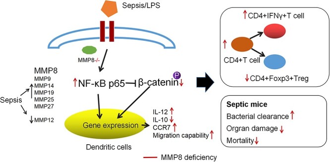
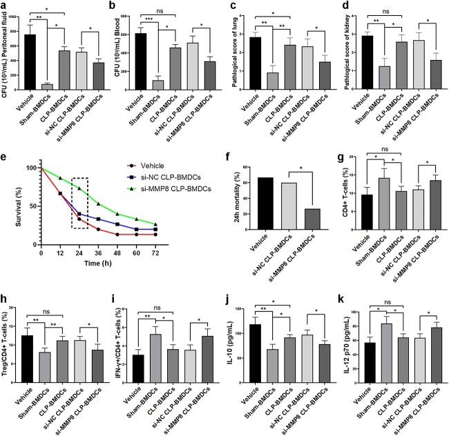
Background: Tolerogenic dendritic cells (DCs) are associated with poor prognosis of sepsis. Matrix metalloproteinases (MMPs) have been shown to have immunomodulatory effects. However, whether MMPs are involved in the functional reprogramming of DCs is unknown. The study aims to investigate the role of MMPs in sepsis-induced DCs tolerance and the potential mechanisms.

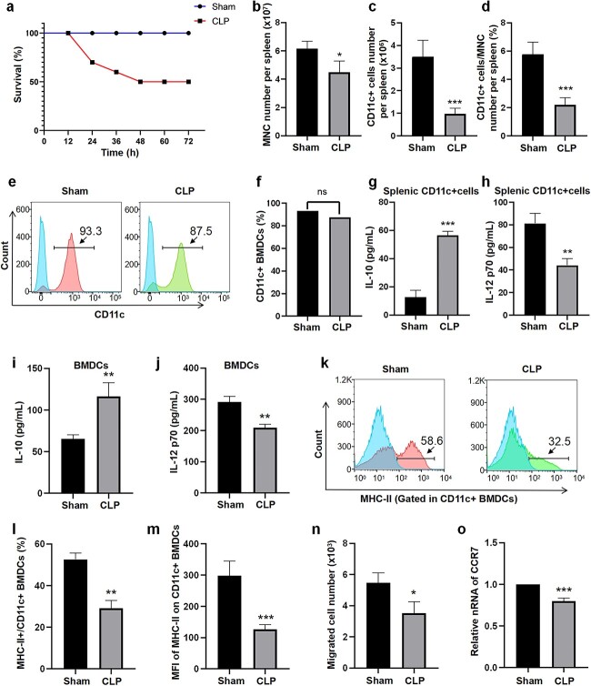
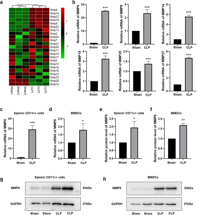
Methods: A murine model of late sepsis was induced by cecal ligation and puncture (CLP). The expression levels of members of the MMP family were detected in sepsis-induced tolerogenic DCs by using microarray assessment. The potential roles and mechanisms underlying MMP8 in the differentiation, maturation and functional reprogramming of DCs during late sepsis were assessed both in vitro and in vivo.

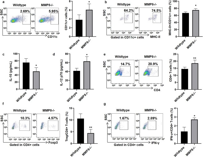
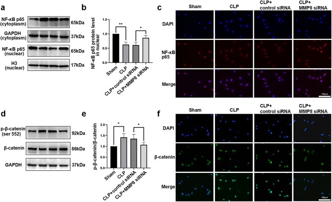
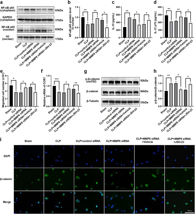
Results: DCs from late septic mice expressed higher levels of MMP8, MMP9, MMP14, MMP19, MMP25 and MMP27, and MMP8 levels were the highest. MMP8 deficiency significantly alleviated sepsis-induced immune tolerance of DCs both in vivo and in vitro. Adoptive transfer of MMP8 knockdown post-septic bone marrow-derived DCs protected mice against sepsis-associated lethality and organ dysfunction, inhibited regulatory T-cell expansion and enhanced Th1 response. Furthermore, the effect of MMP8 on DC tolerance was found to be associated with the nuclear factor kappa-B p65/β-catenin pathway.

Conclusions: Increased MMP8 levels in septic DCs might serve as a negative feedback loop, thereby suppressing the proinflammatory response and inducing DC tolerance.

Keywords: Dendritic cells; Immune tolerance; Matrix metalloproteinases; Nuclear factor kappa-B; Sepsis; Signaling pathway; β-catenin.


Conflict of interest statement

None declared.

Figures

Graphical Abstract
Graphical Abstract
Figure 1
Figure 1
Phenotype and function of splenic and bone marrow (BM)-derived dendritic cells (DCs) in septic mice. (a) Mortality in the different experimental groups (10 mice/group) was recorded every 12 h. (b, c) The absolute numbers of mononuclear cells (MNCs) and CD11c-positive cells per spleen. (d) The percentages of CD11c-positive cells in MNCs per spleen. (e, f) Representative flow cytometric analysis was performed to determine the percentages of CD11c-positive cells in BM-derived cells. Cytokine production (IL-10 and IL-12p70) by splenic CD11c-positive cells (g, h) and BMDCs (i, j) was measured following 24 h of treatment with LPS. (k, l) Representative flow cytometric analysis was performed to determine the percentages of MHCII+ cells among BMDCs. (m) The median fluorescence intensity (MFI) of MHC-II on BMDCs. (n) The number of migrated BM-derived CD11c-positive cells was determined by flow cytometry. (o) The mRNA levels of CCR7 were tested by RT-PCR. Data are shown as the mean ± SD (n = 3); ***p < 0.001, **p < 0.01 and *p < 0.05; ns, not significant. CLP cecal ligation and puncture, BMDCs bone marrow derived dendritic cells, CCR7 chemokine C-C-motif receptor 7, MHC major histocompatibility complex class, RT-PCR reverse transcription-polymerase chain reaction
Figure 2
Figure 2
Expression patterns of MMPs in tolerogenic DCs during late sepsis. (a) Heatmaps depicting the expression levels of MMPs in splenic DCs from septic and sham mice. (b) Histogram depicting the levels of MMPs that increased in splenic DCs 3 days post-CLP. The mRNA levels of MMP8 in splenic CD11c-positive cells (c) and BMDCs (d). Representative immunoblots and densitometric values of MMP8 in splenic CD11c-positive cells (e, g) and BMDCs (f, h) are shown; β-actin was used as the loading control. n = 3, ***p < 0.001, **p < 0.01 and *p < 0.05. CLP cecal ligation and puncture, MMP matrix metalloproteinase, BMDCs bone marrow-derived dendritic cell, MHC major histocompatibility complex class
Figure 3
Figure 3
MMP8 knockdown on sepsis-induced immune tolerance of DCs in vitro. (a, b) Western blot analysis was used to determine the expression levels of MMP8 in BMDCs. (c) The mRNA levels of MMP8 in BMDCs were determined by RT-PCR. (d, e) Representative flow cytometric analysis was performed to determine the percentage of MHCII+ cells among CD11c-positive BMDCs. (f) The median fluorescence intensity (MFI) of MHC-II on CD11c-positive BMDCs. IL-10 (g) and IL-12p70 (h) levels in the supernatants of BMDCs were detected by ELISA. (i) The number of migrated CD11c-positive BMDCs was measured by flow cytometry. (j) mRNA levels of CCR7 were determined by RT-PCR. Data are shown as the mean ± SD (n = 3); ***p < 0.001, **p < 0.01 and *p < 0.05. CLP cecal ligation and puncture, MMP8 matrix metalloproteinase 8, BMDCs bone marrow-derived dendritic cell, MHC major histocompatibility complex class, siRNA small interfering RNA, CCR7 chemokine C-C-motif receptor 7
Figure 4
Figure 4
MMP8 deficiency alleviates sepsis-induced immune tolerance of DCs and Treg expansion in a ‘two-hit’ mouse model. The percentages of CD11c+ (a) and CD11c+MHCII+ (b) cells in the spleen were determined by flow cytometric analysis. The protein levels of IL-10 (c) and IL-12p70 (d) in the plasma were tested by ELISA. Flow cytometric analysis was used to assess the percentages of CD4+ (e), CD4+Foxp3+ (f) and CD4+IFN-γ+ (g) T cells in the spleen. n = 6, **p < 0.01 and *p < 0.05. CLP cecal ligation and puncture, MMP8 matrix metalloproteinase 8, IL interleukin, IFN interferon, Foxp3 forkhead box P3, MHC major histocompatibility complex class
Figure 5
Figure 5
Adoptive transfer of post-septic BMDCs transfected with MMP8 siRNA protects mice from fatal outcomes. Peritoneal fluid (a) and peripheral blood (b) at 24 h after CLP were collected for culturing. The number of bacterial colonies was counted (n = 3). Pathological score of lung (c) and renal tissues (d) in each group (n = 6). (e) Mortality in the different groups (15 mice/group) was recorded every 12 h. (f) Mortality differences between the groups were compared at the 24-h time point after surgery. Flow cytometric analysis was performed to determine the percentages of CD4+ (g), CD4+Foxp3+ (h) and CD4+IFN-γ+ (i) T cells in the spleen. The levels of IL-10 (j) and IL-12p70 (k) in the plasma were measured by ELISA. Data are expressed as the mean ± SD (n = 5); **p < 0.01 and *p < 0.05; ns, not significant. CLP cecal ligation and puncture, BMDCs bone marrow-derived dendritic cells, MMP8 matrix metalloproteinase 8, si-NC negative control small interfering RNA (siRNA), si-MMP8 MMP8 small interfering RNA (siRNA), MMP8 matrix metalloproteinase 8, Treg regulatory T cell, IL interleukin, IFN-γ interferon-γ, ELISA enzyme-linked immunosorbent assay
Figure 6
Figure 6
MMP8 knockdown enhances NF-kB p65 nuclear translocation and suppresses β-catenin activity in BMDCs. Western blotting was used to detect the nuclear and cytoplasmic protein levels of NF-κB p65 (a) and phospho-β-catenin (d) in BMDCs. The relative nuclear protein levels of NF-κB p65 (b) and ratios of β-catenin to total β-catenin (e) are shown (n = 4 or 5). Nuclear translocation of p65 (c) and β-catenin (f) was analyzed by immunofluorescence (n = 3). Scale bar: 100 µm. Data are shown as the mean ± SD; **p < 0.01 and *p < 0.05. CLP cecal ligation and puncture, MMP8 matrix metalloproteinase 8, siRNA small interfering RNA, NF-κB nuclear factor kappa-B, GAPDH glyceraldehyde-3-phosphate dehydrogenase, H3 histone 3
Figure 7
Figure 7
Inhibition of NF-kB suppresses the function of DCs and promotes β-catenin activation in response to LPS after MMP8 knockdown. (a, b) Western blotting was used to determine nuclear and cytoplasmic protein levels of p65. ELISA was performed to test the levels of IL-10 (c) and IL-12p70 (d) secreted by CD11c + BMDCs. (e) The number of migrated CD11c-positive BMDCs was assessed by flow cytometry. (f) mRNA levels of CCR7 were determined by RT–PCR. (g, h) The protein levels of phospho-β-catenin were measured by western blotting. (i) Nuclear translocation of β-catenin analyzed by immunofluorescence. Scale bar: 100 µm. Data are shown as the mean ± SD, n = 3; ***p < 0.001, **p < 0.01 and *p < 0.05. CLP cecal ligation and puncture, MMP8 matrix metalloproteinase 8, siRNA small interfering RNA, NF-κB nuclear factor kappa-B, GAPDH glyceraldehyde-3-phosphate dehydrogenase, H3 histone 3, IL interleukin, CCR7 chemokine C-C-motif receptor 7, LPS lipopolysaccharide

Similar articles

Cited by

References

    1. Wang LX, Ren C, Yao RQ, Luo YN, Yin Y, Wu Y, et al. . Sestrin2 protects against lethal sepsis by suppressing the pyroptosis of dendritic cells. Cell Mol Life Sci. 2021;78:8209–27. - PMC - PubMed
    1. Zhang Y, Chen L, Luo Y, Wang K, Liu X, Xiao Z, et al. . Pink1/parkin-mediated Mitophagy regulated the apoptosis of dendritic cells in sepsis. Inflammation. 2022;45:1374–87. - PubMed
    1. Delano MJ, Ward PA. Sepsis-induced immune dysfunction: can immune therapies reduce mortality? J Clin Invest. 2016;126:23–31. - PMC - PubMed
    1. Bouras M, Asehnoune K, Roquilly A. Contribution of dendritic cell responses to sepsis-induced immunosuppression and to susceptibility to secondary pneumonia. Front Immunol. 2018;9:2590. - PMC - PubMed
    1. Venet F, Demaret J, Gossez M, Monneret G. Myeloid cells in sepsis-acquired immunodeficiency. Ann N Y Acad Sci. 2021;1499:3–17. - PubMed