Review
. 2024 Feb 15:12:1342905.
doi: 10.3389/fcell.2024.1342905. eCollection 2024.

How great thou ART: biomechanical properties of oocytes and embryos as indicators of quality in assisted reproductive technologies

Affiliations
Review

How great thou ART: biomechanical properties of oocytes and embryos as indicators of quality in assisted reproductive technologies

Monika Fluks et al. Front Cell Dev Biol. .

Abstract

Assisted Reproductive Technologies (ART) have revolutionized infertility treatment and animal breeding, but their success largely depends on selecting high-quality oocytes for fertilization and embryos for transfer. During preimplantation development, embryos undergo complex morphogenetic processes, such as compaction and cavitation, driven by cellular forces dependent on cytoskeletal dynamics and cell-cell interactions. These processes are pivotal in dictating an embryo's capacity to implant and progress to full-term development. Hence, a comprehensive grasp of the biomechanical attributes characterizing healthy oocytes and embryos is essential for selecting those with higher developmental potential. Various noninvasive techniques have emerged as valuable tools for assessing biomechanical properties without disturbing the oocyte or embryo physiological state, including morphokinetics, analysis of cytoplasmic movement velocity, or quantification of cortical tension and elasticity using microaspiration. By shedding light on the cytoskeletal processes involved in chromosome segregation, cytokinesis, cellular trafficking, and cell adhesion, underlying oogenesis, and embryonic development, this review explores the significance of embryo biomechanics in ART and its potential implications for improving clinical IVF outcomes, offering valuable insights and research directions to enhance oocyte and embryo selection procedures.

Keywords: assisted reproductive technologies; biomechanics; cytoskeleton; embryo; mouse; oocyte; preimplantation development; quality assessment.


Conflict of interest statement

AA is a co-inventor in the patent “Methods for predicting mammalian embryo viability” (patent no. US 9 410 939 B2) on the application of cytoplasmic movement analysis in evaluation of embryo quality. The remaining authors declare that the research was conducted in the absence of any commercial or financial relationships that could be construed as a potential conflict of interest.

Figures
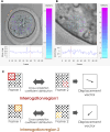
FIGURE 1
FIGURE 1
Cytoplasmic movement velocity (CMV) assessment by Particle Image Velocimetry (PIV). (A, B) Images from the PIV software used by some of the authors (Ajduk et al., 2011). PIV analysis was conducted for high-resolution time-lapse images of (A) mouse metaphase II oocyte, (B) polar trophectoderm cell in a mouse blastocyst. Both the length and the color of the vectors visible inside the cells reflect the cytoplasmic velocity. Cyan represents the slowest-moving vectors. Magenta represents the fastest-moving vectors. The graphs (below) show the mean cytoplasmic velocity in the analyzed region over time. The direction of the vectors indicates the direction of cytoplasm displacement between frames. (C) Schematic representation of the PIV algorithm. The algorithm divides the images into small interrogation windows, identifying the pattern of pixels in each window, and calculating the displacement of particles between frames. This information is used to generate a map of velocity vectors representing the cytoplasmic flow and to calculate mean cytoplasmic velocity.
FIGURE 2
FIGURE 2
Cortical tension (CT, γ) analysis. CT analysis can be conducted by micropipette aspiration. Assessment of CT requires the measurement of cell curvature radius and aspiration pressure (Ps) when the deformation length (L) becomes equal to the micropipette radius (Rp) and utilizes the Young–Laplace equation.
FIGURE 3
FIGURE 3
Atomic force spectroscopy. (A) Atomic force spectroscopy setup diagram. Details in the main text. (B) Force-distance curve analysis to measure the viscoelastic properties of a material. 1) The cantilever tip approaches the sample until it makes contact, and the force at the interaction between the tip and the sample is measured; 2) the tip is further compressed into the sample, deforming its surface, the forces acting on the tip during compression are recorded, allowing for calculation of the surface stiffness; 3) the tip is retracted from the sample surface. Forces acting on the tip as it moves away from the sample are measured allowing for calculation of surface adhesion.
FIGURE 4
FIGURE 4
Dynamics of blastocyst cavitation. The equatorial area of blastocysts (dashed line) is measured at (A) the onset of cavitation, (B) the time-point of maximum expansion just before the first contraction, (C) the last phase of the first contraction, (D) the time-point of maximum expansion before the next contraction, (E) the last phase of the contraction. Time points (hh:mm) indicate the time of imaging and correspond to the graph below (F). (F) A graph representing oscillations of the blastocyst size over time. The arrows indicate the time points corresponding to the measurements in (A–E).

Similar articles

Cited by

References

    1. Aghigh A., Bancelin S., Rivard M., Pinsard M., Ibrahim H., Légaré F. (2023). Second harmonic generation microscopy: a powerful tool for bio-imaging. Biophys. Rev. 15, 43–70. 10.1007/S12551-022-01041-6 - DOI - PMC - PubMed
    1. Ajduk A., Ilozue T., Windsor S., Yu Y., Seres K. B., Bomphrey R. J., et al. (2011). Rhythmic actomyosin-driven contractions induced by sperm entry predict mammalian embryo viability. Nat. Commun. 2, 417–7. 10.1038/ncomms1424 - DOI - PMC - PubMed
    1. Ajduk A., Szkulmowski M. (2019). Light microscopy of mammalian gametes and embryos: methods and applications. Int. J. Dev. Biol. 63, 235–244. 10.1387/ijdb.180300aa - DOI - PubMed
    1. Ajduk A., Zernicka-Goetz M. (2013). Quality control of embryo development. Mol. Asp. Med. 34, 903–918. 10.1016/j.mam.2013.03.001 - DOI - PubMed
    1. Aksenov M. Y., Aksenova M. V., Butterfield D. A., Geddes J. W., Markesbery W. R. (2001). Protein oxidation in the brain in Alzheimer’s disease. Neuroscience 103, 373–383. 10.1016/S0306-4522(00)00580-7 - DOI - PubMed

LinkOut - more resources