

Optic nerve sheath diameter and its association with brain swelling in pediatric cerebral malaria: a retrospective study
- PMID: 38425660
- PMCID: PMC10902095
- DOI: 10.3389/fped.2024.1295254
Optic nerve sheath diameter and its association with brain swelling in pediatric cerebral malaria: a retrospective study
Abstract
Introduction: Mortality in pediatric cerebral malaria (CM) in low- and middle-income countries (LMICs) is associated with brain swelling on magnetic resonance imaging (MRI); however, MRI is unavailable in most LMICs. Optic nerve sheath diameter (ONSD) measurement is an inexpensive method of detecting increased intracranial pressure compared with the invasive opening pressure (OP). Our primary objective was to determine if increased ONSD correlated with brain swelling on MRI in pediatric CM. Our secondary objective was to determine if increased ONSD correlated with increased OP and/or poor neurological outcome in pediatric CM. We hypothesized that increased ONSD would correlate with brain swelling on MRI and increased OP and that ONSD would be higher in survivors with sequelae and non-survivors.
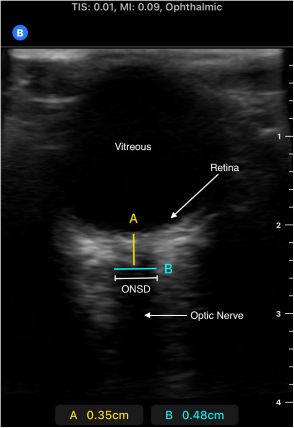
Methods: We performed a retrospective chart review of children aged 0-12 years in Blantyre, Malawi, from 2013 to 2022 with CM as defined by the World Health Organization. Brain swelling on admission MRI was characterized by brain volume scores (BVS); severe swelling was scored as 7-8, mild-to-moderate as 4-6, normal as 3. The admission ONSD was measured via ultrasound; it was defined as abnormal if it was >4.5 mm in children >1 year and >4 mm in children <1 year. Favorable outcome was defined as a normal neurological exam on discharge in survivors. The primary and secondary objectives were evaluated using Spearman's correlation; and the demographics were compared using chi-square and the Kruskal-Wallis test (Stata, College Station, TX, USA).
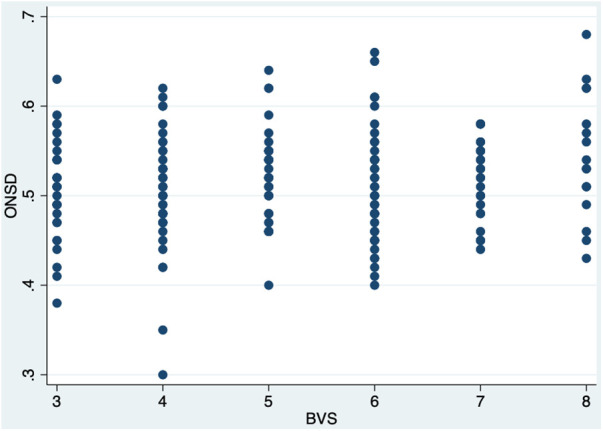
Results: Median age of the 207-patients cohort was 50 months [interquartile range (IQR) 35-75]; 49% (n = 102) were female. Of those, 73% (n = 152) had a favorable outcome, and 14% (n = 30) died. Twenty-nine (14%) had a normal BVS, 134 (65%) had mild-to-moderate swelling, and 44 (21%) had severe swelling. ONSD was elevated in 86% (n = 178) of patients, while 12% of patients had increased OP. There was a weakly positive correlation between BVS and ONSD (r = 0.14, p = 0.05). The median ONSD was not significantly different compared by discharge outcome (p = 0.11) or by BVS (p = 0.18).
Conclusion: ONSD was not a reliable tool to correlate with BVS, neurological outcome, or OP in children with CM. Future studies to identify alternative methods of early identification of CM patients at highest risk for morbidity and mortality are urgently needed.
Keywords: Africa South of the Sahara; brain edema; cerebral malaria; falciparum malaria; pediatrics; point-of-care technology.
© 2024 Raees, Gushu, Taylor, Seydel, Wynkoop and O'Brien.
Conflict of interest statement
The authors declare that the research was conducted in the absence of any commercial or financial relationships that could be construed as a potential conflict of interest.
Figures

Similar articles
-
Optic nerve sheath diameter and pulsatility index for the diagnosis and follow-up in pediatric traumatic brain injury: a prospective observational cohort study.Childs Nerv Syst. 2023 Sep;39(9):2467-2477. doi: 10.1007/s00381-023-05959-4. Epub 2023 Apr 26. Childs Nerv Syst. 2023. PMID: 37099137
-
Relationship between optic nerve sheath diameter measured by magnetic resonance imaging, intracranial pressure, and neurological outcome in cardiac arrest survivors who underwent targeted temperature management.Resuscitation. 2019 Dec;145:43-49. doi: 10.1016/j.resuscitation.2019.10.004. Epub 2019 Oct 16. Resuscitation. 2019. PMID: 31628979
-
Pediatric optic nerve and globe measurements on magnetic resonance imaging: establishing norms for children.Acta Radiol. 2023 Jun;64(6):2162-2169. doi: 10.1177/02841851231169176. Epub 2023 Apr 25. Acta Radiol. 2023. PMID: 37097831
-
The role of optic nerve sheath diameter ultrasound in brain infection.eNeurologicalSci. 2021 Feb 22;23:100330. doi: 10.1016/j.ensci.2021.100330. eCollection 2021 Jun. eNeurologicalSci. 2021. PMID: 33728383 Free PMC article. Review.
-
The magnetic resonance imaging assessment of optic nerve sheath diameter in pediatric acute liver failure patients.Eur Rev Med Pharmacol Sci. 2022 Feb;26(3):853-859. doi: 10.26355/eurrev_202202_27993. Eur Rev Med Pharmacol Sci. 2022. PMID: 35179751
Cited by
-
Point-of-care ultrasound use in austere environments: A scoping review.PLoS One. 2024 Dec 5;19(12):e0312017. doi: 10.1371/journal.pone.0312017. eCollection 2024. PLoS One. 2024. PMID: 39636834 Free PMC article.
References
-
- World Health Organization. Malaria. (nd). Available online at: https://www.who.int/news-room/fact-sheets/detail/malaria (accessed June 5, 2023).
LinkOut - more resources
Full Text Sources
Miscellaneous