. 2024 Feb 15:11:1295075.
doi: 10.3389/fbioe.2023.1295075. eCollection 2023.

Reconstruction of the human nipple-areolar complex: a tissue engineering approach

Affiliations

Reconstruction of the human nipple-areolar complex: a tissue engineering approach

Louis Maistriaux et al. Front Bioeng Biotechnol. .

Abstract

Introduction: Nipple-areolar complex (NAC) reconstruction after breast cancer surgery is challenging and does not always provide optimal long-term esthetic results. Therefore, generating a NAC using tissue engineering techniques, such as a decellularization-recellularization process, is an alternative option to recreate a specific 3D NAC morphological unit, which is then covered with an in vitro regenerated epidermis and, thereafter, skin-grafted on the reconstructed breast. Materials and methods: Human NACs were harvested from cadaveric donors and decellularized using sequential detergent baths. Cellular clearance and extracellular matrix (ECM) preservation were analyzed by histology, as well as by DNA, ECM proteins, growth factors, and residual sodium dodecyl sulfate (SDS) quantification. In vivo biocompatibility was evaluated 30 days after the subcutaneous implantation of native and decellularized human NACs in rats. In vitro scaffold cytocompatibility was assessed by static seeding of human fibroblasts on their hypodermal side for 7 days, while human keratinocytes were seeded on the scaffold epidermal side for 10 days by using the reconstructed human epidermis (RHE) technique to investigate the regeneration of a new epidermis. Results: The decellularized NAC showed a preserved 3D morphology and appeared white. After decellularization, a DNA reduction of 98.3% and the absence of nuclear and HLA staining in histological sections confirmed complete cellular clearance. The ECM architecture and main ECM proteins were preserved, associated with the detection and decrease in growth factors, while a very low amount of residual SDS was detected after decellularization. The decellularized scaffolds were in vivo biocompatible, fully revascularized, and did not induce the production of rat anti-human antibodies after 30 days of subcutaneous implantation. Scaffold in vitro cytocompatibility was confirmed by the increasing proliferation of seeded human fibroblasts during 7 days of culture, associated with a high number of living cells and a similar viability compared to the control cells after 7 days of static culture. Moreover, the RHE technique allowed us to recreate a keratinized pluristratified epithelium after 10 days of culture. Conclusion: Tissue engineering allowed us to create an acellular and biocompatible NAC with a preserved morphology, microarchitecture, and matrix proteins while maintaining their cell growth potential and ability to regenerate the skin epidermis. Thus, tissue engineering could provide a novel alternative to personalized and natural NAC reconstruction.

Keywords: ECM; decellularization; extracellular matrix; nipple–areolar complex; nipple–areolar complex reconstruction; recellularization; reconstructive surgery; tissue engineering.


Conflict of interest statement

The authors declare that the research was conducted in the absence of any commercial or financial relationships that could be construed as a potential conflict of interest. The authors declare that they were editorial board members of Frontiers at the time of submission. This had no impact on the peer review process and the final decision.

Figures
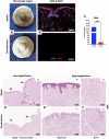
FIGURE 1
FIGURE 1
Human NAC decellularization and cell clearance. (A) Macroscopic aspect of the harvested native (top) and decellularized (bottom) NAC, which appears white and is entirely de-epithelialized. (B) DAPI (blue) and MHC class I + HLA A+ HLA B (red) immunofluorescence staining confirms the total decellularization by the absence of nuclei and MHC-I + HLA A + HLA B antigens in decellularized tissues (bottom) compared to the native tissues (top) (scale bar = 100 μm). (C) DNA quantification in native NAC (n-NAC) (n = 5) versus decellularized NAC (d-NAC) (n = 11) scaffolds shows a significant DNA reduction (98%) after decellularization. The DNA concentration is expressed in ng/mg dry weight. Error bars: SD; ****p < 0.001. (D–F) H&E-stained sections of n-NAC (top) and d-NAC (bottom) at low magnification (D) with the areolar part (AP) and nipple part (NP) of the NAC. High magnification of H&E-stained sections focused on the epidermis (E) and nervous ramifications (F) of native (top) and decellularized (bottom) NACs. Both magnifications confirm the complete decellularization (scale bar for D = 400 μm and for E–F = 50 μm).
FIGURE 2
FIGURE 2
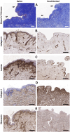
Extracellular matrix microarchitecture preservation. (A) Masson’s trichrome staining of native (left) and decellularized (right) NACs at low magnification, with the AP and NP, shows the preservation of the NAC microarchitecture and collagen fibers in decellularized tissues (scale bar = 100 μm). (B–E) Immunohistochemistry stainings of the main ECM proteins evaluating the preservation of type IV collagen (B), laminin (C), type I collagen (D), and fibronectin (E) in native tissues (left) compared with decellularized tissues (right) (scale bar = 200 μm).
FIGURE 3
FIGURE 3
Extracellular matrix component preservation. (A–C) Main ECM protein quantification in n-NACs and d-NACs. (A) Collagen quantification shows the preservation of collagen in d-NACs. (B) GAG and (C) elastin quantifications highlight a significant reduction in decellularized scaffolds compared to the native tissue. Collagen, GAG, and elastin concentrations are expressed in μg/mg dry weight. Error bars: SD; ****p < 0.001, ns = not significant. (D) Residual SDS quantification in d-NAC shows a significantly very low amount of SDS residues in acellular scaffolds after the last washing step of the decellularization protocol, confirming the non-toxicity of the decellularized scaffolds. Residual SDS is expressed in μg/mg dry weight. Error bar: SD; ****p < 0.001, n = 3. (E) Quantification of human growth factors: All 41 human GFs were detected before (n = 3) and after decellularization (n = 4), with a significant reduction in 38/41 GFs and an insignificant reduction in 3/41 GFs. The results are expressed as the mean density. Error bar: SD; ****p < 0.0001, ***p < 0.001, **p < 0.01, *p < 0.05, and ns = not significant.
FIGURE 4
FIGURE 4
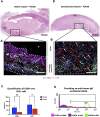
In vivo biocompatibility of decellularized NACs subcutaneously implanted in rats. (A) H&E-stained native NAC implant on POD30, surrounded by a thick vascularized purple circumferential PIC composed of immune cells and fibrosis with no implant infiltration. * = implant; scale bar = 400 μm. (B) H&E-stained decellularized NAC implant on POD30, with complete cell infiltration associated with a thin peripheral vascularized cell layer. * = implant; scale bar = 400 μm. (A’, B’) Multiplex immunofluorescence for CD31 (red), CD68 (purple), and CD3 (green) cells in n-NAC (A’) and d-NAC (B’), showing the formation of a thick peripheral immune cell layer without the penetration of the native implant compared to the complete infiltration of the decellularized scaffold by host cells. Moreover, vessels (CD31+, red staining) were found in the n-NAC peripheral layer without the penetration of the implants while they were around and infiltrating the entire thickness of the decellularized scaffolds after 30 days of implantation (scale bars = 50 μm, * = implant, dotted lines = delimitation of the implant, and white arrow = neo-vessels). (C) Quantification of positive CD68- and CD3-stained cells in n-NACs (blue) and d-NACs (red) on multiplex immunofluorescence on POD30. The results are expressed in the amount of positive stained cells (CD3+ or CD68+) per mm2. Error bars: SD; **p < 0.001; ns = not significant. (D) Flow cytometry of circulating rat anti-human IgG on POD30 showing total immunization after the implantation of native scaffolds (5/5) and the absence of IgG after the implantation of decellularized scaffolds (4/5). Image of flow cytometry is a summary picture of the assay: POD0, both tissues (rose); POD30, n-NACs (green); and POD30, d-NACs (purple).
FIGURE 5
FIGURE 5
Human fibroblast seeding on the hypodermal side of d-NACs. (A) H&E staining of adherent human fibroblasts (arrows) on the acellular hypodermal side and, in some locations, adherent to the epidermal side of d-NACs after 7 days of static culture (scale bar = 200 μm). (A’) Higher magnification of the H&E-stained section highlighting adherent fibroblasts forming several cell layers on the hypodermal side of the scaffold (scale bar = 100 μm). (B) Live/dead staining of seeded fibroblasts shows a high viability on day 7 of the culture on the scaffold (living cells = green and dead cells = red) (scale bar = 500 μm). (C) Cell viability of the seeded dermis: ECM- red and control wells- blue. The results are expressed as the mean cell viability. Error bars: SD; ns = not significant. (D) A PrestoBlue cell viability assay realized on seeded d-NACs (red, n = 3) and control culture wells (blue, n = 3) attests the biocompatibility of the produced scaffolds by the increase in metabolic activity during the 7 days of culture. The results are expressed as the mean fluorescence intensity. Error bars: SD; **p < 0.01; ns = not significant.
FIGURE 6
FIGURE 6
Regeneration of an epidermis using the RHE technique. (A) H&E staining of human keratinocytes seeded on the epidermal side of a d-NAC scaffold shows the formation of a stratified epithelium with a superficial squamous layer (arrow) using the RHE technique (scale bar = 200 μm). ((A), right insert) Higher magnification of the regenerated epidermis on d-NAC after 3 days of culture in the medium and then lifted onto an air–liquid interface for 7 days (arrow = desquamating layer) (scale bar = 100 μm). (B, C) Pancytokeratin immunofluorescence of the regenerated epidermis on different samples of discs from d-NACs (B in green and C in red), showing different thicknesses of RHE and confirming the expression of cutaneous keratin markers by seeded keratinocytes (scale bar = 200 μm).

Similar articles

Cited by

References

    1. Alizadeh M., Rezakhani L., Soleimannejad M., Sharifi E., Anjomshoa M., Alizadeh A. (2019). Evaluation of vacuum washing in the removal of SDS from decellularized bovine pericardium: method and device description. Heliyon 5, e02253. 10.1016/j.heliyon.2019.e02253 - DOI - PMC - PubMed
    1. Allman A. J., McPherson T. B., Badylak S. F., Merrill L. C., Kallakury B., Sheehan C., et al. (2001). Xenogeneic extracellular matrix grafts elicit a th2-restricted immune response. Transplantation 71, 1631–1640. 10.1097/00007890-200106150-00024 - DOI - PubMed
    1. Alshaikh A. B., Padma A. M., Dehlin M., Akouri R., Song M. J., Brännström M., et al. (2019). Decellularization of the mouse ovary: comparison of different scaffold generation protocols for future ovarian bioengineering. J. Ovarian Res. 12, 58. 10.1186/s13048-019-0531-3 - DOI - PMC - PubMed
    1. American Society of Plastic Surgery (2020). Plastic surgery statistic report. Available at: https://www.plasticsurgery.org/documents/News/Statistics/2020/reconstruc... (Accessed January 3, 2023).
    1. Andrée B., Bela K., Horvath T., Lux M., Ramm R., Venturini L., et al. (2014). Successful re-endothelialization of a perfusable biological vascularized matrix (BioVaM) for the generation of 3D artificial cardiac tissue. Basic Res. Cardiol. 109, 441. 10.1007/s00395-014-0441-x - DOI - PubMed