

Can99mTc-MDP-SPECT/CT Differentiate Loosening and Infection After Hip and Knee Replacements?
- PMID: 38425818
- PMCID: PMC10899129
- DOI: 10.1007/s43465-024-01095-6
Can99mTc-MDP-SPECT/CT Differentiate Loosening and Infection After Hip and Knee Replacements?
Abstract
Background: Prosthetic loosening and infection are still common complications after joint replacement. Over the past few years, single-photon emission computed tomography-computed tomography (SPECT/CT) was widely used and showed unique value based on the combination of anatomic and metabolic information of foci. However, its performance in differentiating between prosthetic loosening and periprosthetic infection after joint replacement is still the focus of clinicians and deserves further investigation.
Purpose: This retrospective study was aimed to determine whether bone scintigraphy combined with SPECT/CT still can differentiate prosthetic infection from loosening in patients after joint replacement. The differential efficacy in hip and knee prosthesis was also analyzed. Blood biomarkers for the diagnosis of periprosthetic infection were also evaluated.
Patients and methods: Data sets of 74 prosthetic joints (including knees and hips), with suspected prosthetic loosening or infection between 2015 and 2021, were evaluated. Besides the results of nuclear imaging, X-ray images and serum biomarker were also recorded. Telephone follow-up and revision surgery after SPECT/CT were used as a gold standard. The sensitivity and accuracy of different imaging modalities were calculated by Chi-square test. The diagnostic efficacy of imaging methods and serum biomarkers were then analyzed by the area under curve (receiver operating characteristic curves, ROC) in SPSS 26.
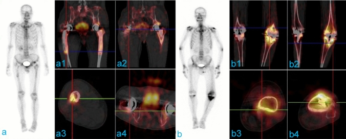
Results: In all, 47 joints (14 knees and 33 hips) were confirmed as aseptic loosening, while 25 joints (18 knees and 7 hips) were confirmed as infection. The sensitivity and accuracy of SPECT combined with SPECT/CT imaging were the highest (92.86% and 87.84%, respectively). The differential efficacy of bone scintigraphy combined with SPECT/CT imaging was also better than any other single imaging modality. In the analysis of involved prosthesis, prosthetic loosening occurred more in hip prosthesis and knee prosthesis was easily infected (P < 0.05). Finally, the sensitivity of ESR and CRP were 80% and 84%, respectively.
Conclusions: Bone scintigraphy with hybrid SPECT/CT remains encouraging in differentiating prosthetic infection from loosening after joint replacement. The diagnostic efficacy of differentiation in hip prosthesis was better than knee. Serum biomarkers cannot be used alone to differentiate prosthetic infection from loosening.
Keywords: 99mTc-MDP–SPECT/CT; Differentiate; Infection; Loosening; Prosthesis.
© Indian Orthopaedics Association 2024. Springer Nature or its licensor (e.g. a society or other partner) holds exclusive rights to this article under a publishing agreement with the author(s) or other rightsholder(s); author self-archiving of the accepted manuscript version of this article is solely governed by the terms of such publishing agreement and applicable law.
Conflict of interest statement
Conflict of interestsOn behalf of all authors, the corresponding author states that there is no conflict of interest.
Figures


Similar articles
-
Application of 68Ga-citrate PET/CT for differentiating periprosthetic joint infection from aseptic loosening after joint replacement surgery.Bone Joint Res. 2022 Jun;11(6):398-408. doi: 10.1302/2046-3758.116.BJR-2021-0464.R1. Bone Joint Res. 2022. PMID: 35731211 Free PMC article.
-
The role of bone SPECT/CT in the evaluation of painful joint prostheses.Nucl Med Commun. 2015 Sep;36(9):931-40. doi: 10.1097/MNM.0000000000000348. Nucl Med Commun. 2015. PMID: 26049374
-
The Diagnostic Characteristic and Reproducibility of Bone Scintigraphy Single-Photon Emission Computed Tomography/Computed Tomography for Diagnosing Aseptic Loosening of Uncemented Total Knee Arthroplasty.J Arthroplasty. 2024 Jul;39(7):1707-1713.e1. doi: 10.1016/j.arth.2024.01.005. Epub 2024 Jan 11. J Arthroplasty. 2024. PMID: 38218556
-
Prosthetic joint infections: radionuclide state-of-the-art imaging.Eur J Nucl Med Mol Imaging. 2012 May;39(5):892-909. doi: 10.1007/s00259-012-2062-7. Epub 2012 Feb 24. Eur J Nucl Med Mol Imaging. 2012. PMID: 22361912 Review.
-
Bone scan in painful knee arthroplasty: obsolete or actual examination?Acta Biomed. 2017 Jun 7;88(2S):68-77. doi: 10.23750/abm.v88i2-S.6516. Acta Biomed. 2017. PMID: 28657567 Free PMC article. Review.
References
LinkOut - more resources
Full Text Sources
Research Materials
Miscellaneous