

Hand Reconstruction Using Anterolateral Thigh Free Flap by Terminal Perforator-to-Digital Artery Anastomosis: Retrospective Analysis
- PMID: 38425858
- PMCID: PMC10901603
- DOI: 10.1055/a-2161-7419
Hand Reconstruction Using Anterolateral Thigh Free Flap by Terminal Perforator-to-Digital Artery Anastomosis: Retrospective Analysis
Abstract
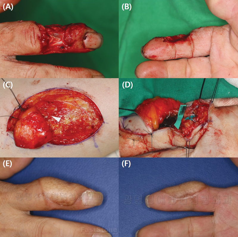
This study aimed to analyze cases of anterolateral thigh (ALT) free flap used for hand reconstruction with terminal perforator-to-digital artery anastomosis. Patients who underwent ALT free flap placement with terminal perforator-to-digital artery anastomosis for hand reconstruction between January 2011 and August 2021 were included. The number, length, and diameter of the perforators and veins, flap size, and operative time were investigated through a retrospective review of charts and photographs. The occurrences of arterial thrombosis, venous thrombosis, arterial spasm, and flap necrosis were analyzed. In total, 50 patients were included in this study. The mean diameter and length of the perforators were 0.68 mm and 3.25 cm, respectively, and the mean number of veins anastomosed was 1.88, with a mean diameter of 0.54 mm. Complications included four cases of arterial thrombosis, one case of venous thrombosis, seven cases of partial necrosis, and one case of total flap failure. Regression analysis showed that a longer perforator was associated with arterial thrombosis whereas larger flap size and number of anastomosed veins were associated with partial necrosis ( p < 0.05). The terminal perforator-to-digital artery anastomosis offers advantages in using compact free flaps with short pedicle lengths to cover small hand defects.
Keywords: hand injuries; microsurgical free flap; perforator flaps; reconstructive surgical procedures.
The Author(s). This is an open access article published by Thieme under the terms of the Creative Commons Attribution License, permitting unrestricted use, distribution, and reproduction so long as the original work is properly cited. ( https://creativecommons.org/licenses/by/4.0/ ).
Conflict of interest statement
Conflict of Interest J.S.K. and D.C.L. are editorial board member of the journal but were not involved in the peer reviewer selection, evaluation, or decision process of this article. No other potential conflicts of interest relevant to this article were reported.
Figures


Similar articles
-
[Clinical application of lobulated transplantation of free anterolateral thigh perforator flap in the treatment of electric burns of limbs].Zhonghua Shao Shang Za Zhi. 2019 Nov 20;35(11):790-797. doi: 10.3760/cma.j.issn.1009-2587.2019.11.005. Zhonghua Shao Shang Za Zhi. 2019. PMID: 31775467 Chinese.
-
[Combination mode and optimization strategy of harvest procedure of anterolateral thigh chimeric perforator myocutaneous flap].Zhongguo Xiu Fu Chong Jian Wai Ke Za Zhi. 2023 Feb 15;37(2):180-184. doi: 10.7507/1002-1892.202209059. Zhongguo Xiu Fu Chong Jian Wai Ke Za Zhi. 2023. PMID: 36796813 Free PMC article. Chinese.
-
Reconstruction of an upper posterior thigh extensive defect with a free split-anterolateral thigh (s-ALT) flap by perforator-to-perforator anastomosis: A case report.Microsurgery. 2019 Jan;39(1):91-94. doi: 10.1002/micr.30295. Epub 2018 Jan 17. Microsurgery. 2019. PMID: 29341249
-
[Outcome of relaying anterolateral thigh perforator flap in resurfacing the donor site wound following free anteromedial thigh perforator flap transfer for reconstruction of defect after oral tumor radical resection].Zhonghua Shao Shang Za Zhi. 2017 Feb 20;33(2):72-76. doi: 10.3760/cma.j.issn.1009-2587.2017.02.003. Zhonghua Shao Shang Za Zhi. 2017. PMID: 28219139 Chinese.
-
One Versus Two Veins in Free Anterolateral Thigh Flap Reconstruction: A Systematic Review and Meta-Analysis.Cureus. 2022 Dec 9;14(12):e32358. doi: 10.7759/cureus.32358. eCollection 2022 Dec. Cureus. 2022. PMID: 36628050 Free PMC article. Review.
References
-
- Holm A, Zachariae L. Fingertip lesions. An evaluation of conservative treatment versus free skin grafting. Acta Orthop Scand. 1974;45(03):382–392. - PubMed
-
- Kozin E D, Sethi R K, Herr M et al.Comparison of perioperative outcomes between the supraclavicular artery island flap and fasciocutaneous free flap. Otolaryngol Head Neck Surg. 2016;154(01):66–72. - PubMed
-
- Doh G, Kim B, Lee D et al.Hemodynamic principles in free tissue transfer: Vascular changes at the anastomosis site. Arch Hand Microsurg. 2021;26:285–292.
-
- Valdatta L, Tuinder S, Buoro M, Thione A, Faga A, Putz R. Lateral circumflex femoral arterial system and perforators of the anterolateral thigh flap: an anatomic study. Ann Plast Surg. 2002;49(02):145–150. - PubMed
-
- Yang J W, Kim J S, Lee D C et al.The radial artery superficial palmar branch flap: a modified free thenar flap with constant innervation. J Reconstr Microsurg. 2010;26(08):529–538. - PubMed
LinkOut - more resources
Full Text Sources