. 2024 Feb 23:39:101066.
doi: 10.1016/j.ymgmr.2024.101066. eCollection 2024 Jun.

Induced pluripotent stem cell-derived hepatocytes reveal TCA cycle disruption and the potential basis for triheptanoin treatment for malate dehydrogenase 2 deficiency

Affiliations

Induced pluripotent stem cell-derived hepatocytes reveal TCA cycle disruption and the potential basis for triheptanoin treatment for malate dehydrogenase 2 deficiency

Déborah Mathis et al. Mol Genet Metab Rep. .

Abstract

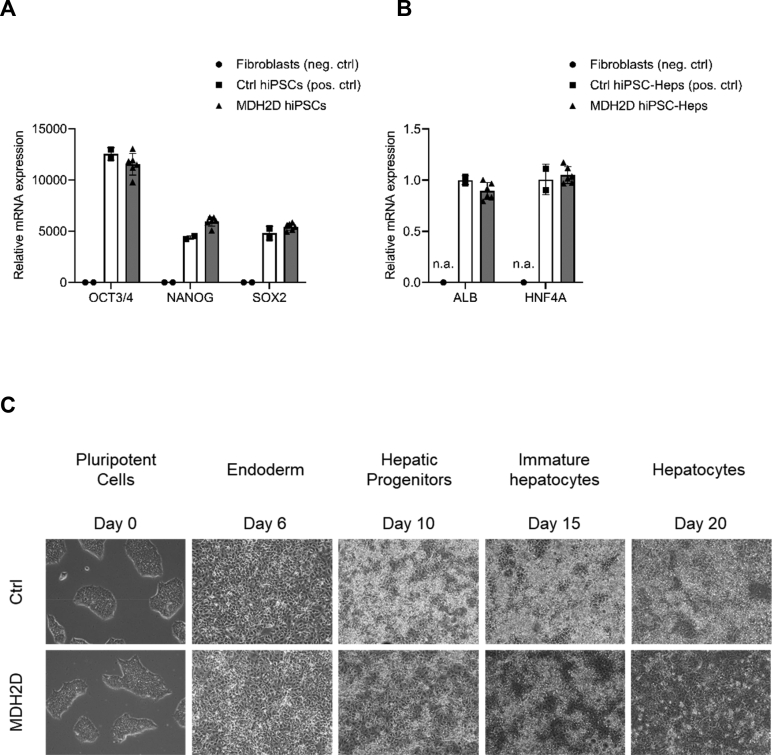
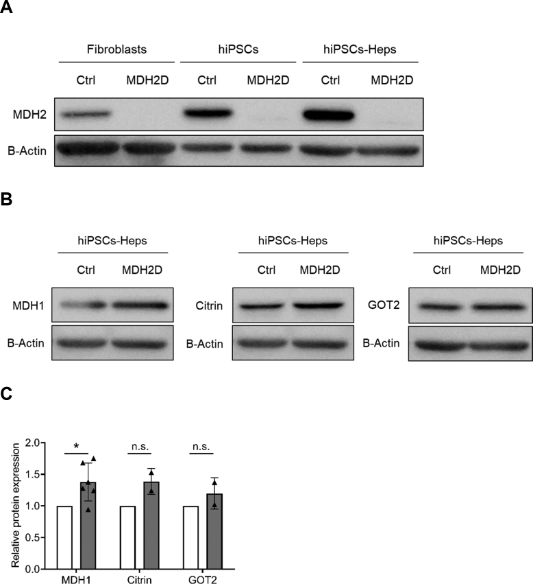
Mitochondrial malate dehydrogenase 2 (MDH2) is crucial to cellular energy generation through direct participation in the tricarboxylic acid (TCA) cycle and the malate aspartate shuttle (MAS). Inherited MDH2 deficiency is an ultra-rare metabolic disease caused by bi-allelic pathogenic variants in the MDH2 gene, resulting in early-onset encephalopathy, psychomotor delay, muscular hypotonia and frequent seizures. Currently, there is no cure for this devastating disease. We recently reported symptomatic improvement of a three-year-old girl with MDH2 deficiency following treatment with the triglyceride triheptanoin. Here, we aimed to better characterize this disease and improve our understanding of the potential utility of triheptanoin treatment. Using fibroblasts derived from this patient, we generated induced pluripotent stem cells (hiPSCs) and differentiated them into hepatocytes (hiPSC-Heps). Characterization of patient-derived hiPSCs and hiPSC-Heps revealed significantly reduced MDH2 protein expression. Untargeted proteotyping of hiPSC-Heps revealed global dysregulation of mitochondrial proteins, including upregulation of TCA cycle and fatty acid oxidation enzymes. Metabolomic profiling confirmed TCA cycle and MAS dysregulation, and demonstrated normalization of malate, fumarate and aspartate following treatment with the triheptanoin components glycerol and heptanoate. Taken together, our results provide the first patient-derived hiPSC-Hep-based model of MDH2 deficiency, confirm altered TCA cycle function, and provide further evidence for the implementation of triheptanoin therapy for this ultra-rare disease.

Synopsis: This study reveals altered expression of mitochondrial pathways including the tricarboxylic acid cycle and changes in metabolite profiles in malate dehydrogenase 2 deficiency and provides the molecular basis for triheptanoin treatment in this ultra-rare disease.

Keywords: Human induced pluripotent stem cell technology; Malate aspartate shuttle; Malate dehydrogenase 2 deficiency; Metabolic profiling; Proteomics; Triheptanoin; hiPSC-derived hepatocytes.


Conflict of interest statement

All authors declare no conflicts of interest.

Figures

Fig. 1
Fig. 1
Scheme showing energy metabolism pathways, the MDH2 enzyme and the rational for triheptanoin treatment as alternative energy fuel. FA: fatty acid; C7: heptanoate; Glu: glutamate; 2-KG: 2-ketoglutarate.
Fig. 2
Fig. 2
hiPSCs and hiPSC-Heps generation and characterization. (A) Relative mRNA expression levels of pluripotency markers OCT3/4, NANOG and SOX2 in MDH2D hiPSCs and in fibroblasts which were used as negative control (neg. ctrl) and in previously generated and characterized Ctrl hiPSCs which were used as positive control (pos. ctrl) for pluripotency marker expression. (B) Relative mRNA expression levels of hepatocyte-specific markers ALB and HNF4A in MDH2 hiPSC-Heps and the same control samples as described in (A). n.a.; no amplification. (C) Representative images of Ctrl (upper) and MDH2D (lower) hiPSCs through-out the differentiation process to hiPSC-Heps, illustrating cell morphological changes.
Fig. 3
Fig. 3
Western blot analysis of MDH2 and additional components of the malate aspartate shuttle. (A) MDH2 protein levels in MDH2D fibroblasts, induced pluripotent stem cells (hiPSCs) and differentiated hepatocytes (hiPSC-Heps) along with corresponding Ctrl, as determined by western blot. Two further replicates are presented in Supp. Fig. 1A. (B, C) MDH1, Citrin and GOT2 protein levels in MDH2D and Ctrl hiPSC-Heps as determined (B) and quantified (C) by western blot. A further replicate as well as western blot of each protein in fibroblasts and hiPSCs are presented in Supp. Fig. 1B. For both (A) and (B), B-Actin was used as loading control/housekeeping protein. For (B), MDH1 and Citrin was analyzed on the same membrane, therefore an identical image for B-Actin is shown here.
Fig. 4
Fig. 4
Untargeted proteotyping reveals altered mitochondrial protein levels in MDH2-deficient patient-derived hiPSC-Heps. (A) Principal component analysis (PCA) revealing clear separation of MDH2D and Ctrl hiPSC-Heps. (B) Volcano plot demonstrating differential protein expression in MDH2D and Ctrl hiPSC-Heps. Mitochondrial proteins, as defined by MitoCarta3.0 are in orange, MDH2 in teal. Two-sided t-test was used to calculate p-values. (C and D) Sample clustering of mitochondrial (C) and specifically TCA cycle enzymes (D) in MDH2D and Ctrl hiPSC-Heps. Three replicates per cell line are represented.
Fig. 5
Fig. 5
Untargeted metabolite profiling of MDH2-deficient patient-derived hiPSC-Heps. (A) Sample clustering of metabolites determined in MDH2D and Ctrl hiPSC-Heps. (B) Differential metabolite concentrations in untreated MDH2D and Ctrl hiPSC-Heps. (C) Principal component analysis (PCA) of metabolite concentrations showing separation of MDH2D and Ctrl hiPSC-Heps upon treatments. Treatments were either heptanoate alone (Hept) or heptanoate and glycerol combined (HeptGlyc) or solvent only (=DMSO; none). (D) Response of fumarate, malate and aspartate to treatments. Other measured metabolites are provided in Suppl. Fig. 3. Three replicates per cell line are represented.

Similar articles

Cited by

References

    1. Borst P. The malate-aspartate shuttle (Borst cycle): how it started and developed into a major metabolic pathway. IUBMB Life. 2020;72(11):2241–2259. - PMC - PubMed
    1. Broeks M.H., et al. Inborn disorders of the malate aspartate shuttle. J. Inherit. Metab. Dis. 2021;44(4):792–808. - PMC - PubMed
    1. Minarik P., et al. Malate dehydrogenases--structure and function. Gen. Physiol. Biophys. 2002;21(3):257–265. - PubMed
    1. Ait-El-Mkadem S., et al. Mutations in MDH2, encoding a Krebs cycle enzyme, cause early-onset severe encephalopathy. Am. J. Hum. Genet. 2017;100(1):151–159. - PMC - PubMed
    1. Laemmle A., et al. Triheptanoin - novel therapeutic approach for the ultra-rare disease mitochondrial malate dehydrogenase deficiency. Mol. Genet. Metab. Rep. 2021;29 - PMC - PubMed