. 2024 Jan 25;4(2):432-440.
doi: 10.1021/jacsau.3c00572. eCollection 2024 Feb 26.

Peptide-Based Covalent Inhibitors Bearing Mild Electrophiles to Target a Conserved His Residue of the Bacterial Sliding Clamp

Affiliations

Peptide-Based Covalent Inhibitors Bearing Mild Electrophiles to Target a Conserved His Residue of the Bacterial Sliding Clamp

Guillaume Compain et al. JACS Au. .

Abstract

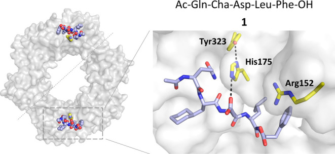
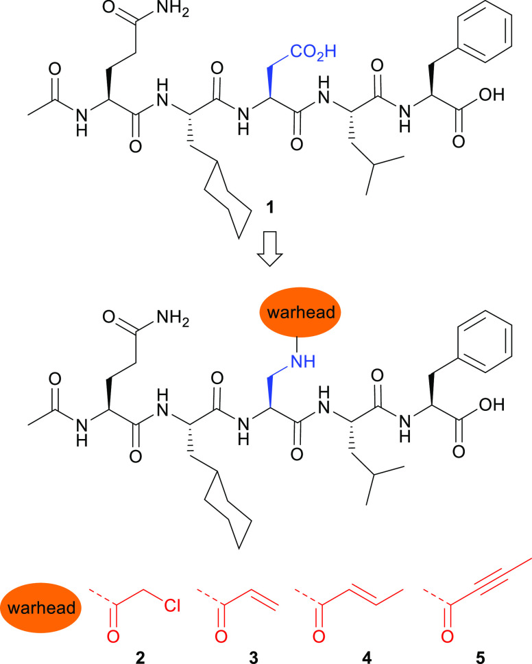
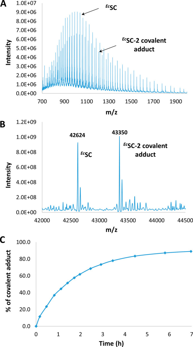
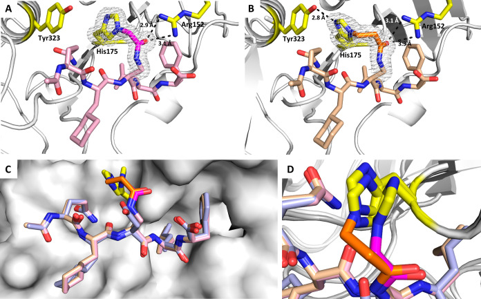
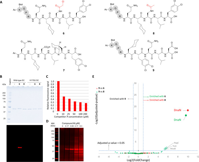
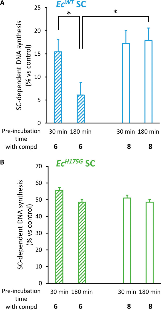
Peptide-based covalent inhibitors targeted to nucleophilic protein residues have recently emerged as new modalities to target protein-protein interactions (PPIs) as they may provide some benefits over more classic competitive inhibitors. Covalent inhibitors are generally targeted to cysteine, the most intrinsically reactive amino acid residue, and to lysine, which is more abundant at the surface of proteins but much less frequently to histidine. Herein, we report the structure-guided design of targeted covalent inhibitors (TCIs) able to bind covalently and selectively to the bacterial sliding clamp (SC), by reacting with a well-conserved histidine residue located on the edge of the peptide-binding pocket. SC is an essential component of the bacterial DNA replication machinery, identified as a promising target for the development of new antibacterial compounds. Thermodynamic and kinetic analyses of ligands bearing different mild electrophilic warheads confirmed the higher efficiency of the chloroacetamide compared to Michael acceptors. Two high-resolution X-ray structures of covalent inhibitor-SC adducts were obtained, revealing the canonical orientation of the ligand and details of covalent bond formation with histidine. Proteomic studies were consistent with a selective SC engagement by the chloroacetamide-based TCI. Finally, the TCI of SC was substantially more active than the parent noncovalent inhibitor in an in vitro SC-dependent DNA synthesis assay, validating the potential of the approach to design covalent inhibitors of protein-protein interactions targeted to histidine.


Conflict of interest statement

The authors declare no competing financial interest.

Figures

Figure 1
Figure 1
X-ray cocrystal structure of EcSC-1 complex (PDB ID:6FVL).
Figure 2
Figure 2
Chemical structures of noncovalent inhibitor 1 and covalent inhibitors 25.
Figure 3
Figure 3
(A) Representative example of a mass spectrum obtained after incubating ECSC with a covalent inhibitor (here, compound 2 was incubated at 37 °C for 49 min). (B) Corresponding deconvoluted mass spectrum. (C) Percentage of covalent adduct formed over time determined from deconvoluted spectra (see SI.5).
Figure 4
Figure 4
Crystal structures of EcSC-2 adduct (A) solved at 1.22 Å resolution (PDB ID: 8PAY) and EcSC-3 adduct (B) solved at 1.37 Å resolution (PDB ID: 8PAT); the electron density is shown in gray around the covalent bond formed with His175; the carbon atoms from the warheads are highlighted in magenta and orange, respectively. (C) Superimposition of the crystal structures of EcSC-1 complex (light blue), EcSC-2 (pink, RMSD of atomic positions with EcSC-1 complex = 0.498 Å (316 to 316 atoms)), and EcSC-3 (beige, RMSD of atomic positions with EcSC-1 = 0.461 Å (334 to 334 atoms)) adducts. (D) Zoom on the covalent bonds showing the rotation of His175 on EcSC-2 compared to EcSC-1 and EcSC-3 crystal structures.
Figure 5
Figure 5
(A) Structure of compounds 69. The spacer Gly-Pro-Arg was used for biotinylated peptides 6, 8, and 9. (B) Histidine 175 is essential for the formation of a covalent adduct with 6. His-tag purified wild-type or H175G EcSC were incubated with or without compound 6 or the cognate biotin-labeled noncovalent peptide ligand 8. SDS-PAGE electrophoresis of the incubated SC and Coomassie blue staining (top) reveals a mobility shift only when 6 is incubated with the wild-type SC, but not with the H175G mutant. Staining of biotinylated products (bottom) identifies the shifted band as the covalent 6-EcwtSC complex (SI. 7 and Figure S12). (C) SC-binding competition assay. Purified His-Tag-SC was incubated with a fixed amount of 6 (10 μM) and increasing concentrations (0–200 μM) of the noncovalent high-affinity SC binder 7 as indicated. Covalently bound biotinylated peptides were revealed after SDS-PAGE separation and infrared dye coupled streptavidin labeling (see SI. 8). The relative fluorescence detected in each lane was plotted against the noncovalent competitor concentration. (D) Pull-down experiments following co-incubation of E. coli whole cell lysate with compound 6. Covalent reaction products were captured onto streptavidin-coated magnetic beads and analyzed as described above. The Mw of the most intense band detected (around 40 kDa) indicates that the major reaction product formed is likely the covalent 6-SC adduct. (E) Spectral-count-based proteomic analysis of pull-down experiments in whole cell lysate. The volcano plot displays the log2 fold change (x axis) against the–log10 adjusted p-value (y axis) for all proteins enriched when using peptide 9 (control, left part of the graph) compared with SC-binding peptides 6 (green circles) or 8 (red circles). Cut-offs (dashed lines) of log2 FC > 1 (ratio >2) and −log10 adj p value >1.3 (adj p value <0.05) were applied to highlight significantly enriched proteins (see SI.9 and Tables S10).
Figure 6
Figure 6
Time-dependent increased inhibitory effect on SC-dependent DNA synthesis of compound 6 as compared to 8 necessitates the presence of H175 on the SC. SC-dependent DNA synthesis was measured after preincubation of the indicated E. coli SC variants (either wild-type or H175G mutant) with or without compound 8 or 6 for the indicated period. Results presented are the mean (±sd) of 3 independent assays. Statistically different results are indicated (*: p value <0.05). See SI. 11 and Figure S16 for experimental details and gel images.

Similar articles

Cited by

References

    1. Singh J.; Petter R. C.; Baillie T. A.; Whitty A. The Resurgence of Covalent Drugs. Nat. Rev. Drug Discovery 2011, 10 (4), 307–317. 10.1038/nrd3410. - DOI - PubMed
    1. Bauer R. A. Covalent Inhibitors in Drug Discovery: From Accidental Discoveries to Avoided Liabilities and Designed Therapies. Drug Discovery Today 2015, 20 (9), 1061–1073. 10.1016/j.drudis.2015.05.005. - DOI - PubMed
    1. Lonsdale R.; Ward R. A. Structure-Based Design of Targeted Covalent Inhibitors. Chem. Soc. Rev. 2018, 47 (11), 3816–3830. 10.1039/C7CS00220C. - DOI - PubMed
    1. Gehringer M.; Laufer S. A. Emerging and Re-Emerging Warheads for Targeted Covalent Inhibitors: Applications in Medicinal Chemistry and Chemical Biology. J. Med. Chem. 2019, 62 (12), 5673–5724. 10.1021/acs.jmedchem.8b01153. - DOI - PubMed
    1. Boike L.; Henning N. J.; Nomura D. K. Advances in Covalent Drug Discovery. Nat. Rev. Drug Discovery 2022, 21, 881–898. 10.1038/s41573-022-00542-z. - DOI - PMC - PubMed

LinkOut - more resources