. 2024 Feb 12;4(2):713-729.
doi: 10.1021/jacsau.4c00040. eCollection 2024 Feb 26.

Proteolysis Targeting Chimera Degraders of the METTL3-14 m6A-RNA Methyltransferase

Affiliations

Proteolysis Targeting Chimera Degraders of the METTL3-14 m6A-RNA Methyltransferase

Francesco Errani et al. JACS Au. .

Abstract

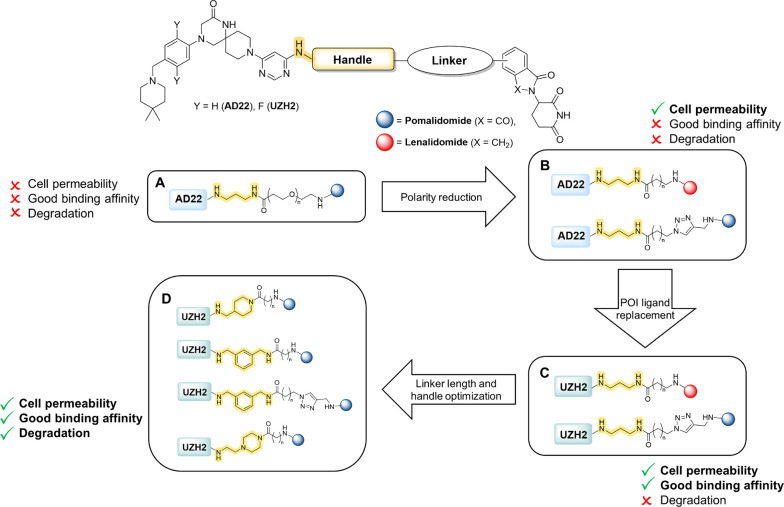
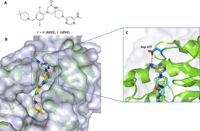
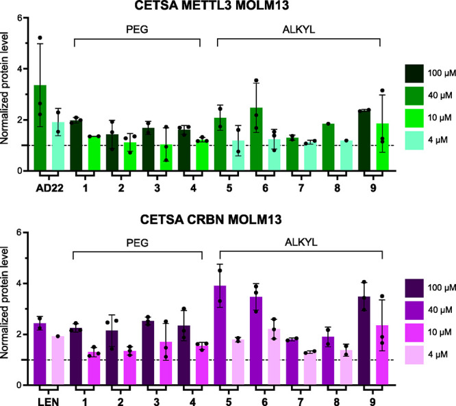
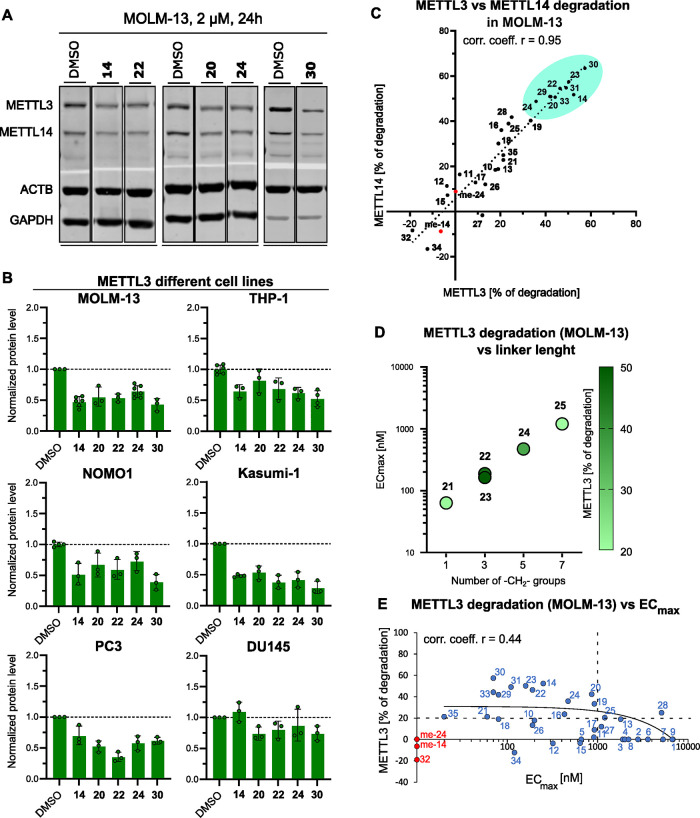
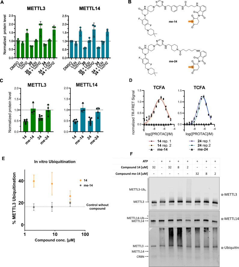
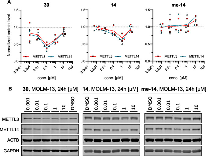
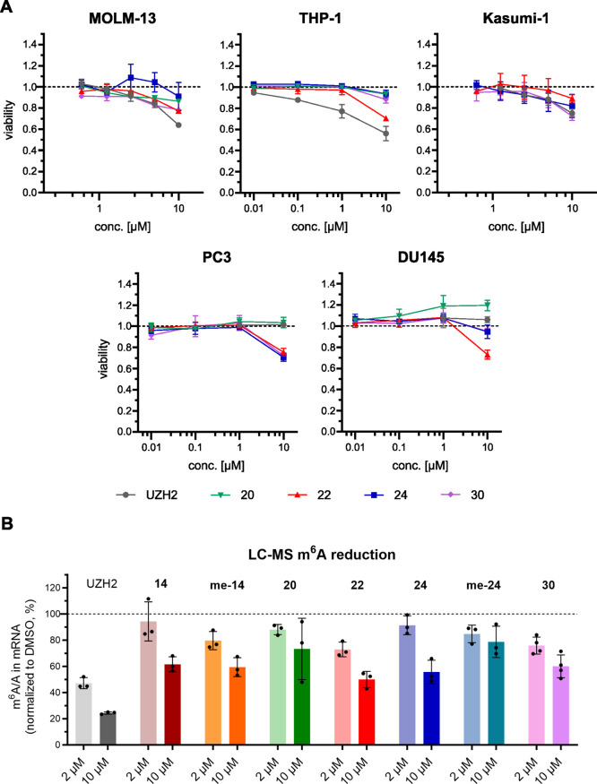
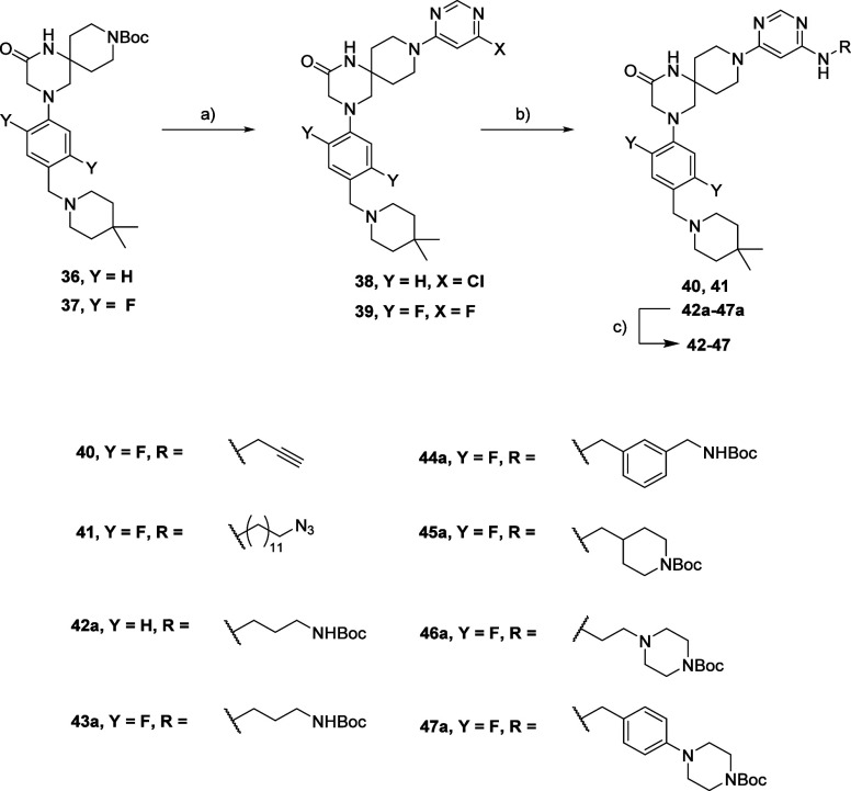
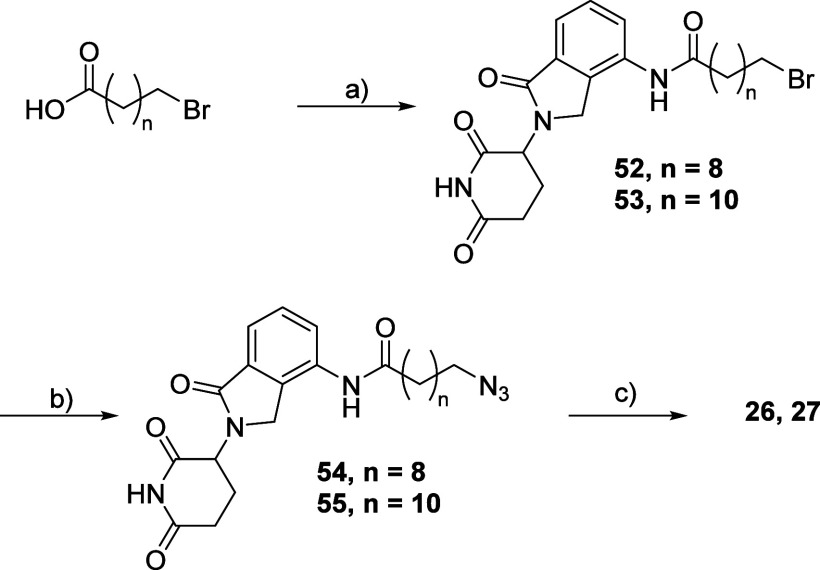
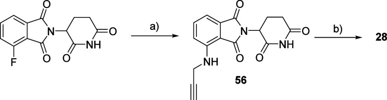
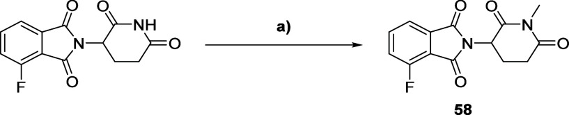
Methylation of adenine N6 (m6A) is the most frequent RNA modification. On mRNA, it is catalyzed by the METTL3-14 heterodimer complex, which plays a key role in acute myeloid leukemia (AML) and other types of blood cancers and solid tumors. Here, we disclose the first proteolysis targeting chimeras (PROTACs) for an epitranscriptomics protein. For designing the PROTACs, we made use of the crystal structure of the complex of METTL3-14 with a potent and selective small-molecule inhibitor (called UZH2). The optimization of the linker started from a desfluoro precursor of UZH2 whose synthesis is more efficient than that of UZH2. The first nine PROTAC molecules featured PEG- or alkyl-based linkers, but only the latter showed cell penetration. With this information in hand, we synthesized 26 PROTACs based on UZH2 and alkyl linkers of different lengths and rigidity. The formation of the ternary complex was validated by a FRET-based biochemical assay and an in vitro ubiquitination assay. The PROTACs 14, 20, 22, 24, and 30, featuring different linker types and lengths, showed 50% or higher degradation of METTL3 and/or METTL14 measured by Western blot in MOLM-13 cells. They also showed substantial degradation on three other AML cell lines and prostate cancer cell line PC3.


Conflict of interest statement

The authors declare no competing financial interest.

Figures

Scheme 1
Scheme 1. General Structure of PROTACs and Optimization Strategy
Figure 1
Figure 1
Protein structure-based design of PROTACs. (a) 2D structures of UZH2 and its desfluoro precursor AD22. (b) Overlap of the crystal structures of METTL3 bound to AD22 (carbon atoms in cyan, PDB 7O0P) and UZH2 (carbon atoms in orange, PDB 7O2F). (c) Zoom in on the hydrogen bond between the side chain of Asp377 and the methylamine of AD22 and UZH2.
Figure 2
Figure 2
Evaluation of AD22-based PROTACS 1-9 in the MOLM-13 (AML) cell line. The stabilization of METTL3 (top) and Cereblon (CRBN, bottom) was quantified by CETSA at 54 °C. The SAM-competitive inhibitor AD22 was employed as a control for METTL3 (top, left), while lenalidomide (LEN) was used as a control for Cereblon (bottom, left). The dashed line represents the protein level of the DMSO control used for normalization (y = 1).
Figure 3
Figure 3
(a) Representative Western blots from a cellular degradation assay with PROTACs 14, 20, 22, 24, and 30 in the MOLM-13 cell line. Full membranes are shown in Figure S2. (b) METTL3 Western blot quantification by densitometry from a cellular degradation assay at PROTAC concentration of 2 μMin the AML cell lines MOLM-13, THP-1, NOMO-1, and KASUMI-1 and in the prostate cancer cell lines PC3 and DU145 (representative Western blots are shown in Figure S3). The dashed line represents the protein level of the DMSO control, used for normalization (y = 1). (c) Correlation of the degradation of METTL3 and METTL14 in MOLM-13 at PROTAC concentration of 2 μM; the dotted black line is a linear regression of all the compounds (corr. coeff. r = 0.95). The nine most active PROTACs are highlighted in turquoise. me-14 and me-24 (in red) are the methylated negative controls of PROTACs 14 and 24 (for structures, see Figure 4B). (d) Correlation between the number of −CH2– groups in the linker and the ECmax value measured in the TCFA. The color of the data points reflects the degradation of METTL3 as measured by Western blot in MOLM-13 (legend on the right). While the ECmax improves until the shortest length of the linker (one −CH2−), the highest degradation is observed at intermediate lengths, i.e., (−CH2−)3. (e) Scatter plot of the degradation of METTL3 in MOLM-13 as a function of the ECmax values measured in the TCFA. The data points on the y-axis (red) did not show any detectable ternary complex formation in the TCFA. The linear regression for the compounds with both degradation and ECmax values (blue) is shown (black continuous line). The horizonal dashed line marks a METTL3 degradation level of 20%, and the vertical dashed line marks ECmax = 1 μM.
Figure 4
Figure 4
Control experiments, TCFA and in vitro ubiquitination. (a) METTL3 and METTL14 levels in the presence of lenalidomide or UZH2 in MOLM-13. The dashed line represents the protein level of the DMSO control, used for normalization (y = 1). (b) Chemical structures of PROTAC negative controls me-14 and me-24. (c) METTL3 and METTL14 degradation by PROTACs 14 and 24 in comparison with their negative controls me-14 and me-24 in MOLM-13. The dashed line represents the protein level of the DMSO control, used for normalization (y = 1). (d) Biochemical FRET-based ternary complex formation assay (TCFA) with PROTACs 14 and 24 and their methylated negative controls me-14 and me-24. (e) In vitro ubiquitination assay results with compound 14 and its negative control me-14. All data originate from biological and biochemical duplicates or more. (f) Western blot analysis of the in vitro ubiquitination assay. The ubiquitination reaction mixture (E1, E2, CUL4A-RBX1, CRBN-DDB1, and METTL3-METTL14 in reaction buffer) was incubated with or without ATP and at different concentrations of compounds 14 and me-14 as indicated at 30 °C for 2 h. The proteins were separated by SDS-PAGE followed by Western blot analysis with α-METTL3, α-METTL14, and α-Ubiquitin antibodies. Shown here is one representative Western blot of three biological replicates of the experiment. For panel (e), densitometry was performed using the α-METTL3 blots of all three replicates. In the α-METTL14 blot, a weak band appears above the METTL14 band at 32, 8, and 2 μM of compound 14, presumably indicating monoubiquitination of METTL14. However, the ubiquitination of METTL3 is clearly more efficient. The band indicated with an asterisk (*) originates from the unspecific detection of METTL3 with the α-METTL14 antibody.
Figure 5
Figure 5
Cellular characterization. (a) Concentration dependence of METTL3 (red) and METTL14 (blue) degradation by PROTACs 30 and 14 and the methylated negative control of 14 (me-14) in MOLM-13 cells. The dashed line represents the protein level of the DMSO control, used for normalization (y = 1). (b) Representative Western blots. Full membranes are shown in Figure S5.
Figure 6
Figure 6
Cellular characterization. (a) Cell viability assay for PROTACs 20, 22, 24,and 30 and the METTL3 catalytic inhibitor UZH2 in AML cell lines (top) and prostate cancer cell lines (bottom). The dashed line represents the protein level of the DMSO control, used for normalization (y = 1). (b) LC-MS quantification of m6A/A levels in polyadenylated RNA in MOLM-13. The dashed line represents the protein level of the DMSO control, used for normalization (y = 1).
Scheme 2
Scheme 2. Synthesis Route for Compounds 4047
Reagents and conditions: (a) (i) HCl aq. 37%, MeOH; (ii) for 38 and 39: 4,6-dichloro pyrimidine (38)/4,6-difluoro pyrimidine (39), TEA, iPrOH; (b) RNH2, TEA, DMSO (40, 41)/EtOH (42a-47a); (c) for 4247: TFA, DCM.
Scheme 3
Scheme 3. Synthesis Route for Compound 48
Reagents and conditions: (a) tert-butyl 4-(5-bromopyrimidin-2-yl) piperazine-1-carboxylate, CuI, (L)-proline, K2CO3, DMSO; (b) HCl 4 M in dioxane, MeOH.
Scheme 4
Scheme 4. Synthesis Route for Compounds 120, 2225, 2931, and 33
Reagents and conditions: (a) HATU (1–4, 8, 9, 29–31)/COMU (5–7, 10–28, 32–35), DIPEA, DMF.
Scheme 5
Scheme 5. Synthesis Route for Compounds 49 and 51
Reagents and conditions: (a) For 49a: Boc-glycine, COMU, DIPEA, DMF; (b) for 50: 2-chloroacetyl chloride, DIPEA, dry THF; (c) for 51a: Boc-piperazine, DMSO, 50 °C; (d) for 49 and 51: TFA, DCM.
Scheme 6
Scheme 6. Synthesis Route for Compounds 21, 34, and 35
Reagents and conditions: (a) For 21, 34, and 35: 4-fluoro thalidomide, TEA, and DMSO.
Scheme 7
Scheme 7. Synthesis Route for Compounds 26 and 27
Reagents and conditions: (a) (i) SOCl2; (ii) lenalidomide, THF; (b) NaN3, DMF; (c) 40, CuSO4, Na ascorbate, THF.
Scheme 8
Scheme 8. Synthesis Route for Compound 28
Reagents and conditions: (a) propargyl amine, TEA, DMSO; (b) 41, CuSO4, Na ascorbate, THF.
Scheme 9
Scheme 9. Synthesis Route for Compound 32
Reagents and conditions: (a) (i) tert-butyl (2-(piperazin-1-yl) ethyl) carbamate, DIPEA; DMSO; (ii) HCl 4 M in dioxane, MeOH; b) 39, DIPEA, DMSO.
Scheme 10
Scheme 10. Synthesis Route for Compound 58
Reagents and conditions: (a) CH3I, K2CO3, DMF.

Similar articles

Cited by

References

    1. Kumar S.; Mohapatra T. Deciphering Epitranscriptome: Modification of mRNA Bases Provides a New Perspective for Post-Transcriptional Regulation of Gene Expression. Front. Cell Dev. Biol. 2021, 9, 62841510.3389/fcell.2021.628415. - DOI - PMC - PubMed
    1. Roignant J. Y.; Soller M. m6A in mRNA: An Ancient Mechanism for Fine-Tuning Gene Expression. Trends Genet. 2017, 33 (6), 380–390. 10.1016/j.tig.2017.04.003. - DOI - PubMed
    1. Boccaletto P.; Stefaniak F.; Ray A.; Cappannini A.; Mukherjee S.; Purta E.; Kurkowska M.; Shirvanizadeh N.; Destefanis E.; Groza P.; Avşar G.; Romitelli A.; Pir P.; Dassi E.; Conticello S. G.; Aguilo F.; Bujnicki J. M.. MODOMICS: A Database of RNA Modification Pathways. 2021 Update. Nucleic Acids Res. 2021, 50. 10.1093/nar/gkab1083. - DOI - PMC - PubMed
    1. Dominissini D.; Moshitch-Moshkovitz S.; Schwartz S.; Salmon-Divon M.; Ungar L.; Osenberg S.; Cesarkas K.; Jacob-Hirsch J.; Amariglio N.; Kupiec M.; Sorek R.; Rechavi G.. Topology of the Human and Mouse m6A RNA Methylomes Revealed by m6A-Seq. 2012, 485 ( (7397), ), 201–206. 10.1038/nature11112. - DOI - PubMed
    1. Alarcón C. R.; Goodarzi H.; Lee H.; Liu X.; Tavazoie S.; Tavazoie S. F. HNRNPA2B1 Is a Mediator of m6A-Dependent Nuclear RNA Processing Events. Cell 2015, 162 (6), 1299–1308. 10.1016/j.cell.2015.08.011. - DOI - PMC - PubMed

LinkOut - more resources