. 2024 Feb 16;4(2):855-864.
doi: 10.1021/jacsau.4c00008. eCollection 2024 Feb 26.

Engineering Bacterial Biofilm Development and Structure via Regulation of Silver Nanoparticle Density in Graphene Oxide Composite Coating

Affiliations

Engineering Bacterial Biofilm Development and Structure via Regulation of Silver Nanoparticle Density in Graphene Oxide Composite Coating

Shanshan Wang et al. JACS Au. .

Abstract

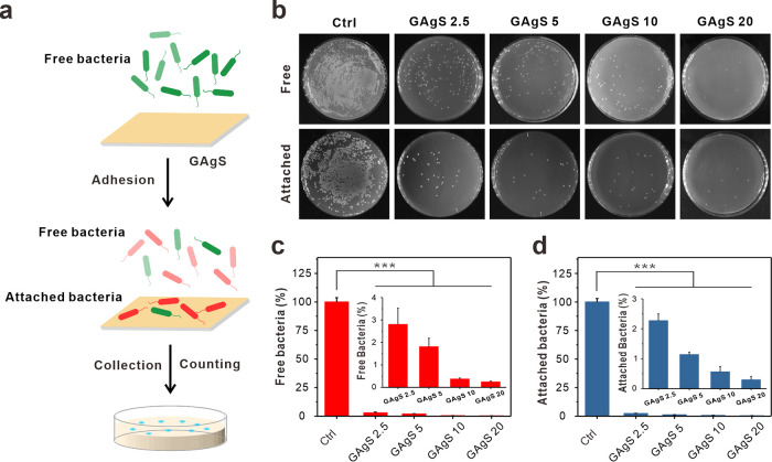
Graphene-based composites have shown significant potential in the treatment of biofilm infections in clinical settings due to their exceptional antimicrobial properties and specific mechanisms. Nevertheless, a comprehensive understanding of the influence exerted by nanoparticles embedded in the composites on the development and structure of biofilms is still lacking. Here, we fabricate different graphene oxide-silver nanoparticle (GAg) composite-modified substrates (GAgS) with varying densities of silver nanoparticles (AgNPs) and investigate their effects on planktonic bacterial adhesion, subsequent biofilm formation, and mature biofilm structure. Our findings indicate that the initial attachment of Pseudomonas aeruginosa cells during biofilm formation is determined by the density of AgNPs on the GAgS surface. In contrast, the subsequent transition from adherent bacteria to the biofilm is determined by GAgS's synergistic antimicrobial effect. There exists a threshold for the inhibitory performance of GAgS, where the 20 μg/cm2 GAg composite completely prevents biofilm formation; below this concentration, GAgS delays the development of the biofilm and causes structural changes in the mature biofilm with enhanced bacterial growth and increased production of extracellular polymeric substance. More importantly, GAgS have minimal impact on mammalian cell morphology and proliferation while not inducing hemolysis in red blood cells. These results suggest that GAg composites hold promise as a therapeutic approach for addressing medical devices and implant-associated biofilm infections.


Conflict of interest statement

The authors declare no competing financial interest.

Figures

Scheme 1
Scheme 1. AgNP Density of GAgS Affects Biofilm Formation and Cell Proliferation
Figure 1
Figure 1
Synthesis and characterization of GAgS.(a) Schematic image of GAg and GAgS preparation. (b) SEM images of GAg substrates. (c) EDS spectra of graphene-based substrates. (d) Density of Ag particles and content of the Ag element on different substrates. (e) Roughness of GAg substrates. Asterisks denote significantly increased roughness as compared with the control group (**P < 0.01, ***P < 0.001).
Figure 2
Figure 2
Adhesion capability of planktonic P. aeruginosa on GAgS. (a) Schematic of bacterial adhesion at the initial stage of biofilm formation. (b) Growth of free and attached P. aeruginosa on GAgS after 2 h. The number of (c) free and (d) attached bacteria on GAgS. Insets provide an enlarged depiction of the bacteria rate of GAgS. Asterisks denote significantly reduced bacteria as compared with the control group (***P < 0.001).
Figure 3
Figure 3
Development of the biofilm on GAgS. (a) Schematic of biofilm development. (b) Effects of different concentrations of the GAg substrate on biofilm formation. (c) 3D confocal images of the biofilm. (d) Total biomass, (e) cell biomass, (f) EPS biomass, and (g) thickness of the biofilm at different culture times. Asterisks denote significantly inhibited biofilm biomass as compared with the control group (*P < 0.05, **P < 0.01, ***P < 0.001).
Figure 4
Figure 4
Structure of the biofilm observed by SEM. (a) Top and (b) lateral images of the biofilm on different substrates for 12 and 24 h. (c) Quantification of biofilm thickness in lateral SEM images.
Figure 5
Figure 5
Biocompatibility of GAgS. (a) Cell viability of MC3T3-E1 cells grown on different substrates. (b) Cellular morphology on the substrate with and without GAgS. (i) Calcein/PI staining. (ii) SEM imaging. (iii) Actin immunofluorescence staining (green) and nuclei (blue). (c) Hemolytic toxicity of different GAgS. Triton as the positive group; Tris buffer as the negative group. Asterisks represent significantly reduced hemolysis as compared with positive control (***P < 0.001).

Similar articles

References

    1. Arciola C. R.; Campoccia D.; Montanaro L. Implant Infections: Adhesion, Biofilm Formation and Immune Evasion. Nat. Rev. Microbiol. 2018, 16 (7), 397–409. 10.1038/s41579-018-0019-y. - DOI - PubMed
    1. Daubert D. M.; Weinstein B. F. Biofilm as a Risk Factor in Implant Treatment. Periodontology 2000 2019, 81 (1), 29–40. 10.1111/prd.12280. - DOI - PubMed
    1. Caldara M.; Belgiovine C.; Secchi E.; Rusconi R. Environmental, Microbiological, and Immunological Features of Bacterial Biofilms Associated with Implanted Medical Devices. Clin. Microbiol. Rev. 2022, 33 (2), 627–635. 10.1128/cmr.00221-20. - DOI - PMC - PubMed
    1. Yuan L.; Straub H.; Shishaeva L.; Ren Q. Microfluidics for Biofilm Studies. Annu. Rev. Anal. Chem. 2023, 16 (1), 139–159. 10.1146/annurev-anchem-091522-103827. - DOI - PubMed
    1. Flemming H.-C.; van Hullebusch E. D.; Neu T. R.; Nielsen P. H.; Seviour T.; Stoodley P.; Wingender J.; Wuertz S. The Biofilm Matrix: Multitasking in a Shared Space. Nat. Rev. Microbiol. 2023, 21 (2), 70–86. 10.1038/s41579-022-00791-0. - DOI - PubMed

LinkOut - more resources