Review
. 2024 May;81(5):991-1007.
doi: 10.1161/HYPERTENSIONAHA.123.21356. Epub 2024 Mar 1.

Hypertension, Neurodegeneration, and Cognitive Decline

Affiliations
Review

Hypertension, Neurodegeneration, and Cognitive Decline

Anthony Pacholko et al. Hypertension. 2024 May.

Abstract

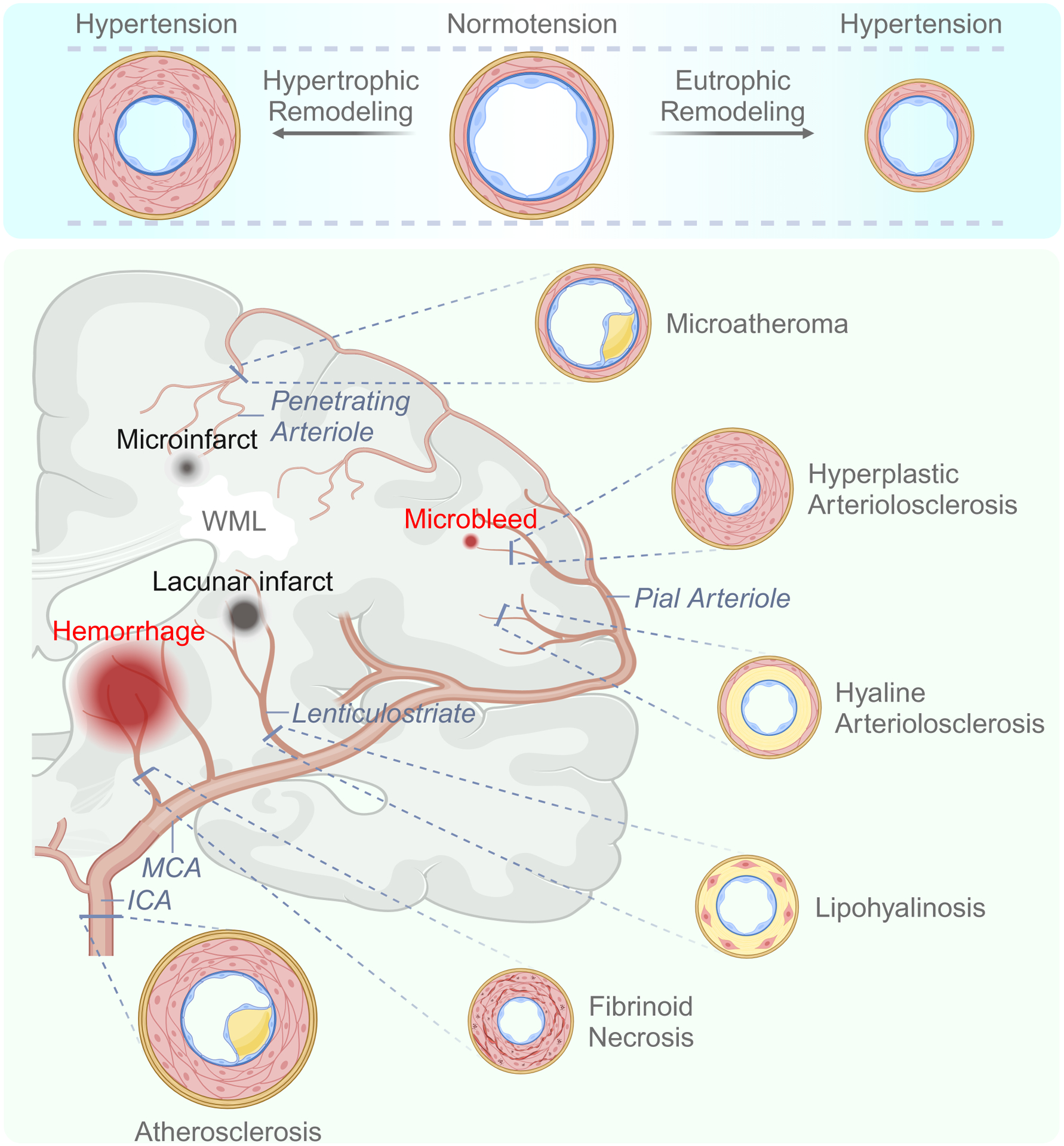
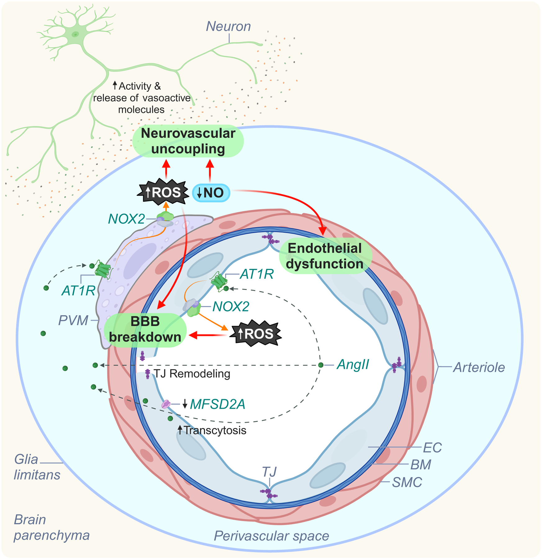
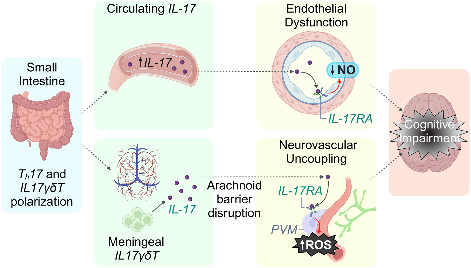
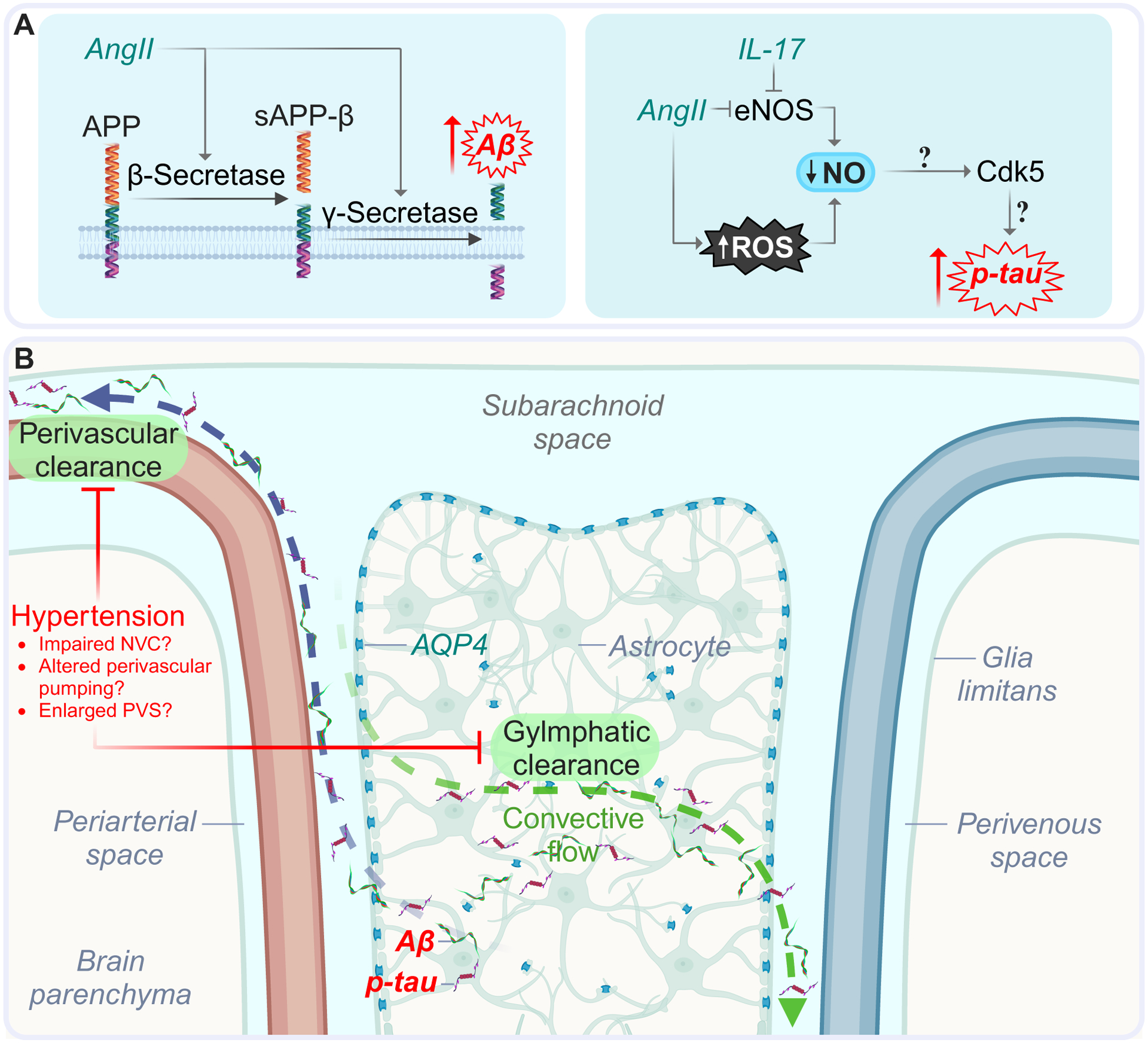
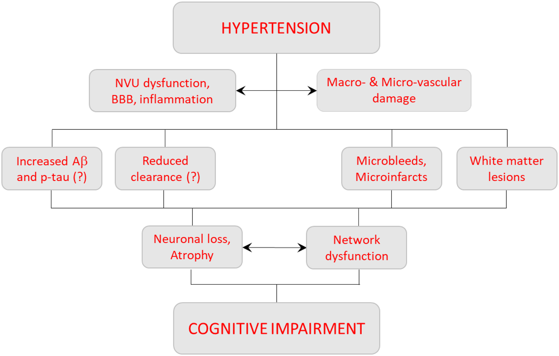
Elevated blood pressure is a well-established risk factor for age-related cognitive decline. Long linked to cognitive impairment on vascular bases, increasing evidence suggests a potential association of hypertension with the neurodegenerative pathology underlying Alzheimer disease. Hypertension is well known to disrupt the structural and functional integrity of the cerebral vasculature. However, the mechanisms by which these alterations lead to brain damage, enhance Alzheimer pathology, and promote cognitive impairment remain to be established. Furthermore, critical questions concerning whether lowering blood pressure by antihypertensive medications prevents cognitive impairment have not been answered. Recent developments in neurovascular biology, brain imaging, and epidemiology, as well as new clinical trials, have provided insights into these critical issues. In particular, clinical and basic findings on the link between neurovascular dysfunction and the pathobiology of neurodegeneration have shed new light on the overlap between vascular and Alzheimer pathology. In this review, we will examine the progress made in the relationship between hypertension and cognitive impairment and, after a critical evaluation of the evidence, attempt to identify remaining knowledge gaps and future research directions that may advance our understanding of one of the leading health challenges of our time.

Keywords: Alzheimer disease; blood pressure; brain; hypertension; stroke.


Conflict of interest statement

Disclosures C. Iadecola serves on the scientific advisory board of Broadview Ventures. The other author reports no conflicts.

Figures

Figure 1:
Figure 1:. Structural alterations and segmental pathology induced by hypertension.
Top: Vascular remodeling proceeds in either an outward or inward manner in response to mechanical, cellular, inflammatory, and oxidative factors. Bottom: The predominant vascular structural alterations associated with hypertension are indicated according to the affected segment of the neurovasculature. Key pathological outcomes are also depicted. Details are provided in the text. Abbreviations: WML, white matter lesion; ICA – internal carotid artery; MCA – middle cerebral artery.
Figure 2:
Figure 2:. Potential mechanisms of neurovascular dysfunction in AngII hypertension.
Circulating AngII interacts with AT1R on endothelial cells to initiate BBB disruption (tight junction remodeling and suppression of MFSD2A, leading to increased transcytosis) and enable its entry into the PVS. Next, circulating and brain-derived AngII engage with AT1R on PVM, leading to NOX2 activation, vascular oxidative (superoxide) and nitrosative (peroxynitrite) stress, reduced NO, further BBB disruption, and neurovascular dysfunction. Abbreviations: NO, nitric oxide; SMC, smooth muscle cell; EC, endothelial cell; PVS, perivascular space; PVM, perivascular macrophage; ROS, reactive oxygen species; BBB, blood-brain-barrier; AngII, angiotensin II; NOX2, NADPH oxidase 2; AT1R, angiotensin II receptor type I; TJ, tight junction.
Figure 3:
Figure 3:. Putative contributions of meningeal immunity to neurovascular dysfunction in salt-sensitive hypertension.
DOCA-salt treatment leads to production of IL-17 from T cells in the small intestine and dural immune compartment. Gut-derived circulating IL-17 interacts with IL-17RA on endothelial cells to disrupt endothelial function. IL17 produced in the meninges enters the subarachnoid space, travels to the PVS, and engages with IL-17RA on PVMs to induce vascular oxidative stress (via NOX2) and neurovascular dysfunction. Abbreviations: NO, nitric oxide; SMC, smooth muscle cell; EC, endothelial cell; PVS, perivascular space; PVM, perivascular macrophage; ROS, reactive oxygen species; IL-17, interleukin 17; Nox2, NADPH oxidase 2; IL-17RA, interleukin 17 receptor A; Th17, T helper 17 cell; IL17γδT – IL-17-producing gamma delta T cell.
Figure 4:
Figure 4:. Potential mechanisms associating hypertension and AD.
The oxidative and inflammatory sequela of hypertension could promote AD pathology by increasing Aβ and p-tau (A), and disrupting perivascular and glymphatic clearance (B). Hypertension may enhance Aβ accumulation through increased processing of APP by secretase enzymes. Tau phosphorylation may be elevated under hypertensive conditions consequent to reductions in endothelial NO bioavailability and attendant activation of cyclin-dependent kinase 5. Abbreviations: SMC, smooth muscle cell; EC, endothelial cell; PVS, perivascular space; AngII, angiotensin II; IL-17, interleukin 17; AQP4, aquaporin 4; Aβ, amyloid-β; APP, amyloid precursor protein; sAPPβ, soluble peptide APPβ; BM, basement membrane; p-tau, phosphorylated tau.
Figure 5:
Figure 5:. Hypertensive sequalae which underlie cognitive impairment.
Hypertension promotes structural alterations to the cerebrovasculature concurrent with NVU functional deficits. The resultant microbleeds, microinfarcts, and local hypoxia-ischemia drive neuronal loss and degradation of white matter tracts (especially in thalamo-cortico circuits), leading to brain atrophy and network disruption. Additionally, hypertension may provoke β-amyloidogenesis (upregulated secretase activity) and disrupt clearance of toxic metabolites, giving rise to Alzheimer’s-associated proteinopathies. Collectively, these events likely contribute to cognitive impairment. Abbreviations: WML, white matter lesion; CBF, cerebral blood flow; NVU, neurovascular unit.

Similar articles

Cited by

References

    1. Swan GE, DeCarli C, Miller BL, et al. Association of midlife blood pressure to late-life cognitive decline and brain morphology. Neurology. 1998;51(4):986–983. - PubMed
    1. Gottesman RF, Schneider AL, Albert M, et al. Midlife hypertension and 20-year cognitive change: the atherosclerosis risk in communities neurocognitive study. JAMA neurology. 2014;71(10):1218–1227. - PMC - PubMed
    1. Gottesman RF, Albert MS, Alonso A, et al. Associations Between Midlife Vascular Risk Factors and 25-Year Incident Dementia in the Atherosclerosis Risk in Communities (ARIC) Cohort. JAMA neurology. 2017;74(10):1246–1254. - PMC - PubMed
    1. Kety SS, Hafkenschiel JH. The blood flow, vascular resistance, and oxygen consumption of the brain in essential hypertension. The Journal of clinical investigation. 1948;27(4):511–514 - PubMed
    1. Leopold IH, Kety SS. Correlation of the cerebrovascular resistance and the grade of hypertensive retinal findings. American journal of ophthalmology. 1949;32(3):365–368. - PubMed

Substances