. 2024 Mar 1;134(5):e174194.
doi: 10.1172/JCI174194.

Stromal Pbrm1 mediates chromatin remodeling necessary for embryo implantation in the mouse uterus

Stromal Pbrm1 mediates chromatin remodeling necessary for embryo implantation in the mouse uterus

Qiliang Xin et al. J Clin Invest. .

Abstract

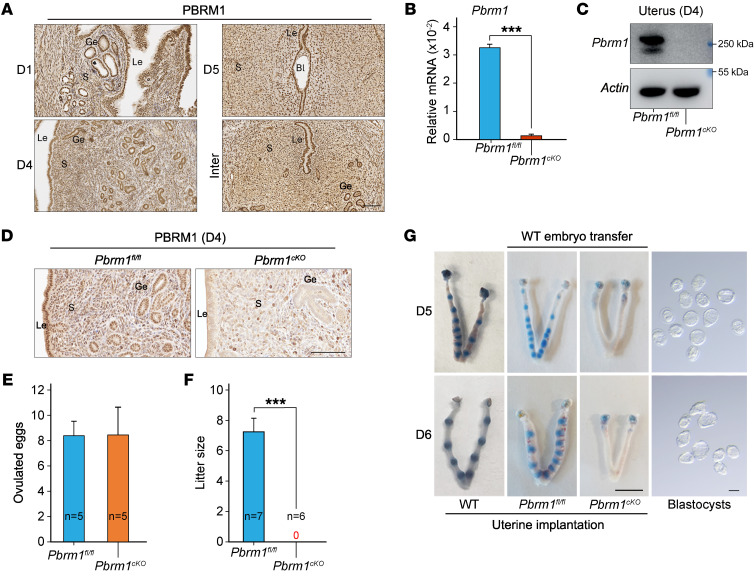
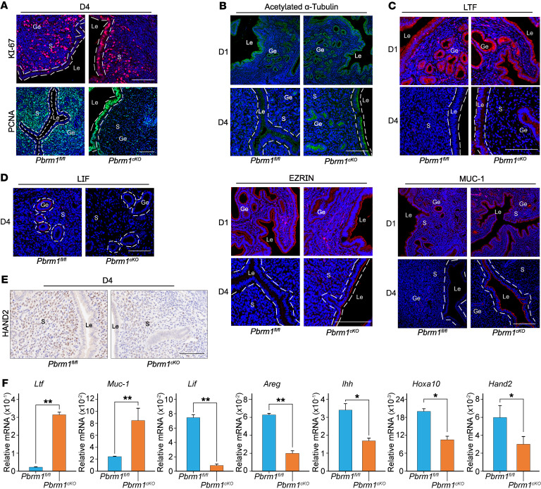
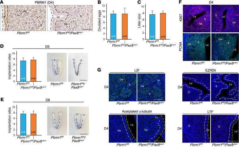
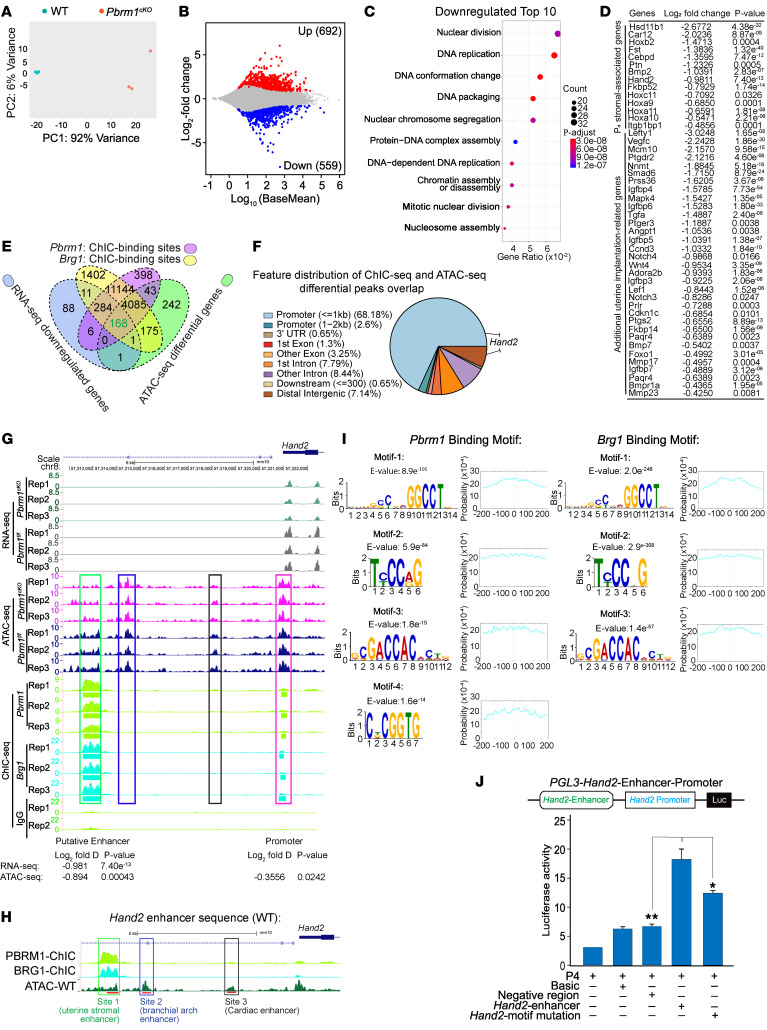
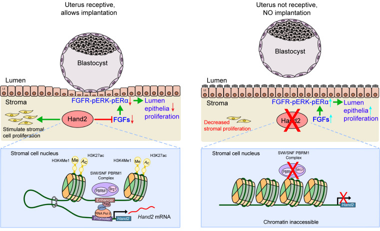
Early gestational loss occurs in approximately 20% of all clinically recognized human pregnancies and is an important cause of morbidity. Either embryonic or maternal defects can cause loss, but a functioning and receptive uterine endometrium is crucial for embryo implantation. We report that the switch/sucrose nonfermentable (SWI/SNF) remodeling complex containing polybromo-1 (PBRM1) and Brahma-related gene 1 (BRG1) is essential for implantation of the embryonic blastocyst on the wall of the uterus in mice. Although preimplantation development is unaffected, conditional ablation of Pbrm1 in uterine stromal cells disrupts progesterone pathways and uterine receptivity. Heart and neural crest derivatives expressed 2 (Hand2) encodes a basic helix-loop-helix (bHLH) transcription factor required for embryo implantation. We identify an enhancer of the Hand2 gene in stromal cells that requires PBRM1 for epigenetic histone modifications/coactivator recruitment and looping with the promoter. In Pbrm1cKO mice, perturbation of chromatin assembly at the promoter and enhancer sites compromises Hand2 transcription, adversely affects fibroblast growth factor signaling pathways, prevents normal stromal-epithelial crosstalk, and disrupts embryo implantation. The mutant female mice are infertile and provide insight into potential causes of early pregnancy loss in humans.

Keywords: Endocrinology; Epigenetics; Fertility; Reproductive biology; Sex hormones.


Conflict of interest statement

Conflict of interest: The authors have declared that no conflict of interest exists.

Figures

Figure 1
Figure 1. Implantation failure and female infertility after uterine-specific Pbrm1 deletion.
(A) Immunohistochemical staining showing the spatiotemporal expression of PBRM1 in WT uteri on D1, D4, and D5 of pregnancy. Bl, blastocyst; S, stromal. Scale bar: 100 mm. (B) RT-qPCR analysis of Pbrm1 mRNA levels in D4 uteri from Pbrm1fl/fl and Pbrm1cKO mice. Values are normalized to Gapdh expression and represented as mean ± SEM (n = 3). ***P < 0.001, independent-sample Student’s t test. (C) Immunoblotting of PBRM1 protein in D4 Pbrm1fl/fl and Pbrm1cKO uteri. β-actin, load control. (D) Immunohistochemistry of PBRM1 protein in D4 Pbrm1fl/fl and Pbrm1cKO uteri. Scale bar: 100 mm. (E) Number of ovulated eggs in Pbrm1fl/fl and Pbrm1cKO mice. Numbers within the bars indicate numbers of mice tested. (F) Average litter sizes of Pbrm1fl/fl and Pbrm1cKO female mice. Numbers within the bars indicate numbers of mice tested. Data are represented as mean ± SEM. ***P < 0.001, independent-sample Student’s t test. (G) Representative images of normal embryo morphology from D5 to D6 pregnant Pbrm1cKO mice, exhibiting embryo implantation failure in the uteri beyond D5. Scale bars: 1 cm (uterus); 100 mm (embryos).
Figure 2
Figure 2. Pbrm1 deficiency disrupts uterine receptivity and dysregulates E2/P4 signaling.
(A) Immunofluorescence staining of KI-67 and PCNA documents an aberrant epithelial proliferation accompanied by a decreased stromal proliferation in Pbrm1cKO mouse uteri on D4. Scale bars: 100 mm. (B) Immunofluorescence staining of cilia marker acetylated α-tubulin (top panels) and microvilli marker EZRIN (bottom panels) in Pbrm1fl/fl and Pbrm1cKO mouse uteri. Scale bars: 100 mm. (CE) Immunofluorescence (C and D) and immunohistochemical staining (E) of receptivity marker genes document impaired uterine receptivity in Pbrm1cKO females on D4. Scale bars: 100 mm. (F) RT-qPCR analysis of implantation-related marker gene expression in D4 Pbrm1fl/fl and Pbrm1cKO uteri. Values are normalized to Gapdh expression level and represented as mean ± SEM (n = 3). *P < 0.05; **P < 0.01, independent-sample Student’s t test.
Figure 3
Figure 3. Epithelium-selective deletion of Pbrm1 leads to normal embryo implantation and female fertility.
(A) Immunohistochemistry documents specific deficiency of epithelial PBRM1 in Pbrm1fl/fl/Pax8cre/+ mouse uteri. Scale bar: 100 mm. (B and C) Number of ovulated eggs (B) and average litter sizes (C) of Pbrm1fl/fl and Pbrm1fl/fl/Pax8cre/+ female mice. Numbers within the bars indicate numbers of mice examined. Data are represented as mean ± SEM. (D and E) Normal implantation in Pbrm1fl/fl/Pax8cre/+ mice compared with Pbrm1fl/fl mice as determined by Chicago Sky Blue dye injection on D5 to D6 of pregnancy. Numbers within the bars indicate numbers of female mice tested. Scale bars: 1 cm. (F and G) Immunofluorescence images of proliferation status and receptivity marker genes document normal uterine receptivity in Pbrm1fl/fl/Pax8cre/+ females on D4. Scale bars: 100 mm.
Figure 4
Figure 4. PBRM1 promotion of Hand2 transcription is dependent on chromatin remodeling.
(A) PCA plot of RNA-Seq results of uterine stromal cells from Pbrm1fl/fl and Pbrm1cKO mice. (B) MA plots of differentially expressed RNAs in Pbrm1fl/fl and Pbrm1cKO mUSCs. Upregulated and downregulated RNAs are shown as red and blue dots, respectively (>1.5-fold, P <0.01, Padj < 0.1). (C) The top 10 GO terms of downregulated transcripts. (D) Genes directly associated with uterine receptivity and implantation that were significantly downregulated (log2 fold change and P value) from Pbrm1fl/fl and Pbrm1cKO uterine stromal cell RNA-Seq. (E) Venn diagram showing overlap among genes (n = 168) with reduction of RNA expression and differential ATAC accessibility by Pbrm1 deletion and genes with PBRM1/BRG1 direct binding. (F) Genomic distribution of overlapping genes (n = 168) that are most likely to be direct targets of the PBRM1/BRG1 complex. (G) Genome browser view of normalized RNA-Seq signals, ATAC-Seq, and PBRM1/BRG1 ChIC-Seq tracks for Hand2 in Pbrm1fl/fl and Pbrm1cKO primary mUSCs. Green rectangle, newly identified uterine specific enhancer regulated by SWI/SNF complex; blue rectangle, known branchial arch enhancer; black rectangle, cardiac-specific enhancer; red rectangle, promoter of Hand2; Rep 1, 2 and 3, 3 biological replicates. (H) Schematic representation of enhancer regions of Hand2 in different tissues. Hand2 uterine (site 1, red), branchial arch enhancer (site 2, green), and cardiac enhancer (site 3, blue). (I) Prediction of DNA-binding site motifs for PBRM1/BRG1 derived from ChIC-Seq data. Graph to right indicates the probability of the binding motif. (J) Renilla-normalized luciferase reporter assay to evaluate Hand2 promoter activation transfected with Hand2 WT or motif mutation vectors. Values are represented as mean ± SEM (n = 3). P values were calculated by post hoc pairwise t test after 1-way ANOVA. *P < 0.05; **P < 0.01.
Figure 5
Figure 5. Uterine-specific depletion of Brg1 results in embryo implantation failure and female infertility.
(A) Immunohistochemical staining documents a spatiotemporal expression of BRG1 in WT uteri on D1, D4, and D5 of pregnancy. Scale bar: 100 mm. (BD) RT-qPCR (B), immunoblotting (C), and immunohistochemical images (D) of Brg1 mRNA and BRG1 protein levels in D4 uteri from Brg1fl/fl and Brg1fl/fl/PRIRES-cre/+ mice. Values are normalized to Gapdh expression and represented as mean ± SEM (n = 4). ***P < 0.001, independent-sample Student’s t test. β-actin, load control. Scale bar: 100 mm. (E) Number of ovulated eggs in Brg1fl/fl and Brg1fl/fl/PRIRES-cre/+ mice. Numbers within the bars indicate numbers of mice tested. (F) Average litter sizes of Brg1fl/fl and Brg1fl/fl/PRIRES-cre/+ female mice. Numbers within the bars indicate numbers of mice tested. Data are represented as mean ± SEM. ***P < 0.001, independent-sample Student’s t test. (G and H) Representative images of normal embryo morphology from D5 (G) and D6 (H) pregnant Brg1fl/fl/PRIRES-cre/+ mice, exhibiting embryo implantation failure in the uteri beyond D5. Numbers within the bars indicate numbers of mice with implantation sites per total tested mice. Data are represented as mean ± SEM. **P < 0.01, independent-samples Student’s t test. Scale bars, 1 cm (uterus); 100 mm (embryos). (I and J) Immunofluorescence staining of PCNA and HAND2 document aberrant epithelial proliferation and impaired uterine receptivity in Brg1fl/fl/PRIRES-cre/+ females on D4. Scale bars: 100 mm.
Figure 6
Figure 6. Loss of PBRM1 decreases enhancer histone modifications and recruitment of transcriptional factors and compromises enhancer/promoter interactions.
(A) ChIP-qPCR for enhancer markers H3K4me1/H3K27ac modifications and recruitment of transcriptional factors Gata4 and P300 to the Hand2 uterine–specific enhancer region (site 1) are reduced in Pbrm1cKO mUSCs. Values are normalized to input. Data are represented as mean ± SEM. *P < 0.05; **P < 0.01, independent-sample Student’s t test. (B) The same as A of the branchial arch (site 2) and (C) cardiac enhancer (site 3) regions showed no differences in the H3K4me1/H3K27ac modifications and transcriptional factor recruitment between Pbrm1fl/fl and Pbrm1cKO uterine stromal cells. (D) 3C interaction frequency (enhancer-promoter looping) between Hand2-specific enhancer and its promoter in Pbrm1fl/fl and Pbrm1cKO mUSCs. LCR serves as anchor. Values are represented as mean ± SEM (n = 3). P values were calculated by Student’s t test. *P < 0.05. (E) ChIP-qPCR result shows enriched binding of PBRM1 to the putative enhancer site upon P4 treatment. P value was calculated by post hoc pairwise t test after 2-way ANOVA. *P < 0.05. (F and G) RT-qPCR analysis of oviductal (F) and uterine (G) Hand2 transcriptional levels in WT and Hand2-specific enhancer knockout mice on D4 of pregnancy. Values are represented as mean ± SEM of 3 biological replicates. *P < 0.05, independent-sample Student’s t test.
Figure 7
Figure 7. Augmented Fgfs/Fgfr-pErk1/2-pErα signaling in Pbrm1-deficient uteri disrupts embryo implantation.
(A) RT-qPCR of FGF family growth factors (FGF1, FGF7, FGF16, FGF17, FGF18, FGF21) in the uterine stroma of Pbrm1fl/fl and Pbrm1cKO mice on D4 of pregnancy. Values are normalized to Gapdh expression and represented as mean ± SEM (n = 3). *P < 0.05; **P < 0.01, independent-sample Student’s t test. (B and C) Immunofluorescence staining of FGFs (FGF1, FGF17, FGF18) and FGFR2 in Pbrm1fl/fl and Pbrm1cKO females on D4. (D) Immunostaining of p-FRS2, p-ERK1/2, and p-ERα documents augmented Fgfs/Fgfr-pErk1/2-pErα signaling in Pbrm1cKO Le on D4 of pregnancy. Scale bars: 100 mm.
Figure 8
Figure 8. Uterine stromal PBRM1 governs uterine receptivity necessary for embryo implantation.
Left: Upon P4 priming on D4, PBRM1 is recruited to the Hand2 enhancer and promoter and facilitates chromatin accessibility and physical interactions (looping) essential for transcription activation in a SWI/SNF complex–dependent manner. This ensures normal stromal-epithelium crosstalk conducive to uterine receptivity and implantation. Right: In the absence of PBRM1 (lower) in the stromal-epithelium, crosstalk does not occur in the absence of Hand2 expression (upper).

Similar articles

Cited by

References

    1. Cha J, et al. Mechanisms of implantation: strategies for successful pregnancy. Nat Med. 2012;18(12):1754–1767. doi: 10.1038/nm.3012. - DOI - PMC - PubMed
    1. Norwitz ER, et al. Implantation and the survival of early pregnancy. N Engl J Med. 2001;345(19):1400–1408. doi: 10.1056/NEJMra000763. - DOI - PubMed
    1. Wang H, Dey SK. Roadmap to embryo implantation: clues from mouse models. Nat Rev Genet. 2006;7(3):185–199. doi: 10.1038/nrg1808. - DOI - PubMed
    1. Xin QL, et al. Transcriptional activation of nuclear estrogen receptor and progesterone receptor and its regulation. Sheng Li Xue Bao. 2016;68(4):435–454. - PubMed
    1. Thompson M. Polybromo-1: the chromatin targeting subunit of the PBAF complex. Biochimie. 2009;91(3):309–319. doi: 10.1016/j.biochi.2008.10.019. - DOI - PMC - PubMed