. 2024 Apr;51(4):59.
doi: 10.3892/or.2024.8718. Epub 2024 Mar 1.

m6A methyltransferase METTL14‑mediated RP1‑228H13.5 promotes the occurrence of liver cancer by targeting hsa‑miR‑205/ZIK1

Affiliations

m6A methyltransferase METTL14‑mediated RP1‑228H13.5 promotes the occurrence of liver cancer by targeting hsa‑miR‑205/ZIK1

Jia Xu et al. Oncol Rep. 2024 Apr.

Abstract

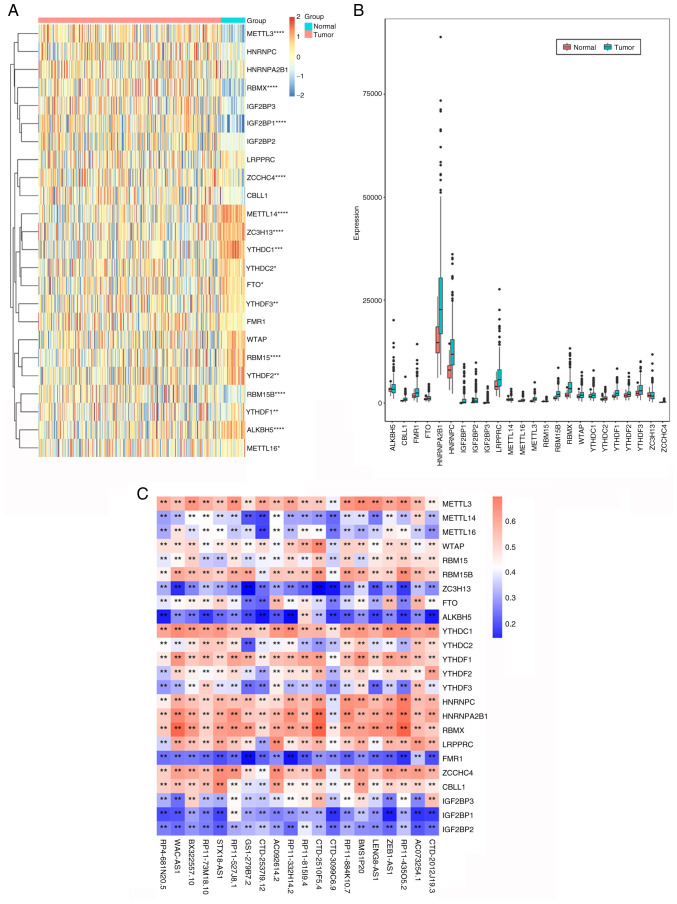
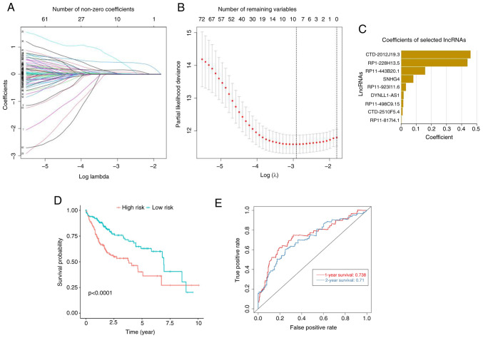
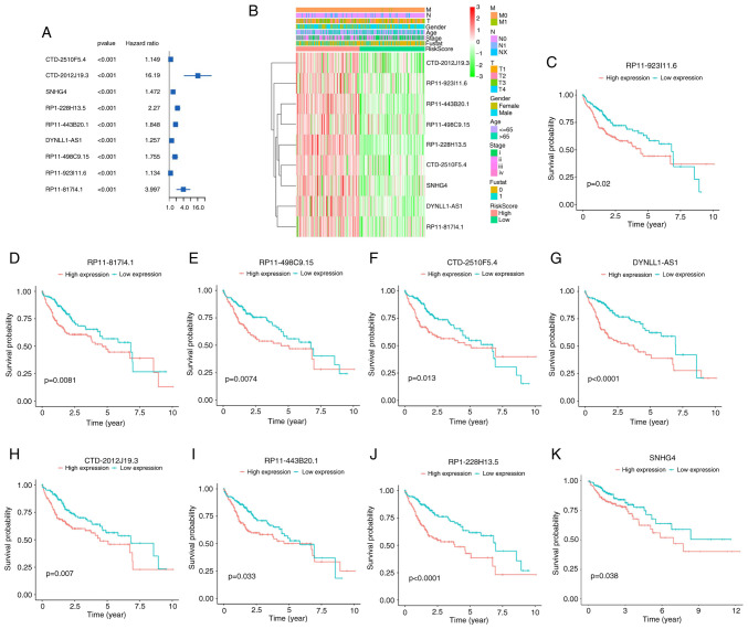
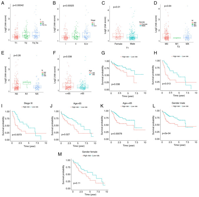
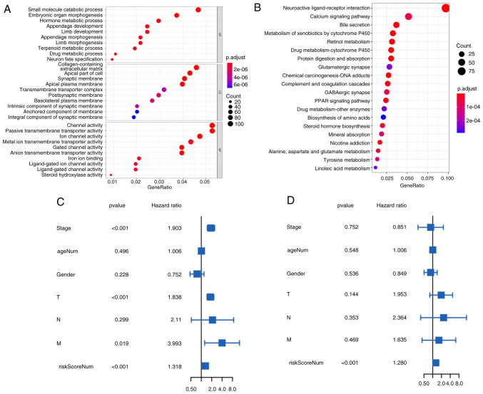
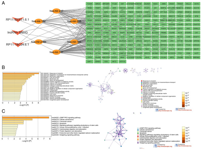
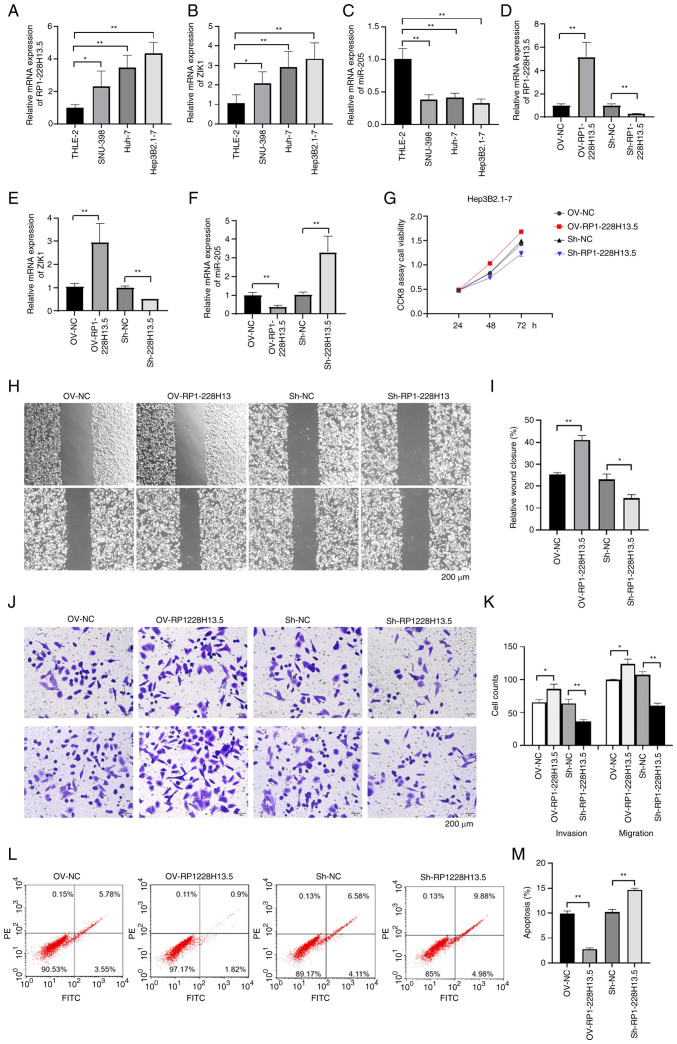
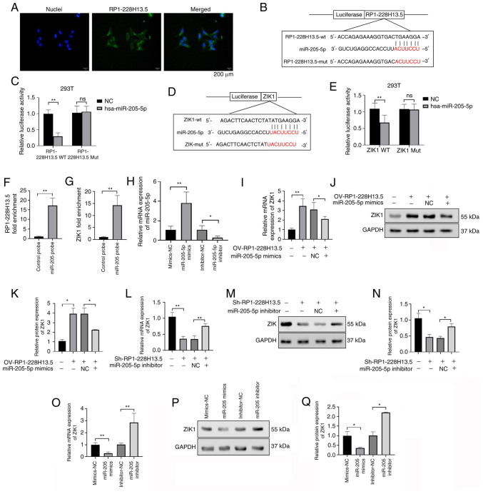
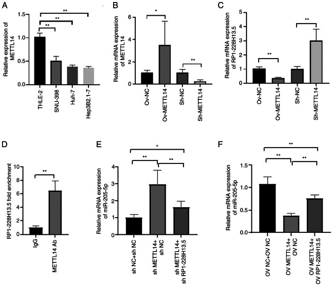
The aim of the present study was to explore the association between N6‑methyladenosine (m6A) modification regulatory gene‑related long noncoding (lnc)RNA RP1‑228H13.5 and cancer prognosis through bioinformatics analysis, as well as the impact of RP1‑228H13.5 on cell biology‑related behaviors and specific molecular mechanisms. Bioinformatics analysis was used to construct a risk model consisting of nine genes. This model can reflect the survival time and differentiation degree of cancer. Subsequently, a competing endogenous RNA network consisting of 3 m6A‑related lncRNAs, six microRNAs (miRs) and 201 mRNAs was constructed. A cell assay confirmed that RP1‑228H13.5 is significantly upregulated in liver cancer cells, which can promote liver cancer cell proliferation, migration and invasion, and inhibit liver cancer cell apoptosis. The specific molecular mechanism may be the regulation of the expression of zinc finger protein interacting with K protein 1 (ZIK1) by targeting the downstream hsa‑miR‑205. Further experiments found that the m6A methyltransferase 14, N6‑adenosine‑methyltransferase subunit mediates the regulation of miR‑205‑5p expression by RP1‑228H13.5. m6A methylation regulatory factor‑related lncRNA has an important role in cancer. The targeting of hsa‑miR‑205 by RP1‑228H13.5 to regulate ZIK1 may serve as a potential mechanism in the occurrence and development of liver cancer.

Keywords: METTL14; RP1-228H13.5/hsa‑miR‑205/ZIK1; lncRNA; m6A methylated.


Conflict of interest statement

The authors declare that they have no competing interests.

Figures

Figure 1.
Figure 1.
Prognostic analysis of m6A methylation regulatory factor-related lncRNAs. (A) Heatmap of the correlations between m6A-related genes and the 24 prognostic m6A-related lncRNAs. (B) Violin diagram of mRNA expression distribution of 24 RNA m6A methylation regulators in liver cancer tumor tissues and adjacent normal tissues. Blue represents normal tissues and red represents tumor tissues. (C) Heat map of the association between m6A-related genes and m6A-related lncRNAs. *P<0.05, **P<0.01, ***P<0.001 and ****P<0.0001. lncRNA, long non-coding RNA; m6A, N6-methyladenosine.
Figure 2.
Figure 2.
Least absolute1 shrinkage and selection operator (LASSO) regression was performed, calculating the minimum criteria (A-C) coefficient risk score corresponding to each lncRNA was calculated using LASSO regression analysis. (D) Kaplan-Meier curve indicating that the overall survival rate of the high-risk subgroup was lower than that of the low-risk subgroup. (E) Receiver operating characteristic curve analysis was used to predict the one- and two-year survival rate. LASSO, Least Absolute Shrinkage and Selection Operator; lncRNA, long non-coding RNA.
Figure 3.
Figure 3.
Prognostic analysis of nine lncRNAs associated with the m6A gene. (A) Forest map results of the prognostic ability of nine m6A gene-related lncRNAs. (B) Heatmap of the association between the expression level of nine m6A gene-related lncRNAs and clinicopathological features. (C-H) Kaplan-Meier curves showing survival according to (C) RP11-923I11.6 m6A-related lncRNA expression levels; (D) RP11-817I4.1 m6A-related lncRNA expression levels; (E) RP11-498C9.15 m6A-related lncRNA expression levels; (F) CTD-2510F5.4 m6A-related lncRNA expression levels; (G) DYNLL1-AS1 m6A-related lncRNA expression levels; (H) CTD-2012J19.3 m6A-related lncRNA expression levels; (I) RP11-443B20.1 m6A-related lncRNA expression levels; (J) RP1-228H13.5 m6A-related lncRNA expression levels; and (K) SNHG4 m6A-related lncRNA expression levels. lncRNAs, long non-coding RNA; m6A, N6-methyladenosine.
Figure 4.
Figure 4.
Association between clinicopathological features and risk score. (A-F) Patients with different clinicopathological features have different levels of risk scores: (A) TNM stage, (B) stage, (C) gender, (D) M stage, (E) N stage and (F) age. (G-M) Prognostic analysis of risk model in multiple subgroups of patients with liver cancer: (G) T1, (H) T3, (I) Stage III, (J) age >65 years, (K) age ≤65 years, (L) males and (M) females.
Figure 5.
Figure 5.
Enrichment analysis of differentially expressed genes in low- and high-risk subgroups, and univariate and multivariate Cox regression analysis of risk score. (A) Gene Ontology functional analysis of 2,352 differentially expressed genes. (B) Kyoto Encyclopedia of Genes and Genomes pathway analysis of 2,352 differentially expressed genes. Both (C) Univariate and (D) multivariate regression analysis showed that the risk score of patients was a risk factor.
Figure 6.
Figure 6.
Construction of ceRNA network map and enrichment analysis of target genes. (A) LncRNA-miRNA-mRNA ceRNA network map. (B) GO enrichment analysis of target genes. (C) KEGG enrichment analysis of target genes. ceRNA, competing endogenous RNA; lncRNAs, long non-coding RNA; miRNA/miR, microRNA; GO, gene ontology; KEGG, Kyoto Encyclopedia of Genes and Genomes.
Figure 7.
Figure 7.
RP1-228H13.5 is upregulated in liver cancer and promotes the proliferation, migration and invasion of Hep3B2.1–7 cells, while inhibiting cell apoptosis. (A) RT-qPCR assay was used to detect the expression levels of RP1-228H13.5 in THLE-2, SNU-398, Huh-7 and Hep3B2.1–7 cells. (B) RT-qPCR assay was used to detect the expression levels of ZIK1 in THLE-2, SNU-398, Huh-7 and Hep3B2.1–7 cells. (C) RT-qPCR assay was used to detect the expression levels of miR-205-5p in THLE-2, SNU-398, Huh-7 and Hep3B2.1–7 cells. (D-F) Following the overexpression and knockdown of RP1-228H13.5, RT-qPCR was performed to detect the expression levels of (D) RP1-228H13.5, (E) ZIK1 and (F) miR-205-5p. (G) Following the overexpression and knockdown of RP1-228H13.5, CCK-8 assay was used to detect the cell proliferation ability. (H and I) Following the overexpression and knockdown of RP1-228H13.5, a wound-healing assay was used to detect cell migration. (H) Representative images (scale bar, 200 µm) and (I) quantitative results. (J and K) Following overexpression and knockdown of RP1-228H13.5, a Transwell assay was used to detect cell invasion ability. (J) Representative images (scale bar, 200 µm) and (K) quantitative results. (L and M) Following the overexpression and knockdown of RP1-228H13.5, flow cytometry was used to detect cell apoptosis. (L) Representative graphs and (M) quantitative results. Values are expressed as the mean ± standard error of the mean. An unpaired Student's t-test was used for two-group comparison and one-way ANOVA for multi-group comparisons. *P<0.05, **P<0.01. RT-qPCR, reverse transcription-quantitative PCR; CCK-8, cell-counting kit; NC, negative control; OV, overexpression; Sh, short hairpin RNA; miR, microRNA; ZIK1, zinc finger protein interacting with K protein 1.
Figure 8.
Figure 8.
RP1-228H13.5-targeted miR-205-5p regulates the expression of ZIK1. (A) Fluorescence in situ hybridization detected the localization of RP1-228H13.5 in cells (scale bar, 200 µm). (B) Prediction of the binding sites of RP1-228H13.5 and miR-205-5p through the Starbase 2.0 database. (C) The combination of RP1-228H13.5 and miR-205-5p was verified in a dual luciferase experiment. (D) Prediction of the binding sites of miR-205-5p and ZIK1 through the Starbase 2.0 database. (E) The combination of miR-205-5p and ZIK1 was verified by a dual luciferase experiment. (F) RNA pull-down assay verified the binding of miR-205-5p and RP1-228H13.5. (G) RNA pull-down assay verified the binding of miR-205-5p and ZIK1. (H) RT-qPCR assay detected the expression of miR-205-5p after overexpression and knockdown of miR-205-5p in Hep3B2.1–7 cells. (I) Following the transfection of cells with OV-RP1-228H13.5 alone or with miR-205-5p mimics, RT-qPCR was used to detect the expression of ZIK1. (J and K) Following the transfection of cells with OV-RP1-228H13.5 alone or with miR-205-5p mimics, western blotting was used to detect the expression of ZIK1. (J) Representative western blots and (K) quantified results. (L) Following the transfection of Sh-RP1-228H13.5 alone or with miR-205-5p inhibitor into cells, RT-qPCR were used to detect the expression of ZIK1. (M and N) Following the transfection of Sh-RP1-228H13.5 alone or with miR-205-5p inhibitor into cells, western blotting was used to detect the expression of ZIK1. (M) Representative western blots and (N) quantified results. (O) Following the overexpression and knockdown of miR-205-5p, the expression level of ZIK1 was detected by RT-qPCR. (P and Q) Following the overexpression and knockdown of miR-205-5p, the expression level of ZIK1 was detected by western blotting. (P) Representative western blots and (Q) quantified results. Values are expressed as the mean ± standard error of the mean. An unpaired Student's t-test was used for two-group comparison. *P<0.05, **P<0.01. ns, no significance; RT-qPCR, reverse transcription-quantitative PCR; NC, negative control; OV, overexpression; Sh, short hairpin RNA; miR, microRNA; ZIK1, zinc finger protein interacting with K protein 1; mut, mutant; wt, wild-type.
Figure 9.
Figure 9.
N6-methyladenosine methyltransferase METTL14 mediates the regulation of miR-205-5p expression by RP1-228H13.5. (A) RT-qPCR was used to detect the expression level of METTL14 in normal and liver cancer cells. (B and C) METTL14 overexpression and knockdown were performed, and the expression level of (B) METTL14 and (C) RP1-228H13.5 was detected by RT-qPCR in Hep3B2.1–7 cells. (D) RNA pull-down assay verified the binding of METTL14 with RP1-228H13.5. (E) After co-transfecting cells with sh-METTL14 or sh-METTL14 + sh-RP1-228H13.5, the expression of miR-205-5p was detected using RT-qPCR. (F) After co-transfecting cells with OV-METTL14 or OV-METTL14 + OV-RP1-228H13.5, the expression of miR-205-5p was detected by RT-qPCR. Values are expressed as the mean ± standard error of the mean. An unpaired Student's t-test was used for two-group comparison and one-way ANOVA for multi-group comparisons. *P<0.05, **P<0.01. RT-qPCR, reverse transcription-quantitative PCR; NC, negative control; OV, overexpression; Sh, short hairpin RNA; miR, microRNA; Ab, antibody; METTL14, methyltransferase 14, N6-adenosine-methyltransferase subunit.

Similar articles

Cited by

References

    1. Siegel RL, Miller KD, Jemal A. Cancer statistics, 2019. CA Cancer J Clin. 2019;69:7–34. doi: 10.3322/caac.21551. - DOI - PubMed
    1. Ashtari S, Pourhoseingholi MA, Sharifian A, Zali MR. Hepatocellular carcinoma in Asia: Prevention strategy and planning. World J Hepatol. 2015;7:1708–1717. doi: 10.4254/wjh.v7.i12.1708. - DOI - PMC - PubMed
    1. Gong B, Kao Y, Zhang C, Sun F, Zhao H. Systematic Investigation of scutellariae barbatae herba for treating hepatocellular carcinoma based on network pharmacology. Evid Based Complement Alternat Med. 2018;2018:4365739. doi: 10.1155/2018/4365739. - DOI - PMC - PubMed
    1. Du L, Li Y, Kang M, Feng M, Ren Y, Dai H, Wang Y, Wang Y, Tang B. USP48 is upregulated by Mettl14 to attenuate hepatocellular carcinoma via regulating SIRT6 stabilization. SIRT6 stabilization. Cancer Res. 2012;81:3822–3834. doi: 10.1158/0008-5472.CAN-20-4163. - DOI - PubMed
    1. Cheng X, Li M, Rao X, Zhang W, Li X, Wang L, Huang G. KIAA1429 regulates the migration and invasion of hepatocellular carcinoma by altering m6A modification of ID2 mRNA. Onco Targets Ther. 2019;12:3421–3428. doi: 10.2147/OTT.S180954. - DOI - PMC - PubMed

MeSH terms