. 2024 Apr 16;63(8):958-968.
doi: 10.1021/acs.biochem.3c00481. Epub 2024 Mar 1.

A Novel Dual-Fc Bispecific Antibody with Enhanced Fc Effector Function

Affiliations

A Novel Dual-Fc Bispecific Antibody with Enhanced Fc Effector Function

Fulai Zhou et al. Biochemistry. .

Abstract

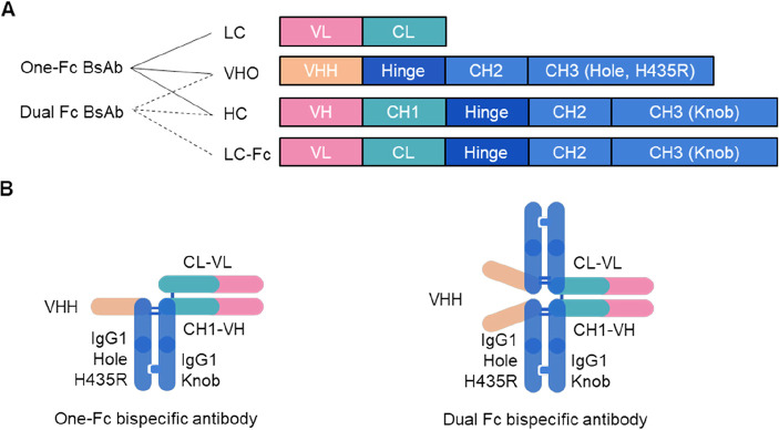
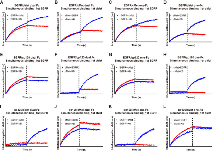
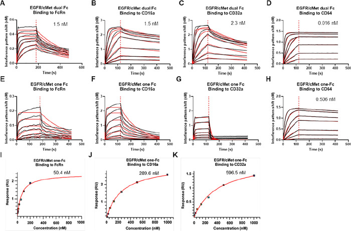
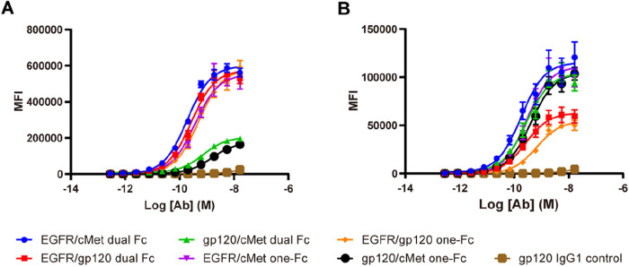
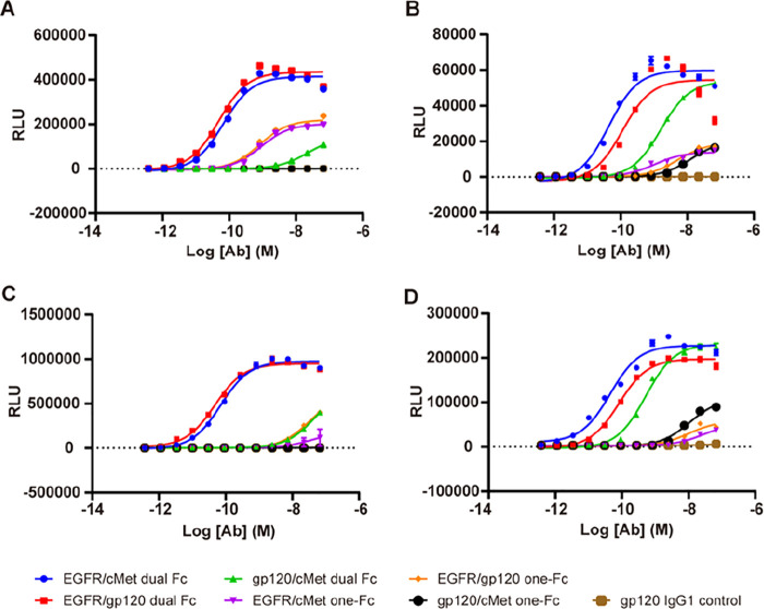
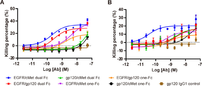
Bispecific antibodies (BsAbs) are undergoing continued development for applications in oncology and autoimmune diseases. While increasing activity by having more than one targeting arm, most BsAb engineering employs single Fc engagement as monoclonal antibodies. Here, we designed a novel immunoglobulin gamma-1 (IgG1)-derived dual-Fc BsAb containing two Fc regions and two distinct asymmetric antigen binding arms comprising a Fab arm and another VHH domain. In conjunction with the knob-into-hole technology, dual-Fc BsAbs could be produced with a high yield and good stability. We explore how Fc engineering effects on dual-Fc constructs could boost the desired therapeutic efficacy. This new format enabled simultaneous bispecific binding to corresponding antigens. Furthermore, compared to the one-Fc control molecules, dual-Fc BsAbs were shown to increase the avidity-based binding to FcγRs to result in higher ADCC and ADCP activities by potent avidity via binding to two antigens and Fc receptors. Overall, this novel BsAb format with enhanced effector functionalities provides a new option for antibody-based immunotherapy.


Conflict of interest statement

The authors declare no competing financial interest.

Figures

Figure 1
Figure 1
Overall strategy for the assembly of the one-Fc or dual-Fc bispecific antibodies. (A) Coexpression of plasmids containing the open reading frame for the variable heavy chain only heavy chain, a normal heavy chain, and the complementary light chain or light chain–Fc fusion to generate the respective one-Fc or dual-Fc bispecific antibody molecules. The knob-into-hole and H435R mutations were indicated in each Fc region. (B) Schematic depiction of a one-Fc or dual-Fc bispecific antibody. The two VHH domains in the dual-Fc bispecific antibody are separate due to lack of interactions. VHH, variable domain of the heavy chain of a heavy chain only antibody; HC, heavy chain; LC, light chain; Fc, crystallizable fragment; VHO, variable heavy chain only antibody; BsAb, bispecific antibody.
Figure 2
Figure 2
Simultaneous binding analyses of the dual-Fc and one-Fc bispecific antibodies by biolayer interferometry. The dual-Fc and one-Fc BsAbs were first tested for binding to EGFR or cMet at a concentration of 50 nM, followed by the second binding to the other antigen at a concentration of 50 nM or KB (control). The EGFR/cMet dual-Fc (A and B) and one-Fc (C and D) BsAbs were first bound to EGFR (blue) and subsequently with cMet (or KB as control, red) (A and C) or first bound to cMet (blue) and subsequently with EGFR (or KB as control, red) (B and D). The EGFR/gp120 dual-Fc (E and F) and one-Fc (G and H) BsAbs were first bound to EGFR (blue) and subsequently with cMet (or KB as control, red) (E and G) or first bound to cMet (blue) and subsequently with EGFR (or KB as control, red) (F and H). The gp120/cMet dual-Fc (I and J) and one-Fc (K and L) BsAbs were first bound to EGFR (blue) and subsequently with cMet (or KB as control, red) (I and K) or first bound to cMet (blue) and subsequently with EGFR (or KB as control, red) (J and L). The y-axis responses were measured as the nanometer shifts in the interference pattern and were proportional to the number of molecules bound to the biosensor. The x-axis indicated the time interval for antigen binding, and the dashed line indicated the time point of association to secondary antigen or KB. The red plots represented binding events between bispecific antibodies with EGFR and cMet. Conversely, the blue plots depicted the binding events between EGFR/cMet and KB. The composition and structure of the dual-Fc and one-Fc bispecific antibodies are provided in Figure S1. BsAbs, bispecific antibodies. KB, kinetics buffer.
Figure 3
Figure 3
Affinity determination of the dual-Fc and one-Fc bispecific antibodies with the Fc receptors. Kinetics analyses of EGFR/cMet bispecific antibodies in dual-Fc format binding to human FcRn (A), CD16a (B), CD32a (C), and CD64 (D). Kinetics analyses of EGFR/cMet bispecific antibodies in one-Fc (E–H) format binding to human FcRn (E), CD16a (F), CD32a (G), and CD64 (H). The representative kinetics and fitting curves were shown and colored in black and red, respectively. The resulting dissociation constant values (KD) were measures of affinity and were given as the mean values from three independent experiments at the top-right corner in each panel, except for the EGFR/cMet one-Fc bispecific antibody binding to human FcRn, CD16a, and CD32a due to their fast dissociation and poor fitting. Representative fitting curves of steady-state affinity analysis for the EGFR/cMet one-Fc bispecific antibody binding to human FcRn (I), CD16a (J), and CD32a (K). The resulting affinity values were given as the mean values from three independent experiments at the top-right corner in each panel. See Table 2 for the detailed kinetics parameters.
Figure 4
Figure 4
Cell binding of the dual-Fc and one-Fc bispecific antibodies. Concentration–response curves of dual-Fc and one-Fc antibodies binding to H292 (A) and MKN45 (B) cells. Data were shown as the mean values ± SEM of at least three independent experiments. Curves for EGFR/cMet, EGFR/gp120, and gp120/cMet dual-Fc bispecific antibodies were colored in blue, red, and green, respectively. Curves for EGFR/cMet, EGFR/gp120, and gp120/cMet one-Fc bispecific antibodies were colored in purple, orange, and black, respectively. The gp120 IgG1 monoclonal antibody (colored in brown) was used as a negative control. MFI, mean fluorescent intensity. SEM, standard error of the mean. Fc, crystallizable fragment. Ab, antibody.
Figure 5
Figure 5
ADCC and ADCP reporter bioassay activity for the dual-Fc and one-Fc bispecific antibodies. The same color scheme was used as Figure 4. (A–D) NFAT activation reflected the induced ADCC or ADCP responses in Jurkat-FcγRIIIa-V158 and Jurkat-FcγRIIa-H131 cell lines, respectively. Concentration–response curves of the ADCC (A and B) response induced by dual-Fc and one-Fc bispecific antibodies in H292 (A) or MKN45 (B) target cells and concentration–response curves of the ADCP (C and D) response induced by dual-Fc and one-Fc bispecific antibodies in H292 (C) or MKN45 (D) target cells. The curves were analyzed using a three-parameter logistic equation. The data shown were the mean values ± SEM of at least three independent experiments. NFAT, nuclear factor of activated T-cells. RLU, relative light unit. SEM, standard error of the mean. Ab, antibody.
Figure 6
Figure 6
Human PBMC ADCC activity for the dual-Fc and one-Fc bispecific antibodies. The same color scheme was used as Figure 4. (A and B) Concentration–response curves of the human PBMC ADCC response induced by dual-Fc and one-Fc bispecific antibodies with H292 (A) or MKN45 (B) target cells (25:1 ratio), analyzed using a three-parameter logistic equation. Data in Table S3 were the mean values ± SEM of at least three independent experiments. SEM, standard error of the mean. huPBMC, human peripheral blood mononuclear cells.

Similar articles

References

    1. Weiner L. M.; Murray J. C.; Shuptrine C. W. Antibody-based immunotherapy of cancer. Cell 2012, 148 (6), 1081–1084. 10.1016/j.cell.2012.02.034. - DOI - PMC - PubMed
    1. Thakur A.; Huang M.; Lum L. G. Bispecific antibody based therapeutics: Strengths and challenges. Blood Rev. 2018, 32 (4), 339–347. 10.1016/j.blre.2018.02.004. - DOI - PubMed
    1. Ait-Oufella H.; Libby P.; Tedgui A. Antibody-based immunotherapy targeting cytokines and atherothrombotic cardiovascular diseases. Arch. Cardiovasc. Dis. 2020, 113 (1), 5–8. 10.1016/j.acvd.2019.11.001. - DOI - PubMed
    1. Bathula N. V.; Bommadevara H.; Hayes J. M. Nanobodies: The future of antibody-based immune therapeutics. Cancer Biother. Radiopharm. 2021, 36 (2), 109–122. 10.1089/cbr.2020.3941. - DOI - PubMed
    1. Vidarsson G.; Dekkers G.; Rispens T. IgG subclasses and allotypes: from structure to effector functions. Front. Immunol. 2014, 5, 520.10.3389/fimmu.2014.00520. - DOI - PMC - PubMed

MeSH terms

Substances

LinkOut - more resources