. 2024 Apr 10;15(4):e0262323.
doi: 10.1128/mbio.02623-23. Epub 2024 Mar 1.

Interferon lambda restricts herpes simplex virus skin disease by suppressing neutrophil-mediated pathology

Affiliations

Interferon lambda restricts herpes simplex virus skin disease by suppressing neutrophil-mediated pathology

Drake T Philip et al. mBio. .

Abstract

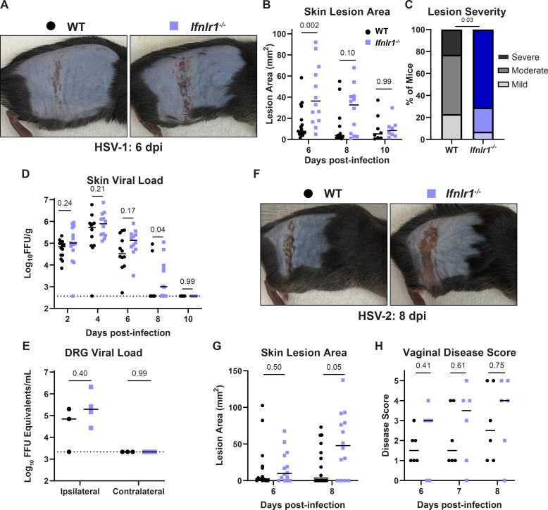
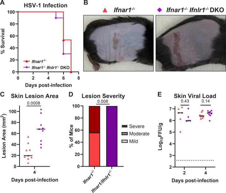
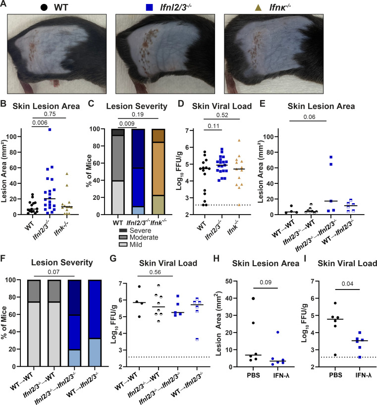
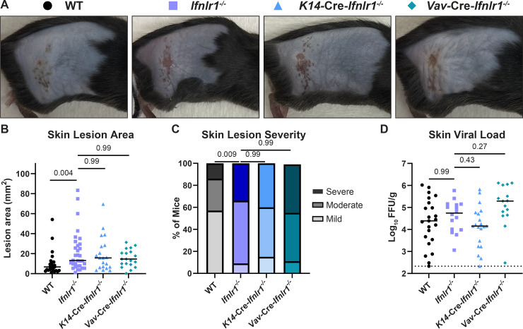
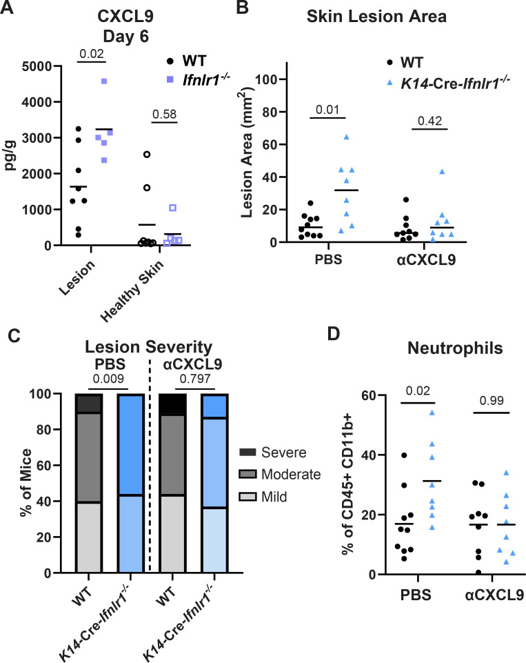
Type III interferons (IFN-λ) are antiviral and immunomodulatory cytokines that have been best characterized in respiratory and gastrointestinal infections, but the effects of IFN-λ against skin infections have not been extensively investigated. We sought to define the skin-specific effects of IFN-λ against the highly prevalent human pathogen, herpes simplex virus (HSV). We infected mice lacking the IFN-λ receptor (Ifnlr1-/-), both the IFN-λ and the IFN-α/β receptors (Ifnar1-/-Ifnlr1-/-), or IFN-λ cytokines (Ifnl2/3-/-) and found that IFN-λ restricts the severity of HSV-1 and HSV-2 skin lesions without affecting viral loads. We used RNAseq to define IFN-λ- and IFN-β-induced transcriptional responses in primary mouse keratinocytes. Using conditional knockout mice, we found that IFN-λ signaling in both keratinocytes and neutrophils was necessary to control HSV-1 skin lesion severity and that IFN-λ signaling in keratinocytes suppressed CXCL9-mediated neutrophil recruitment to the skin. Furthermore, depleting neutrophils or blocking CXCL9 protected against severe HSV-1 skin lesions in Ifnlr1-/- mice. Altogether, our results suggest that IFN-λ plays an immunomodulatory role in the skin that restricts neutrophil-mediated pathology during HSV infection and suggests potential applications for IFN-λ in treating viral skin infections.IMPORTANCEType III interferons (IFN-λ) have been shown to have antiviral and immunomodulatory effects at epithelial barriers such as the respiratory and gastrointestinal tracts, but their effects on the skin have not been extensively investigated. We used mice lacking IFN-λ signaling to investigate the skin-specific effects of IFN-λ against the herpes simplex virus (HSV), which targets epithelial tissues to cause cold sores and genital herpes. We found that IFN-λ limited the severity of HSV skin lesions without affecting viral load and that this protective effect required IFN-λ signaling in both keratinocytes and neutrophils. We found that IFN-λ signaling in keratinocytes suppressed neutrophil recruitment to the skin and that depleting neutrophils protected against severe HSV skin lesions in the absence of IFN-λ. Altogether, our results suggest that IFN-λ plays an immunomodulatory role in the skin that restricts neutrophil-mediated pathology during HSV infection and suggests potential applications for IFN-λ in treating viral skin infections.

Keywords: herpes simplex virus; interferon lambda; keratinocyte; neutrophils; skin.


Conflict of interest statement

The authors declare no conflict of interest.

Figures

Fig 1
Fig 1
IFN-λ signaling protects against severe HSV skin disease without affecting viral replication. (A–E) Eight- to twelve-week-old male and female WT and Ifnlr1−/− mice were infected with 106 FFU of HSV-1 strain NS. (A) Dermatome skin lesions were photographed at 6 dpi. (B) HSV-1 skin lesions were measured from photographs at 6, 8, and 10 dpi using ImageJ. (C) Skin lesion severity was categorized based on the 6 dpi lesion area. (D) Skin viral loads were measured at 2–10 dpi by focus-forming assay. (E) Viral loads in ipsilateral and contralateral dorsal root ganglia (L2–L5 pooled for each mouse) were measured by qPCR. (F–G) Eight- to twelve-week-old male and female WT and Ifnlr1−/− mice were infected with 103 FFU of HSV-2 strain 333. (F) Dermatome lesions were photographed at 8 dpi. (G) HSV-2 skin lesions were measured from photographs at 6 and 8 dpi using ImageJ. (H) 8–12-week-old female WT and Ifnlr1−/− mice were infected with 103 FFU of HSV-2 strain 333 intravaginally and HSV-2 vaginal disease was scored at 6–8 dpi as follows: 1: mild redness and swelling, 2: visible ulceration and fur loss, 3: severe ulceration and signs of sickness, 4: hindlimb paralysis, and 5: moribund/dead. Differences in lesion area and viral load were compared by the Mann-Whitney U test. Differences in categorical skin disease were compared by the Cochran-Armitage test. P values are reported with P < 0.05 considered to be statistically significant. Data are combined from two to three independent experiments.
Fig 2
Fig 2
The protective effect of IFN-λ in the skin does not require IFN-α/β signaling. Eight- to twelve-week-old Ifnar1−/− and Ifnar1−/− Ifnlr1−/− DKO male and female mice were infected with 106 FFU of HSV-1. (A) Lethality was monitored daily (n = 15 Ifnar1−/− and n = 10 DKO mice). (B) Dermatome skin lesions were photographed at 4 dpi. (C) Skin lesion areas were measured from photographs at 4 dpi using ImageJ. (D) Skin lesion severity was categorized based on the 4-dpi lesion area. (E) Skin viral loads were measured at 2 and 4 dpi by focus-forming assay. Survival differences were compared by the Mantel-Cox Log-Rank test. Differences in lesion area and viral load were compared by the Mann-Whitney U test, and differences in categorical skin disease were compared by the Cochran-Armitage test. P values are reported with P < 0.05 considered to be statistically significant. Data are combined from three independent experiments.
Fig 3
Fig 3
IFN-λ cytokines protect against severe HSV-1 skin disease. (A–D) Eight- to twelve-week-old WT, Ifnl2/3−/−, and Ifnk−/− male and female mice were infected with 106 FFU of HSV-1. (A) Dermatome skin lesions were photographed at 6 dpi. (B) Skin lesion areas were measured from photographs at 6 dpi using ImageJ. (C) Skin lesion severity was categorized based on the 6 dpi lesion area. (D) Skin viral loads were measured at 6 dpi by FFA. (E–G) Eight-week-old WT and Ifnl2/3−/− male and female mice were lethally irradiated and transfused with 107 bone marrow cells from WT or Ifnl2/3−/− donors. Ten weeks later, mice were infected with 106 FFU of HSV-1. (E) Skin lesion areas were measured from photographs at 6 dpi using ImageJ. (F) Skin lesion severity was categorized based on the 6 dpi lesion area. (G) Skin viral loads were measured at 6 dpi by FFA. (H–I) Eight- to twelve-week-old WT male and female mice were depilated and then topically treated with 5 µg of recombinant murine IFN-λ3 or PBS. Twenty-four hours later, mice were infected with 106 FFU of HSV-1 at the treated site. (H) Skin lesion areas were measured from photographs at 6 dpi using ImageJ. (I) Skin viral loads were measured at 6 dpi by FFA. Differences in lesion area and viral load were compared by the Mann-Whitney U test, and differences in categorical skin disease were compared by the Cochran-Armitage test. P values are reported with P < 0.05 considered to be statistically significant. Data are combined from two to three independent experiments.
Fig 4
Fig 4
IFN-λ signaling in both keratinocytes and leukocytes is necessary to restrict severe HSV-1 skin disease. Eight- to twelve-week-old male and female WT, Ifnlr1−/−, K14-Cre-Ifnlr1−/−, and Vav-Cre-Ifnlr1−/− mice were infected with 106 FFU of HSV-1. (A) Dermatome skin lesions were photographed at 6 dpi. (B) Skin lesion areas were measured from photographs at 6 dpi using ImageJ. (C) Skin lesion severity was categorized based on the 6 dpi lesion area. (D) Skin viral loads were measured at 6 dpi by FFA. Differences in lesion area and viral load were compared by the Mann-Whitney U test, and differences in categorical skin disease were compared by the Cochran-Armitage test. P values are reported with P < 0.05 considered to be statistically significant. Data are combined from four independent experiments for K14-Cre-Ifnlr1−/− mice and ~10 independent experiments for WT, Ifnlr1−/−, and Vav-Cre-Ifnlr1−/− mice.
Fig 5
Fig 5
IFN-λ signaling in keratinocytes differentially regulates CXCL9 induction. Primary keratinocytes were prepared from WT, Ifnlr1−/−, Ifnar1−/−, and Ifnar1−/−Ifnlr1−/− DKO mice and treated with IFN-λ3 (5 ng/mL), IFN-β (5 ng/mL), or media alone for 24 hours. (A) RNA was extracted, and IFN-stimulated gene expression was measured by RT-qPCR. Ifit1 expression is shown relative to ActB (housekeeping gene) and normalized to expression in media-treated WT cells. Results represent six samples from two independent experiments. (B–F) RNA from three samples per genotype per treatment was analyzed by RNAseq. (B) Principal component analysis for all analyzed samples. (C) Volcano plots showing differentially expressed genes (DEGs) after IFN-λ or IFN-β treatment, compared to media-only treated keratinocytes. Differentially expressed genes were defined as having a |Log2Fold Change| > 1 and an FDR P-value ≤ 0.05. (D) Venn diagram showing DEGs induced by IFN-λ and IFN-β in WT or knockout keratinocytes. (E) Table of IFN-λ-specific DEGs, their Log2 fold change, and FDR P-value for IFN-λ- and IFN-β-treated WT keratinocytes. (F) Volcano plot showing DEGs after IFN-λ treatment compared to IFN-β treatment in WT and receptor knockout keratinocytes.
Fig 6
Fig 6
IFN-λ signaling limits neutrophil abundance and severe skin pathology following HSV-1 infection. Eight- to twelve-week-old male and female WT and Ifnlr1−/− mice were infected with 106 FFU of HSV-1. (A and B) Dermatome lesions and adjacent healthy skin were collected at 4–8 dpi and analyzed by flow cytometry (three independent experiments per time point). (A) Representative histogram of WT and Ifnlr1−/− lesions showing Ly6Ghi neutrophil gating strategy (CD45+, CD11b+, and Ly6Ghi) at 6 dpi. (B) Frequency of neutrophils (Ly6Ghi out of CD45+ CD11b+ live cells) for WT and Ifnlr1−/− dermatome lesions and adjacent healthy skin. Significant differences in neutrophil frequency were determined using an unpaired t test. P values are reported with P < 0.05 considered to be statistically significant. (C and D) Inoculated flank skin was collected at 6 dpi and serial sections of the same skin lesion were processed for histology (six mice per genotype, two independent experiments). (C) H&E staining; the black arrow denotes a neutrophilic pustule, and the red arrow denotes diffuse neutrophilic infiltrate. (D) Anti-HSV-1 immunohistochemistry; viral antigen staining is denoted by black arrows. Scale bars are 100 µm.
Fig 7
Fig 7
IFN-λ signaling suppresses neutrophil-mediated pathology to limit HSV-1 skin disease. (A) Experimental design for neutrophil depletions. (B–D) WT and Ifnlr1−/− mice were infected with 106 FFU of HSV-1. At 0, 2, and 4 dpi, mice were injected intraperitoneally with PBS, 250 µg of isotype control (IgG2a), or 250 µg of αLy6G (Clone 1A8). (B) Spleens were harvested at 6 dpi and analyzed by flow cytometry to confirm neutrophil depletion. (C) Skin lesions were photographed at 6 dpi, and lesion areas were measured using ImageJ. (D) Skin lesion severity was categorized based on the 6 dpi lesion area. (E–G) WT and Ifnlr1−/− mice were infected with 106 FFU of HSV-1, and at 6 dpi, skin lesions were photographed and analyzed by flow cytometry. (E) Representative plots and gating strategy for neutrophil phenotyping markers LFA-1 and CD63 (percentage of CD45+ CD11b+ Ly6G+). (F) Frequency of CD63+ neutrophils. (G) Frequency of LFA-1+ CD63− neutrophils. Differences in lesion area and viral load were compared by the Mann-Whitney U test, and differences in categorical skin disease were compared by the Cochran-Armitage test. Neutrophil frequencies were compared by unpaired t test. P values are reported with P < 0.05 considered to be statistically significant. Data are combined from two to three independent experiments.
Fig 8
Fig 8
IFN-λ signaling in neutrophils contributes to protection from severe HSV-1 skin disease. Eight- to twelve-week-old male and female WT, Ifnlr1−/−, LysM-Cre-Ifnlr1−/−, and Mrp8-Cre-Ifnlr1−/− mice were infected with 106 FFU of HSV-1. (A) Dermatome skin lesions were photographed at 6 dpi. (B) Skin lesion areas were measured from photographs at 6 dpi using ImageJ. (C) Skin lesion severity was categorized based on the 6 dpi lesion area. Differences in lesion area and viral load were compared by the Mann-Whitney U test. Differences in categorical skin disease were compared by the Cochran-Armitage test. P values are reported with P < 0.05 considered to be statistically significant. Data are combined from three to four independent experiments per genotype.
Fig 9
Fig 9
IFN-λ signaling in keratinocytes restricts CXCL9 production to limit neutrophil recruitment and HSV-1 skin disease. Eight- to twelve-week-old male and female mice were infected with 106 FFU of HSV-1 and skin lesions were analyzed at 6 dpi. (A) CXCL9 was measured by ELISA in homogenates of lesion skin or adjacent healthy skin from WT and Ifnlr1−/− mice. (B–D) WT and K14-Cre-Ifnlr1−/− mice were infected with HSV-1 and injected intraperitoneally with anti-CXCL9 or PBS at 0, 2, and 4 dpi. Skin lesion areas were measured from photographs at 6 dpi using ImageJ (B), and lesion severity was categorized (C). (D) Neutrophil frequency in skin lesions was measured by flow cytometry at 6 dpi. Differences in lesion area and viral load were compared by the Mann-Whitney U test. Differences in categorical skin disease were compared by the Cochran-Armitage test. Differences in CXCL9 concentration and neutrophil frequency were compared by unpaired t test. P values are reported with P < 0.05 considered to be statistically significant. Data are combined from two to three independent experiments.

Update of

References

    1. Handfield C, Kwock J, MacLeod AS. 2018. Innate antiviral immunity in the skin. Trends Immunol 39:328–340. doi:10.1016/j.it.2018.02.003 - DOI - PMC - PubMed
    1. Nestle FO, Di Meglio P, Qin J-Z, Nickoloff BJ. 2009. Skin immune sentinels in health and disease. Nat Rev Immunol 9:679–691. doi:10.1038/nri2622 - DOI - PMC - PubMed
    1. Kobayashi T, Naik S, Nagao K. 2019. Choreographing immunity in the skin epithelial barrier. Immunity 50:552–565. doi:10.1016/j.immuni.2019.02.023 - DOI - PMC - PubMed
    1. Mesev EV, LeDesma RA, Ploss A. 2019. Decoding type I and III interferon signalling during viral infection. Nat Microbiol 4:914–924. doi:10.1038/s41564-019-0421-x - DOI - PMC - PubMed
    1. Lazear HM, Schoggins JW, Diamond MS. 2019. Shared and distinct functions of type I and type III Interferons. Immunity 50:907–923. doi:10.1016/j.immuni.2019.03.025 - DOI - PMC - PubMed