. 2024 Sep;81(9-10):436-447.
doi: 10.1002/cm.21847. Epub 2024 Mar 1.

The ability of the LIMD1 and TRIP6 LIM domains to bind strained f-actin is critical for their tension dependent localization to adherens junctions and association with the Hippo pathway kinase LATS1

Affiliations

The ability of the LIMD1 and TRIP6 LIM domains to bind strained f-actin is critical for their tension dependent localization to adherens junctions and association with the Hippo pathway kinase LATS1

Samriddha Ray et al. Cytoskeleton (Hoboken). 2024 Sep.

Abstract

A key step in regulation of Hippo pathway signaling in response to mechanical tension is recruitment of the LIM domain proteins TRIP6 and LIMD1 to adherens junctions. Mechanical tension also triggers TRIP6 and LIMD1 to bind and inhibit the Hippo pathway kinase LATS1. How TRIP6 and LIMD1 are recruited to adherens junctions in response to tension is not clear, but previous studies suggested that they could be regulated by the known mechanosensory proteins α-catenin and vinculin at adherens junctions. We found that the three LIM domains of TRIP6 and LIMD1 are necessary and sufficient for tension-dependent localization to adherens junctions. The LIM domains of TRIP6, LIMD1, and certain other LIM domain proteins have been shown to bind to actin networks under strain/tension. Consistent with this, we show that TRIP6 and LIMD1 colocalize with the ends of actin fibers at adherens junctions. Point mutations in a key conserved residue in each LIM domain that are predicted to impair binding to f-actin under strain inhibits TRIP6 and LIMD1 localization to adherens junctions and their ability to bind to and recruit LATS1 to adherens junctions. Together these results show that the ability of TRIP6 and LIMD1 to bind to strained actin underlies their ability to localize to adherens junctions and regulate LATS1 in response to mechanical tension.

Keywords: Hippo signaling; LIM domain proteins; adherens junctions; f‐actin; mechanical stress.


Conflict of interest statement

CONFLICT OF INTEREST STATEMENT

The authors declare no conflicts of interest.

Figures

Figure 1.
Figure 1.
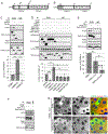
The requirement for α-catenin for TRIP6-vinculin binding and TRIP6 adherens junction localization may be indirect. (A) HEK293A wild type cells (wild type) or HEK293A cells with α-catenin inactivated using CRISPR (α-catenin KO) were transfected with a FLAG-TRIP6 expressing plasmid (TRIP6) and either a GFP-vinculin (vinculin) expressing plasmid or GFP-vinculin-T12 mutant expressing plasmid (vinculin T12). FLAG-TRIP6 immune complexes were isolated from cell lysates and analyzed by western blotting using antibodies against GFP (vinculin (GFP)) and FLAG (TRIP6 (FLAG)). Cell lysates (Inputs) were analyzed using antibodies against GFP (vinculin (GFP)), FLAG (TRIP6 (FLAG)), and α-E catenin(α-catenin). (B) HEK293A (wild type) or HEK293A cells with α-catenin inactivated using CRISPR (α-catenin KO) were stained for TRIP6 and α-catenin. Merged images show TRIP6 (green), α-catenin (red), and DNA (blue).
Figure 2.
Figure 2.
Identification of binding interaction between TRIP6, LATS2, and vinculin. (A,B) Schematic diagram showing different domains of TRIP6 and LIMD1 (NES: nuclear export signal; LIM: LIM domain; PDZ: PDZ domain binding motif). (C) HEK293 cells were transfected with plasmids expressing vinculin and different FLAG-TRIP6 deletion mutants as indicated. FLAG-TRIP6 immune complexes were isolated from cell lysates and analyzed by western blotting using antibodies against vinculin and FLAG (TRIP6 (FLAG)). Cell lysates (Inputs) were analyzed using antibodies against vinculin and FLAG (TRIP6 (FLAG). Quantification of vinculin levels in TRIP6 immune complexes is shown. (D) HEK293 cells were transfected with plasmids expressing GFP-LATS2 and different FLAG-TRIP6 deletion mutants as indicated. FLAG-TRIP6 immune complexes were isolated from cell lysates and analyzed by western blotting using antibodies against GFP (LATS2 (GFP)) and FLAG (TRIP6 (FLAG)). Cell lysates (Inputs) were analyzed using antibodies against GFP (LATS2 (GFP)) and FLAG (TRIP6 (FLAG)). Quantification of LATS2 levels in TRIP6 immune complexes is shown. (Mean ± SD; n=3; *P≤0.05, ***P≤0.001, T-test). (E) HEK293 cells were transfected with plasmids expressing vinculin and different FLAG-TRIP6 deletion mutants as indicated. FLAG-TRIP6 immune complexes were isolated from cell lysates and analyzed by western blotting using antibodies against vinculin and FLAG (TRIP6 (FLAG)). Cell lysates (Inputs) were analyzed using antibodies against vinculin and FLAG (TRIP6 (FLAG)). Quantification of vinculin levels in TRIP6 immune complexes is shown. (Mean ± SD; n=3; *P≤0.05, ***P≤0.001, T-test). (F) HEK293 cells were transfected with plasmids expressing GFP-LATS2 and different FLAG-TRIP6 deletion mutants as indicated. FLAG-TRIP6 immune complexes were isolated from cell lysates and analyzed by western blotting using antibodies against GFP (LATS2) and FLAG (TRIP6 (FLAG)). Cell lysates (Inputs) were analyzed using antibodies against GFP (LATS2) and FLAG (TRIP6 (FLAG)). (G) Lentiviral infection was used to stably express GFP fusions of wild-type TRIP6 and TRIP6-Δ252-277 in TRIP6 knockout (TRIP6-KO) MCF10A cells. The indicated cell lines were stained using anti-GFP and anti-vinculin antibodies. Merged image shows TRIP6 (green), vinculin (red), and DNA (blue). Scale bar =50microns.
Figure 3.
Figure 3.
The TRIP6 and LIMD1 LIM domains are necessary and sufficient for tension dependent localization to adherens junctions. Lentiviral infection was used to stably express GFP fusions of wild-type TRIP6, LIMD1 and various deletion mutants of TRIP6 and LIMD1 in TRIP6 knockout (TRIP6-KO) and LIMD1 knockout (LIMD1-KO) MCF10A cells. For (A) and (B), adherens junction localization of indicated TRIP6 constructs was visualized by co-immunostaining using anti-GFP and anti-vinculin antibody. For (C) and (D), junction localization of indicated LIMD1 constructs was visualized using GFP fluorescence and immunostaining with anti-TRIP6 antibody. Merged images show TRIP6/LIMD1 (green), vinculin/TRIP6 (red), and DNA (blue). For (B) and (D), indicated cell lines were treated with DMSO (solvent control) or with blebbistatin before fixation and staining. Scale bar =50microns.
Figure 4.
Figure 4.
Tension dependent localization of TRIP6 and LIMD1 depends on the ability of their LIM domains to bind f-actin under strain. (A-B) TRIP6 and LIMD1 localize to the ends of actin filaments at adherens junctions. F-actin was visualized by staining with fluorescently tagged phalloidin and localization of GFP constructs was visualized by either using anti-GFP antibodies (for TRIP6 GFP constructs) or using GFP fluorescence (for LIMD1 GFP constructs). Top panels (A,B) scale bars =50 microns. Bottom panels (A’,B’) scale bars = 10 microns, represent blow up views of adherens junctions marked by dashed white box in respective top panel. (C-D) Mutations predicted to reduce tension dependent binding to F-actin reduce adherens junction localization of TRIP6 and LIMD1. The indicated cell lines expressing GFP fusions of wild-type or tension sensing mutants of TRIP6 (C) and LIMD1 (D) were stained for GFP for TRIP6 constructs or visualized using GFP fluorescence for LIMD1 constructs and either vinculin or TRIP6 antibody to mark adherens junctions as indicated. Merged images shows TRIP6/LIMD1(green), vinculin/TRIP6 (red), and DNA (blue). Scale bars = 50 microns.
Figure 5.
Figure 5.
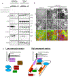
Tension dependent association of TRIP6 with LATS1 and LATS1 recruitment to adherens junctions by LIMD1 depends on the ability each protein’s LIM domains to bind f-actin under strain. (A) Protein lysates were prepared from TRIP6 knockout (TRIP6-KO) MCF10A cells expressing TurboID fusions to wild type TRIP6 or indicated TRIP6 constructs along with control wild-type MCF10A cells (WT). Biotinylated protein fractions were pulled down using streptavidin-agarose beads. Protein complexes on beads were analyzed by Western blotting using antibodies against the indicated proteins. Levels of total proteins were analyzed by blotting whole-cell lysates. (B) The indicated cell lines expressing GFP fusions of wild-type or the tension sensing mutant of LIMD1 (LIMD1-mut1,2,3) were imaged for LATS1 immunostaining signal, GFP fluorescence and DNA. Merged images show LIMD1 (green), LATS1 (red), and DNA (blue). Scale bar=50microns. (C) Model depicting tension dependent changes at adherens junctions based on this and previous studies. Mechanical tension induced by the actomyosin cytoskeleton promotes recruitment of various proteins to adherens junctions. In response to contractile forces from the actomyosin cytoskeleton alpha-catenin and vinculin become activated and bind actomyosin filaments. Strain at the ends of the filaments creates binding sites for the LIMD1 and TRIP6 LIM domains, which then recruit LATS kinase. Remaining questions (?) include 1) what is the function of the binding interaction between vinculin and TRIP6, 2) does the LIMD1 N-terminus also associate with adherens junctions proteins, and 3) what is the relationship between LIMD1 and TRIP6 and do they both contribute to LATS localization to adherens junctions.

References

    1. Bays JL, & DeMali KA (2017). Vinculin in cell-cell and cell-matrix adhesions. Cell Mol Life Sci. doi: 10.1007/s00018-017-2511-3 - DOI - PMC - PubMed
    1. Branon TC, Bosch JA, Sanchez AD, Udeshi ND, Svinkina T, Carr SA, … Ting AY (2018). Efficient proximity labeling in living cells and organisms with TurboID. Nat Biotechnol, 36(9), 880–887. doi: 10.1038/nbt.4201 - DOI - PMC - PubMed
    1. Cho KF, Branon TC, Udeshi ND, Myers SA, Carr SA, & Ting AY (2020). Proximity labeling in mammalian cells with TurboID and split-TurboID. Nat Protoc, 15(12), 3971–3999. doi: 10.1038/s41596-020-0399-0 - DOI - PubMed
    1. Choi HJ, Pokutta S, Cadwell GW, Bobkov AA, Bankston LA, Liddington RC, & Weis WI (2012). alphaE-catenin is an autoinhibited molecule that coactivates vinculin. Proc Natl Acad Sci U S A, 109(22), 8576–8581. doi: 10.1073/pnas.1203906109 - DOI - PMC - PubMed
    1. Cohen DM, Chen H, Johnson RP, Choudhury B, & Craig SW (2005). Two distinct head-tail interfaces cooperate to suppress activation of vinculin by talin. J Biol Chem, 280(17), 17109–17117. doi: 10.1074/jbc.M414704200 - DOI - PubMed

MeSH terms

LinkOut - more resources