. 2024 Apr 12;52(6):3419-3432.
doi: 10.1093/nar/gkae144.

Conserved structures and dynamics in 5'-proximal regions of Betacoronavirus RNA genomes

Affiliations

Conserved structures and dynamics in 5'-proximal regions of Betacoronavirus RNA genomes

Tales Rocha de Moura et al. Nucleic Acids Res. .

Abstract

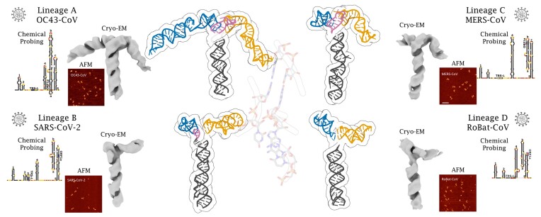
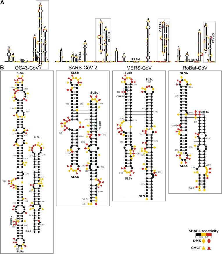
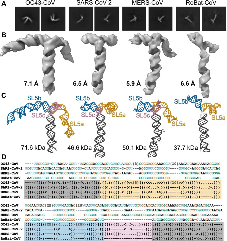
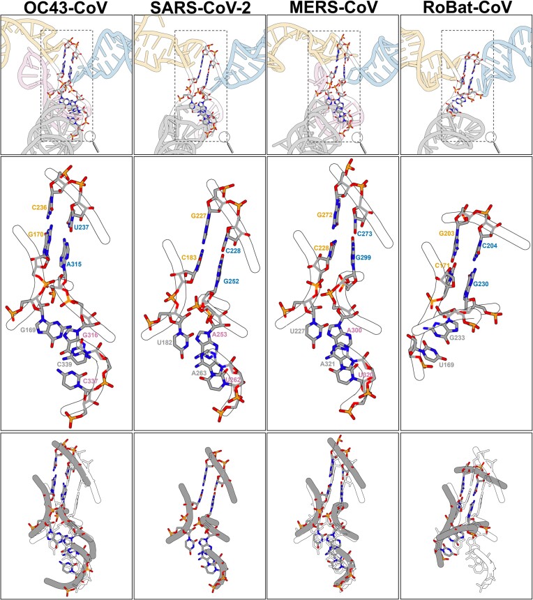
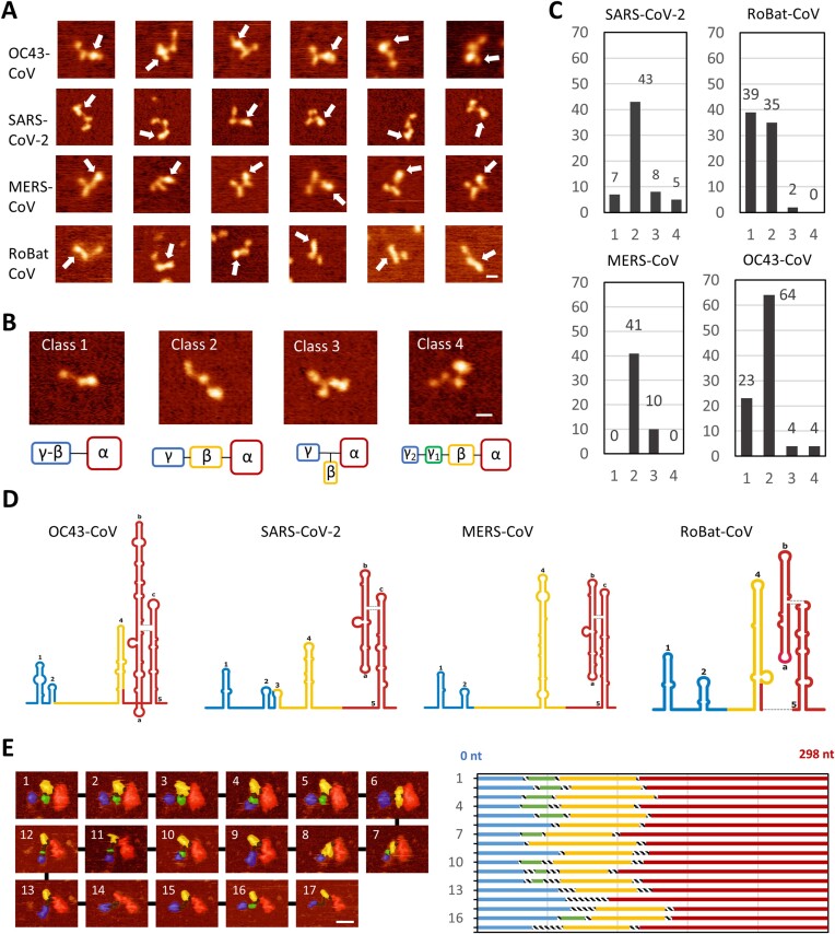
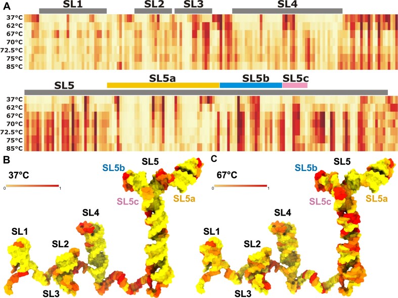
Betacoronaviruses are a genus within the Coronaviridae family of RNA viruses. They are capable of infecting vertebrates and causing epidemics as well as global pandemics in humans. Mitigating the threat posed by Betacoronaviruses requires an understanding of their molecular diversity. The development of novel antivirals hinges on understanding the key regulatory elements within the viral RNA genomes, in particular the 5'-proximal region, which is pivotal for viral protein synthesis. Using a combination of cryo-electron microscopy, atomic force microscopy, chemical probing, and computational modeling, we determined the structures of 5'-proximal regions in RNA genomes of Betacoronaviruses from four subgenera: OC43-CoV, SARS-CoV-2, MERS-CoV, and Rousettus bat-CoV. We obtained cryo-electron microscopy maps and determined atomic-resolution models for the stem-loop-5 (SL5) region at the translation start site and found that despite low sequence similarity and variable length of the helical elements it exhibits a remarkable structural conservation. Atomic force microscopy imaging revealed a common domain organization and a dynamic arrangement of structural elements connected with flexible linkers across all four Betacoronavirus subgenera. Together, these results reveal common features of a critical regulatory region shared between different Betacoronavirus RNA genomes, which may allow targeting of these RNAs by broad-spectrum antiviral therapeutics.


Figures

Graphical Abstract
Graphical Abstract
Figure 1.
Figure 1.
Secondary structure elements within the 5′-proximal regions of βCoVs from A, B, C, and D subgenera. (A) Schematic representations of the secondary structure of the full-length 5′-proximal regions. (B) Details of the secondary structure predictions for the SL5 element. Normalized reactivities across three biological replicates for the in vitro probing experiments are shown. Highly (red) and moderately (yellow) reactive residues from in vitro SHAPE (circles), DMS (diamonds), and CMCT (triangles) experiments are indicated. Highlighted features include stem-loops 1–5 (SL1–SL5), the leader transcriptional regulatory sequence (TRS-L) and the start codon of ORF1a. Substructures within SL5 are labeled as a, b, c. In SL5, the individual base pairs were manually edited to position them according to the 3D model (see below).
Figure 2.
Figure 2.
Structure conservation in the SL5 element in βCoVs: OC43-CoV, SARS-CoV-2, MERS-CoV, and RoBat-CoV. (A) Example reference-free 2D-class averages. (B) Cryo-EM maps. (C) Structural models. (D) Structure-based sequence alignment. Canonical and non-canonical base pairs are indicated by () and <> pairs, respectively, unpaired residues are indicated by dots, deletions are indicated by dashes. Significant insertions in OC43-CoV have been excluded for clarity and are indicated by the number of undisclosed residues in brackets {N}. Images rendered using Chimera (20) and Protein Imager (27).
Figure 3.
Figure 3.
SL5 junction comparison. Stems are distinguished by colors: SL5 basal stem in grey, SL5a in yellow, SL5b in blue, and SL5c in pink. Upper panel: SL5 cartoon representation and junction residues in all-atom representation. Middle panel: SL5 junctions in all atom representation. Lower panel: OC43-CoV, MERS-CoV, and RoBat-CoV SL5 junctions (in grey) superimposed on the SARS-CoV-2 SL5 junction (in white).
Figure 4.
Figure 4.
AFM analysis of the 5′-proximal regions of genomic RNAs from different βCoVs. (A) Representative air AFM images of individual RNA molecules for the four βCoVs analyzed in this study. White arrows indicate the brightest region. Noticeable conformational differences were observed between samples. (B) Representative examples of the four classes of molecules observed, categorized by their region arrangement. The α-domain is assigned to the brightest region, likely representing the SL5 element. (C) Distribution of molecules in different classes of domain arrangement. (D) Volume analysis of the images provides an estimate of the nucleotide sequence range covered by each region. Average nucleotide ranges for each sample resulted in the three regions (Supplementary Figure S15, Supplementary Table S3). (E) Liquid AFM imaging captured domain rearrangements. A series of consecutive images of the SARS-CoV-2 RNA serves as a representative example (Supplementary Video S1). Volume analysis indicates rearrangements of the SL2 and SL3 elements. The bar size in all images is 16 nm. The color gradient, from dark to bright, spans 1.5 nm in air AFM images and 3.5 nm in liquid AFM images. Additional liquid AFM imaging of other βCoV RNAs is shown in Supplementary Figure S16.
Figure 5.
Figure 5.
Thermal stability of the 5′-proximal region of SARS-CoV-2 as probed by SHAPE. (A) Heatmap of SHAPE reactivity level variations when probed in the 37–85°C temperature range. (B) SARS-CoV-2 5′-proximal region model colored according to SHAPE reactivity at 37°C. (C) A model of the SARS-CoV-2 5′-proximal region model 3D structure colored according to SHAPE reactivity at 67°C. Individual structural elements SL1–SL5 were arranged spatially to facilitate visualization in 2D (detailed in Supplementary Methods). Detailed values of reactivities are shown as bar plots in Supplementary Figure S18.

References

    1. Cui J., Li F., Shi Z.-L.. Origin and evolution of pathogenic coronaviruses. Nat. Rev. Micro. 2019; 17:181–192. - PMC - PubMed
    1. Brüssow H., Brüssow L.. Clinical evidence that the pandemic from 1889 to 1891 commonly called the Russian flu might have been an earlier coronavirus pandemic. Microb. Biotechnol. 2021; 14:1860–1870. - PMC - PubMed
    1. Wu Z., Yang L., Ren X., He G., Zhang J., Yang J., Qian Z., Dong J., Sun L., Zhu Y.et al. .. Deciphering the bat virome catalog to better understand the ecological diversity of bat viruses and the bat origin of emerging infectious diseases. ISME J. 2016; 10:609–620. - PMC - PubMed
    1. Ruiz-Aravena M., McKee C., Gamble A., Lunn T., Morris A., Snedden C.E., Yinda C.K., Port J.R., Buchholz D.W., Yeo Y.Y.et al. .. Ecology, evolution and spillover of coronaviruses from bats. Nat. Rev. Micro. 2022; 20:299–314. - PMC - PubMed
    1. Liu P., Li L., Millership J.J., Kang H., Leibowitz J.L., Giedroc D.P.. A U-turn motif-containing stem-loop in the coronavirus 5’ untranslated region plays a functional role in replication. RNA. 2007; 13:763–780. - PMC - PubMed