. 2024 May 1;30(9):1945-1958.
doi: 10.1158/1078-0432.CCR-23-3206.

Radiotherapy Enhances Metastasis Through Immune Suppression by Inducing PD-L1 and MDSC in Distal Sites

Affiliations

Radiotherapy Enhances Metastasis Through Immune Suppression by Inducing PD-L1 and MDSC in Distal Sites

Yuzhu Hou et al. Clin Cancer Res. .

Abstract

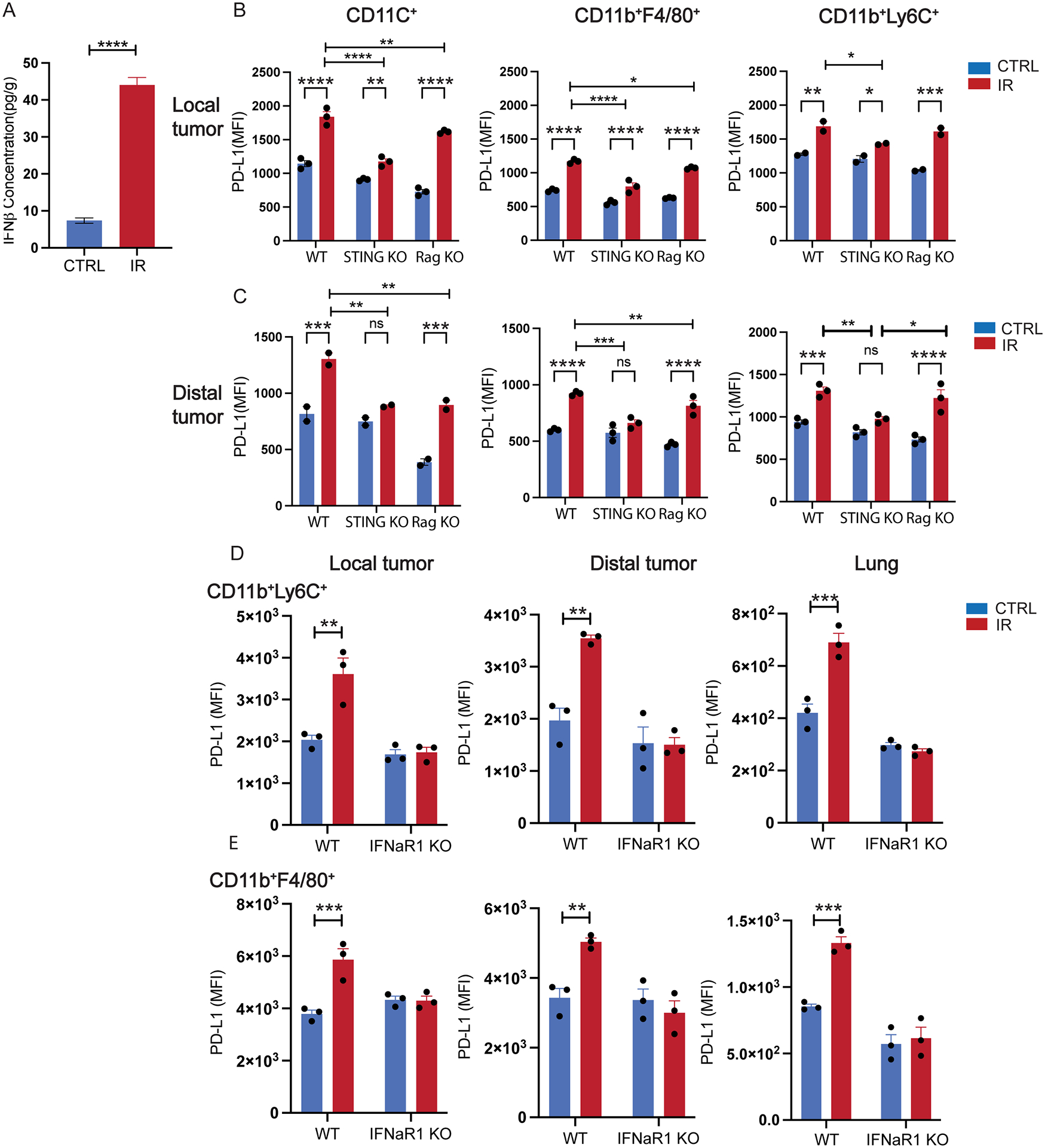
Purpose: Radiotherapy (RT) is a widely employed anticancer treatment. Emerging evidence suggests that RT can elicit both tumor-inhibiting and tumor-promoting immune effects. The purpose of this study is to investigate immune suppressive factors of radiotherapy.

Experimental design: We used a heterologous two-tumor model in which adaptive concomitant immunity was eliminated.

Results: Through analysis of PD-L1 expression and myeloid-derived suppressor cells (MDSC) frequencies using patient peripheral blood mononuclear cells and murine two-tumor and metastasis models, we report that local irradiation can induce a systemic increase in MDSC, as well as PD-L1 expression on dendritic cells and myeloid cells, and thereby increase the potential for metastatic dissemination in distal, nonirradiated tissue. In a mouse model using two distinct tumors, we found that PD-L1 induction by ionizing radiation was dependent on elevated chemokine CXCL10 signaling. Inhibiting PD-L1 or MDSC can potentially abrogate RT-induced metastasis and improve clinical outcomes for patients receiving RT.

Conclusions: Blockade of PD-L1/CXCL10 axis or MDSC infiltration during irradiation can enhance abscopal tumor control and reduce metastasis.


Conflict of interest statement

Conflict-of-interest statement:

RRW has stock and other ownership interests with Boost Therapeutics, Immvira, Reflexion Pharmaceuticals, Coordination Pharmaceuticals, Magi Therapeutics, Oncosenescence. He has served in a consulting or advisory role for Aettis, Astrazeneca, Coordination Pharmaceuticals, Genus, Merck Serono S.A., Nano proteagen, NKMax America, Shuttle Pharmaceuticals, and Persona Dx. He has a patent pending entitled “Methods and Kits for Diagnosis and Triage of Patients with Colorectal Liver Metastases” (PCT/US2019/028071). He has received research grant funding from Varian and Regeneron. He has received compensation including travel, accommodations, or expense reimbursement from Astrazeneca, Boehringer Ingelheim, and Merck Serono S.A. RRW and HLL have a patent pending: PCT/US23/81165 “METHODS FOR TREATING CANCER WITH IMMUNOTHERAPY”.

The remaining authors declare no conflicts of interest.

Figures

Figure 1.
Figure 1.
Abscopal effect of ionizing radiation. A-D. Phenotype of MC38/MC38 tumors. Growth of irradiated local tumors (A) and untreated distal tumors (B). C. IFNγ producing CD8+ T cells from local and distal draining lymph nodes in ELISPOT assays. D. Frequencies of IFNγ and granzyme B expressing CD8+ T cells in tumors. E. Number of spontaneous lung metastasis nodules when primary LLC tumors received indicated treatments. F. Tumor grwth curve of MC38 (received treatments). G. Tumor growth curve of distal tumors (LLC). H. Spontaneous metastasis in lung of LLC mice. I. Quantification of area of metastasis in lungs 30 days after cancer cell transplantation. *, p<0.05; **, p<0.01; ***, p<0.001, ****, p<0.0001. A, B, F, G, N=5, 2-way ANOVA; C-D, N=3–4, t-test assuming unequal variance. E, N=11–13; I, N=5–10, t-test assuming unequal variance; Experiments were repeated 3 times.
Figure 2.
Figure 2.
MDSC frequencies in myeloid cells and PD-L1 expression in irradiated local tumors, unirradiated distal tumors, and lungs compared with unirradiated local control. A, B. RNAseq analysis of CD45+ cells isolated from lungs of animals treated as indicated 3 days after irradiation of local tumors (MC38). A. Representative enriched pathways in CD45+ cells of lungs of irradiated animals. B. Differential expression of genes (DEG) of MSDC scores in different treatments. X-axis: Log2 of fold change (FC) between each DEG. Y-axis: DEGs. Gene (Y-axis): DEGs among MDSC signature genes. C, D. MDSC population of local and distal tumors. E. MDSC population in lung 7 days post-IR in homologous 2-tumor models. F-H. PD-L1 expression of CD11C+DC, macrophage, M-MDSCs in irradiated (local) tumors, untreated (distal) tumors, and lungs in homologous 2-tumor models. I. PD-L1 expression in MDSCs and DCs of patients’ PBMC pre- and post- SBRT in clinical trial NCT03223155. C, D, N=4; E, N=6–8; F-H, N=3. Experiments were repeated 3 times. *, P<0.05; **, P<0.01; ****, P<0.0001. Statistics: C-H, Mann-Whitney test. I, N=27, paired t-test.
Figure 3.
Figure 3.
RT-induced systemic PD-L1 expression in myeloid cells depends on STING and type I IFN signaling but not T cells. A. IFNβ concentration in irradiated (local) tumors post 20 Gy of local irradiation. B. PD-L1 expression on various myeloid cells in irradiated local tumor in WT, STING KO, and RAG KO mice. C. PD-L1 expression on various myeloid cells in un-irradiated distal tumor in WT, STING KO, and RAG KO mice. PD-L1 expression of Ly6C+ cells (D) and macrophages (E) of local tumors, distal tumors, and lung tissue in WT and IFNαR KO mice. N=3–4; *, P<0.05; **, P<0.01; ***, P<0.001; ****, P<0.0001. A, B, C, analyzed by 2-way ANOVA. B, D, E were analyzed by t-test assuming unequal variance. Experiments were repeated 3 times.
Figure 4.
Figure 4.
CXCL10 is the key regulator of PD-L1 expression in myeloid cells in distal tissue. A. CXCL10 concentration in mouse serum 3 days post-IR. B. Serum CXCL10 concentration pre- vs. post-radiotherapy in lung cancer patients. C. Serum CXCL10 concentration in mice 3 days post local tumor irradiation. D. DC PD-L1 expression in lungs of WT and CXCR3 KO mice 3 days after primary tumor irradiation. E. PD-L1 expression in WT or CXCR3 KO BMDC co-cultured with 0 and 5ng/ml concentration of rCXCL10. F. qPCR quantification of mRNA level of CXCL10 in tumor-associated immune cells 2 days post-IR. G. PD-L1 expression post-IR on CD11C+MHCII+ DC cells of lungs as fold over un-treated control. A, N=6–7; Mann-Whitney test. C-F, N=3–6; 2-way ANOVA. B, N=13; Wilcoxon test. *, P<0.05; **, P<0.01; ***, P<0.001; ***, P<0.0001. Mouse experiments were repeated 3 times.
Figure 5.
Figure 5.
Neutralizing CXCL10 inhibits tumor growth and lung metastasis of untreated tumors. A. Growth of treated MC38. B. Growth of distal tumor (LLC). C, D. Number of spontaneous lung metastasis of distal LLC tumor. E. Number of lung metastasis. N=5 for A, B. The experiments were repeated 3 times. N=9–10 for D. *, p<0.05; **, p<0.01; ***, P<0.001; A, B, 2-way ANOVA; D, Mann-Whitney test.
Figure 6.
Figure 6.
PD-L1 and CCR2 blockade abrogates local IR-induced systemic pro-tumor responses. A. Growth curve of distal tumor (LLC) in heterogeneous 2-tumor model. B. H&E staining of lungs of which primary MC38 tumors were treated as indicated. C. Number of metastasis in a lung of which primary MC38 tumors were treated with IR and/or anti-PD-L1. D. Growth of LLC distal tumors with indicated treatments. E. H&E staining of lungs of which primary MC38 tumors were treated with IR and/or anti-CCR2. F. Number of metastasis in a lung. G, H, I. LLC primary tumors were treated as indicated and lung nodules were counted via H&E staining. A and D, N=5. A, D, F and I, experiments were repeated 3–4 times. *, P<0.05; **, P<0.01; ***, P<0.001. ****, P<0.0001. A, and D by 2-way ANOVA analysis; C, F, G, and I by Mann-Whitney test.

References

    1. North RJ. Radiation-induced, immunologically mediated regression of an established tumor as an example of successful therapeutic immunomanipulation. Preferential elimination of suppressor T cells allows sustained production of effector T cells. J Exp Med 1986;164(5):1652–66 doi 10.1084/jem.164.5.1652. - DOI - PMC - PubMed
    1. Teitz-Tennenbaum S, Li Q, Okuyama R, Davis MA, Sun R, Whitfield J, et al. Mechanisms involved in radiation enhancement of intratumoral dendritic cell therapy. J Immunother 2008;31(4):345–58 doi 10.1097/CJI.0b013e318163628c. - DOI - PMC - PubMed
    1. Diamond JM, Vanpouille-Box C, Spada S, Rudqvist NP, Chapman JR, Ueberheide BM, et al. Exosomes Shuttle TREX1-Sensitive IFN-Stimulatory dsDNA from Irradiated Cancer Cells to DCs. Cancer Immunol Res 2018;6(8):910–20 doi 10.1158/2326-6066.CIR-17-0581. - DOI - PMC - PubMed
    1. Lugade AA, Sorensen EW, Gerber SA, Moran JP, Frelinger JG, Lord EM. Radiation-induced IFN-gamma production within the tumor microenvironment influences antitumor immunity. J Immunol 2008;180(5):3132–9 doi 10.4049/jimmunol.180.5.3132. - DOI - PubMed
    1. Walle T, Kraske JA, Liao B, Lenoir B, Timke C, von Bohlen Und Halbach E, et al. Radiotherapy orchestrates natural killer cell dependent antitumor immune responses through CXCL8. Sci Adv 2022;8(12):eabh4050 doi 10.1126/sciadv.abh4050. - DOI - PMC - PubMed

Publication types