. 2024 Mar 1;19(3):e0298208.
doi: 10.1371/journal.pone.0298208. eCollection 2024.

4-aminopyridine improves evoked potentials and ambulation in the taiep rat: A model of hypomyelination with atrophy of basal ganglia and cerebellum

Affiliations

4-aminopyridine improves evoked potentials and ambulation in the taiep rat: A model of hypomyelination with atrophy of basal ganglia and cerebellum

Jose R Eguibar et al. PLoS One. .

Abstract

The taiep rat is a tubulin mutant with an early hypomyelination followed by progressive demyelination of the central nervous system due to a point mutation in the Tubb4a gene. It shows clinical, radiological, and pathological signs like those of the human leukodystrophy hypomyelination with atrophy of the basal ganglia and cerebellum (H-ABC). Taiep rats had tremor, ataxia, immobility episodes, epilepsy, and paralysis; the acronym of these signs given the name to this autosomal recessive trait. The aim of this study was to analyze the characteristics of somatosensory evoked potentials (SSEPs) and motor evoked potentials (MEPs) in adult taiep rats and in a patient suffering from H-ABC. Additionally, we evaluated the effects of 4-aminopyridine (4-AP) on sensory responses and locomotion and finally, we compared myelin loss in the spinal cord of adult taiep and wild type (WT) rats using immunostaining. Our results showed delayed SSEPs in the upper and the absence of them in the lower extremities in a human patient. In taiep rats SSEPs had a delayed second negative evoked responses and were more susceptible to delayed responses with iterative stimulation with respect to WT. MEPs were produced by bipolar stimulation of the primary motor cortex generating a direct wave in WT rats followed by several indirect waves, but taiep rats had fused MEPs. Importantly, taiep SSEPs improved after systemic administration of 4-AP, a potassium channel blocker, and this drug induced an increase in the horizontal displacement measured in a novelty-induced locomotor test. In taiep subjects have a significant decrease in the immunostaining of myelin in the anterior and ventral funiculi of the lumbar spinal cord with respect to WT rats. In conclusion, evoked potentials are useful to evaluate myelin alterations in a leukodystrophy, which improved after systemic administration of 4-AP. Our results have a translational value because our findings have implications in future medical trials for H-ABC patients or with other leukodystrophies.


Conflict of interest statement

The authors have declared that no competing interest exist.

Figures

Fig 1
Fig 1. Somatosensory evoked potentials produced by peroneal nerve stimulation at different spinal cord levels.
A) In wild type Sprague-Dawley (WT) male rats the somatosensory evoked potentials have a characteristic negative wave (upward deflection, black arrow) followed by a single positive (downward deflection), being maximum at T12-L1 and L2-L1 interspinal levels. B) Male taiep rats have two negative peaks (upward deflections, black arrows) being the second (N2) clearly delayed followed by positive evoked wave that is similar to that obtained in WT rats.
Fig 2
Fig 2. Somatosensory evoked potentials in taiep and wild type Sprague-Dawley male rats.
A) Iterative stimulation on the peroneal nerve at 30 Hz produced a progressive delayed N2 wave of the evoked potentials in taiep from 1.65 to 2.31 msec (blue squares) with respect to WT male rats (1.55 msec, empty circles). B) The iterative stimulation with 60 Hz produced a rapid increase in the intervals among successive N2 evoked potential in taiep rats (2.15 msec), but it is not the case in WT because they have a stable N2 evoked potential around 1.72 msec. C) The increase in successive N2 evoked potential was faster in taiep with 2.33 msec intervals with respect to WT with 1.65 msec intervals when peroneal nerve stimulation is 80 Hz. The data is the mean ± E.E.M. of eight subjects.
Fig 3
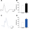
Fig 3. The systemic administration of 4-aminopyridine increased the somatosensory evoked potentials in taiep rats.
A) The somatosensory evoked N2 evoked potential increased after systemic administration of 4-aminopyridine (4-AP, dotted lines) with respect to control evoked potentials (continuous lines) in WT male rats with a shorter latency. B) The somatosensory evoked potentials significantly increase after intravenous administration of 1 mg/Kg of 4-AP (U = 62; P< 0.05, Mann-Whitney U test) with respect to saline-treated rats (empty bar). C) The somatosensory N2 evoked potential increased after systemic administration of 4-aminopyridine (4-AP, blue dotted lines) with respect to control evoked potentials (blue continuous lines) in taiep rats. D) The N2 evoked response significantly increased its amplitude and reduced its latency after the administration of this potassium channel blocker (blue bar; t (29) = 4.3, P< 0.001) with respect to control conditions. The data is the mean ± E.E.M. of eight subjects.
Fig 4
Fig 4. Number of ambulation, rearing, and tremor bouts in control and after 4-aminopyridine administration in male taiep rats.
In the novelty-induced locomotor test, 4-aminopyridine (4-AP) i.p. injection of 1 mg/Kg significantly increased the ambulation bouts (t (29) = 3.6; P< 0.05), but not rearing and tremor bouts. The data is the mean ± E.E.M. of eight subjects.
Fig 5
Fig 5. Motor evoked responses differ in taiep and wild type Sprague-Dawley male rats.
A) The grey area is the hindlimb area of stimulation which produce maximum motor-evoked potentials in lumbar spinal cord. B) In Sprague-Dawley (WT) male rats have a direct wave (D) followed by two indirect waves (I-1 and I-2) due to reverberating activity in the pyramidal pathway. Downward arrow is the stimulus artifact. C) In taiep rats, direct and indirect waves are fused, which produced a characteristic motor-evoked response in this tubulinopathy rat model. The data is the mean ± E.E.M. of eight subjects.
Fig 6
Fig 6. The corticospinal pathway is more susceptible to iterative stimulation and have lower conduction velocity in taiep rats.
A) Motor-evoked potentials (MEPs) induced by iterative electrical stimulation on the cerebral cortex showed a decrease in their amplitudes when increasing the stimulation rate from 5 to 30 Hz in taiep (blue squares), but not in WT rats (empty circles). B) The conduction velocity of the corticospinal tract significantly decreased in taiep to 13.7 m/s (blue bar) with respect to WT with 42.9 m/s (empty bar; t (12), = 42; **P< 0.05). The data is the mean ± E.E.M. of eight subjects of each group of rats.
Fig 7
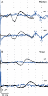
Fig 7. Defects in the conduction of somatosensory evoked potentials of median and tibial nerves in a healthy child and in H-ABC patient.
A) Somatosensory evoked potentials of left and right median nerves showing an N19 latency at the cortex with 50.9 and 51.1 msec delay, respectively, and P22 latencies with 58.0 and 59.6 msec, respectively (blue traces). B) Tibial nerves did not evoke any potentials in both hindlimb nerves (blue traces) with respect to healthy children (dark traces). Recordings were made at a rate of 4 Hz, with a stimulus intensity of 12.5 mA with a square pulse duration of 2 msec. The SSEPs from a healthy patient, recorded with the same equipment are shown in black traces for comparison.
Fig 8
Fig 8. Hypomyelination and demyelination in the spinal cord of taiep rats.
Representative confocal images of transversal sections of spinal cords at the lumbar level in the ventral and dorsal funiculi (as shown in the drawings) of wild type Sprague-Dawley (WT; panels A and C) and taiep rats (panels B and D). The myelin staining (red) is even throughout the section, while neurofilament staining (green) shows a spotty appearance. In the taiep rat, the myelin staining is drastically reduced (B and D). E) In the dorsal column a significant decrease in red the myelin fluorescence intensity in taiep rats (blue bar; P< 0.0001, Mann-Whitney U test) with respect to WT rats (black bar). F) In the ventral funiculus there is also a significant reduction of red myelin fluorescence intensity in taiep (blue bar) with respect to WT male rats (black bar; P< 0.0001, Mann-Whitney U test). Nerve fibers devoid of myelin sheaths fully display their NF content (green). Images of individual channels corresponding to the white dotted squares are shown below each image and allow us to appreciate the severe demyelination in the dorsal and ventral funiculi. Green: neurofilaments, red: myelin, blue: DAPI. Scale bar 50 μm. Myelin fluorescence levels were measured in 6 regions of interest. The data is the mean ± S.E.M. of six subjects of WT and taiep rats.
Fig 9
Fig 9. Taiep rats meet criteria as an adequate model of H-ABC.
Taiep rats is an adequate model of the human leukodystrophy H-ABC because they have face validity in base of similar MRI findings, they had a point mutation in tubulin β 4A gene that altered structurally the corresponding protein (TUBB4A). Additionally, the construct validity is supported by similar somatosensory and motor-evoked potentials and with their behavioral responses in a novelty-induced locomotion measured through ambulation bouts.

Similar articles

References

    1. Cracco JB, Cracco RQ. Spinal somatosensory evoked potentials: maturational and clinical studies. Annals of the New York Academy of Sciences. 1982;388(1):526–537. doi: 10.1111/j.1749-6632.1982.tb50814.x - DOI - PubMed
    1. Nezu A, Kimura S, Takeshita S, Osaka H, Tanaka M. Magnetic stimulation of the corticospinal tracts in in Pelizaeus-Merzbacher disease. Electroencephalography and Clinical Neurophysiology. 1998;108(5):446–448. doi: 10.1016/s0168-5597(98)00018-5 - DOI - PubMed
    1. Holmgren B, Urbá-Holmgren R, Riboni L, Vega-SaenzdeMiera EC. Sprague Dawley rat mutant with tremor, ataxia, tonic immobility episodes, epilepsy and paralysis. Laboratory Animal Science. 1989;39(3):226–228. - PubMed
    1. Urbá-Holmgren R, Santos A, Holmgren B, Eguíbar JR. Two inbred rat sublines that differ in spontaneous yawning behavior also differ in their responses to cholinergic and dopaminergic drugs. Behavioural Brain Research. 1993;56(2):155–159. doi: 10.1016/0166-4328(93)90033-m - DOI - PubMed
    1. Couve E, Cabello JF, Krsulovic J, Roncagliolo M. Binding of microtubules to transitional elements in oligodendrocytes of the myelin mutant taiep rat. Journal of Neuroscience Research. 1997;47(6):573–581. doi: 10.1002/(SICI)1097-4547(19970315)47:6&lt;573::AID-JNR2&gt;3.0.CO;2-A - DOI - PubMed

Substances

Supplementary concepts