. 2024 Mar;10(9):eadn0042.
doi: 10.1126/sciadv.adn0042. Epub 2024 Mar 1.

Epistatic pathways can drive HIV-1 escape from integrase strand transfer inhibitors

Affiliations

Epistatic pathways can drive HIV-1 escape from integrase strand transfer inhibitors

Yuta Hikichi et al. Sci Adv. 2024 Mar.

Abstract

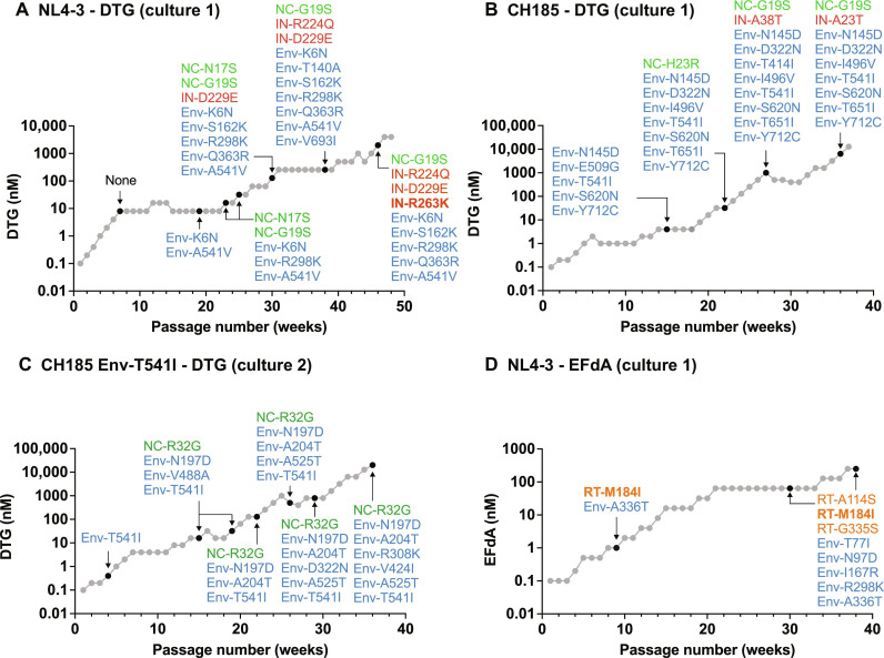
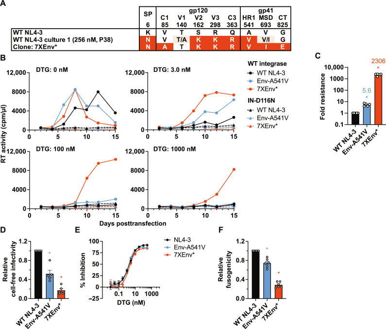
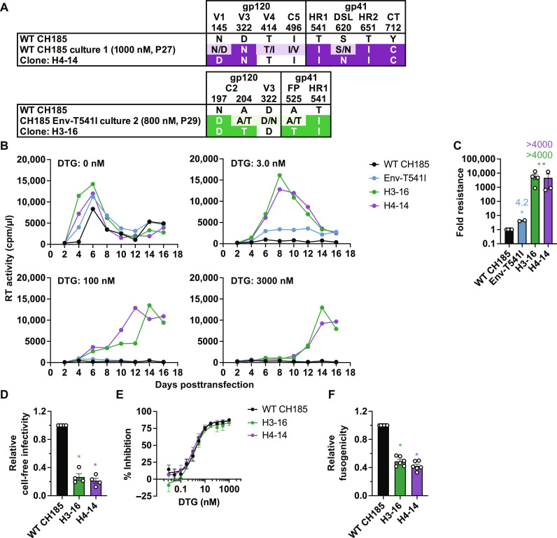
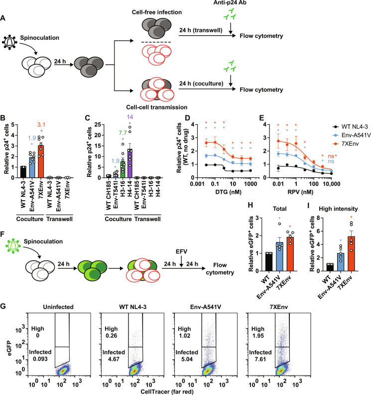
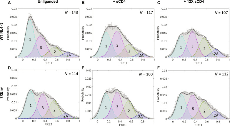
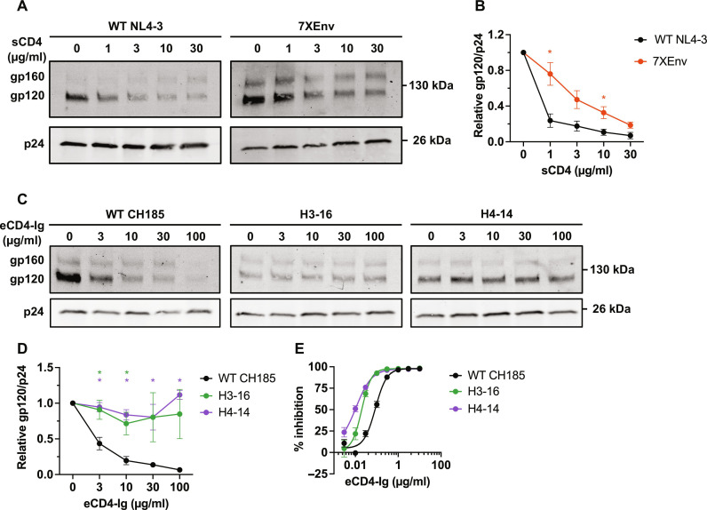
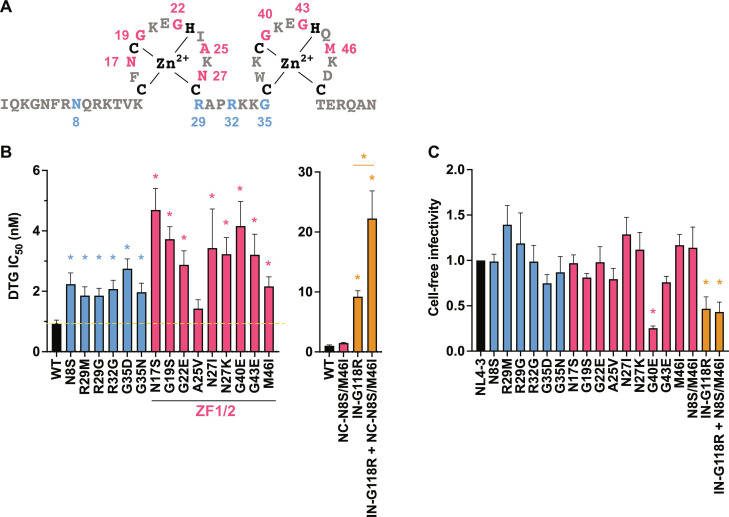
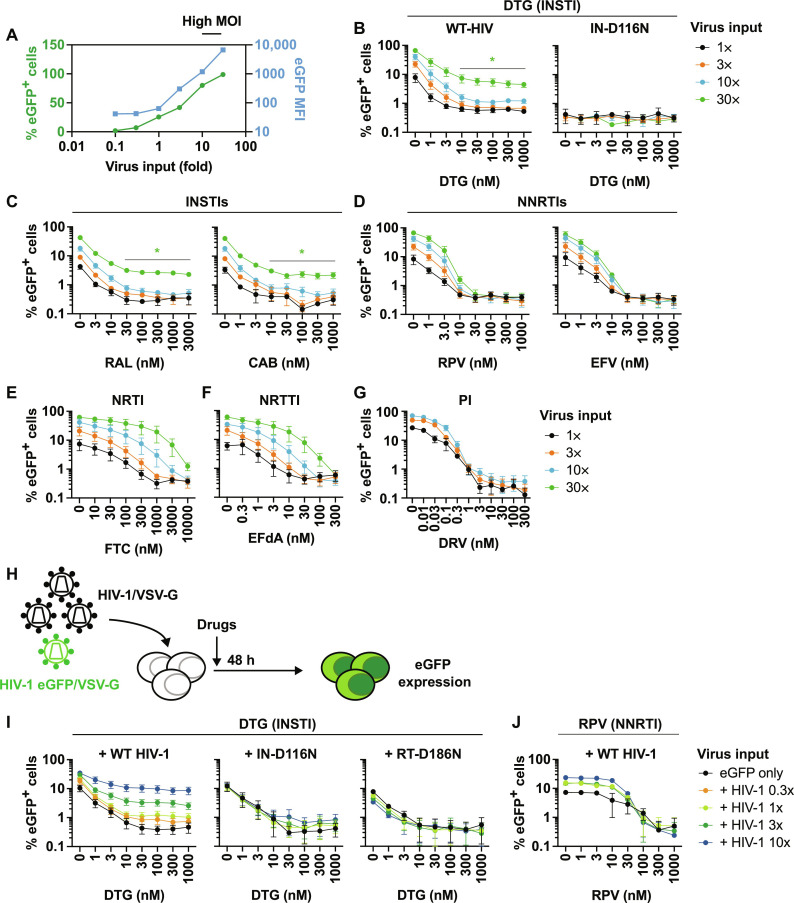
People living with human immunodeficiency virus (HIV) receiving integrase strand transfer inhibitors (INSTIs) have been reported to experience virological failure in the absence of resistance mutations in integrase. To elucidate INSTI resistance mechanisms, we propagated HIV-1 in the presence of escalating concentrations of the INSTI dolutegravir. HIV-1 became resistant to dolutegravir by sequentially acquiring mutations in the envelope glycoprotein (Env) and the nucleocapsid protein. The selected Env mutations enhance the ability of the virus to spread via cell-cell transfer, thereby increasing the multiplicity of infection (MOI). While the selected Env mutations confer broad resistance to multiple classes of antiretrovirals, the fold resistance is ~2 logs higher for INSTIs than for other classes of drugs. We demonstrate that INSTIs are more readily overwhelmed by high MOI than other classes of antiretrovirals. Our findings advance the understanding of how HIV-1 can evolve resistance to antiretrovirals, including the potent INSTIs, in the absence of drug-target gene mutations.


Figures

Fig. 1.
Fig. 1.. In vitro selection of drug-resistant variants.
Long-term passaging of (A) WT NL4-3 (culture 1), (B) WT CH185 (culture 1), and (C) CH185 Env-T541I (culture 2) in the presence of the INSTI DTG. (D) Long-term passaging of WT NL4-3 (culture 1) in the presence of the NRTTI EFdA. SupT1 [(A) and (D)] or SupT1huR5 [(B) and (C)] T cell lines were infected with the indicated viruses to initiate the passaging experiments. At the time points indicated by the arrows, genomic DNA was extracted from the drug-treated cultures, and the Gag-, Pol-, and Env-coding regions were sequenced. Mutations detected at frequencies greater than 25% in the bulk sequencing are shown. Mutations highlighted in bold are established resistance mutations to DTG (A) or EFdA (D).
Fig. 2.
Fig. 2.. The NL4-3 7XEnv* variant exhibits high-level DTG resistance in the context of spreading, multi-round infection but not in cell-free, single-round infection.
(A) An infectious molecular clone with seven Env mutations (7XEnv) and a Vpu mutation (denoted by the *) derived from the long-term passaging experiment was constructed by transferring the env amplicon from NL4-3 culture 1 (passage 38, 256 nM DTG; Fig. 1A) into pNL4-3. The locations of mutations in Env are indicated: C1 and C3, first and third conserved domains of gp120, respectively; V2 and V3, second and third variable domains of gp120, respectively; HR1, heptad repeat 1 of gp41; MSD, membrane-spanning domain; CT, cytoplasmic tail. (B) Replication kinetics of the NL4-3 Env variants in the SupT1 T cell line in the absence or presence of DTG. Replication curves obtained in the presence of 0, 3, 100, and 1000 nM DTG are shown. Data are representative of at least three independent experiments. (C) Fold changes in IC50 were calculated compared to that for the WT over a range of DTG concentrations (0.01 to 3000 nM). IC50 values were calculated on the basis of the AUC of the replication kinetics. (D) Single-round, cell-free viral infectivity of the Env variants. Relative infectivity is shown, normalized to 1 for WT NL4-3. (E) DTG sensitivity in the context of cell-free infection. TZM-bl cells were incubated with 100 TCID50 of WT virus or the Env mutants in the presence of various concentrations of DTG. (F) Cell-cell fusion activity of the NL4-3 Env variants. The transfected HEK293T cells were cocultured with TZM-bl cells in the presence of a cocktail of RPV and DTG to prevent productive infection of the TZM-bl cells. Data from at least three independent experiments are shown as means ± SEM. *P < 0.05, unpaired t test.
Fig. 3.
Fig. 3.. The heavily mutated CH185 Env variants exhibit high-level DTG resistance in the context of spreading, multi-round infection but not in cell-free, single-round infection.
(A) Infectious molecular clones with the indicated Env mutations in the context of CH185. H3-16 and H4-14 clones were constructed by transferring the env amplicons from CH185 culture 1 (passage 27, 1000 nM DTG; Fig. 1B) and CH185 Env-T541I culture 2 (passage 29, 800 nM DTG; Fig. 1C), respectively, into CH185. V1 and V3, first and third variable domain of gp120, respectively; C2, second conserved domain; HR1 and HR2, heptad repeat 1 and 2 of gp41, respectively; FP, fusion peptide; DSL, disulfide loop; CT, cytoplasmic tail. (B) Replication kinetics of the CH185 Env variants in the SupT1 huR5 T cell line in the absence or presence of DTG. Replication curves obtained in the presence of 0, 3, 100, and 3000 nM DTG are shown. Data are representative of at least three independent experiments. (C) Fold changes in IC50 were calculated compared to that for the WT, based on the AUC of the replication kinetics. (D) Single-cycle, cell-free viral infectivity of the Env variants. RT-normalized virus stocks were used to infect TZM-bl cells. Luciferase activity was measured at 48 hours after infection. Relative infectivity is shown, normalized to 1 for WT CH185. (E) DTG sensitivity in the context of cell-free infection. TZM-bl cells were exposed to 100 TCID50 of WT or the Env mutants in the presence of various concentrations of DTG. (F) Cell-cell fusion activity of the CH185 Env variants. The transfected HEK293T cells were cocultured with TZM-bl cells in the presence of a cocktail of RPV and DTG to prevent productive infection of the TZM-bl cells. Data from at least three independent experiments are shown as means ± SEM. *P < 0.05, unpaired t test.
Fig. 4.
Fig. 4.. ARV sensitivity of the NL4-3 7XEnv* variant in the context of spreading infection.
Fold resistance of 7XEnv* to multiple classes of ARVs. The SupT1 T cell line was transfected with WT NL4-3 or 7XEnv* proviral clones in the absence or presence of a range of ARV concentrations. Virus replication kinetics were monitored by measuring RT activity. Fold changes in IC50 relative to WT were calculated. IC50 values were calculated on the basis of the AUC of the replication kinetics. IC50 values were calculated for INSTIs (DTG, RAL, and CAB), NRTI (FTC), NNRTI (EFV and RPV), NRTTI (EFdA), PI (DRV and NFV), entry inhibitors (T-20 and BMS-806), ALLINI (BI-224436), and CA inhibitor (LEN). Data from at least two independent experiments are shown as means ± SEM. *P < 0.05, unpaired t test.
Fig. 5.
Fig. 5.. The heavily mutated Env variants exhibit increased cell-cell transfer capacity.
(A) Experimental design of cell-cell transfer assay. Virus-producer SupT1 cells were spinoculated with the indicated viruses 24 hours before the assay. Target SupT1 cells were labeled with cell proliferation dye. Before the coculture, the number of infected donor cells was normalized by intracellular p24-FITC signals. The donor and the target SupT1 cells were cocultured at a 1:1 ratio in the absence or presence of ARVs. Twenty-four hours after coculture, intracellular p24 antigen was detected by staining with FITC-conjugated anti-p24 Ab. Virus-producing cells were also seeded in a transwell insert to monitor cell-free viral infection. (B) Cell-cell transfer of NL4-3 Env mutants. Fold change in the numbers of p24-positive target cells relative to WT. (C) Cell-cell transfer of CH185 variants. Fold change in the numbers of p24-positive target cells relative to WT. (D and E) Viral sensitivity to DTG and RPV, respectively, in the context of cell-cell transfer. Data are shown as the number of p24-positive cells relative to WT NL4-3 in the absence of drugs. (F) Experimental design of cell-cell transfer assay using pBR43IeG constructs (HIV eGFP). After 24 hours of coculture between infected and target cells, the second round of infection was blocked by adding 1 μM EFV, and then the cells were incubated for 24 hours. (G) Representative plots of eGFP expression after coculture. High–fluorescence intensity populations were defined as containing ~0.25% of the population showing a high-intensity eGFP signal in WT-infected target cells. (H) Relative numbers of eGFP-positive target cells in the absence of DTG. (I) Relative numbers of high–fluorescence intensity eGFP-positive target cells . Data from at least three independent experiments are shown as means ± SEM. *P < 0.05, unpaired t test.
Fig. 6.
Fig. 6.. Sensitivity of the heavily mutated Env variants to ligand binding and neutralization.
Sensitivity of NL4-3 7XEnv to (A) NAbs recognizing the CD4-bound conformation and (B) sCD4. TZM-bl cells were exposed to 100 TCID50 of viruses in the presence of various concentrations of NAbs or sCD4. Luciferase activity was measured at 48 hours after infection. (C to E) NAb/CD4-Ig binding to Env on the cell surface. 293T cells transfected with the indicated WT or 7XEnv pBR43IeG clones were preincubated with (C) CD4-Ig, (D) VRC03, and (E) 17b at 37°C for 30 min. The cells were washed, and Alexa Fluor 647–conjugated anti-human IgG was used to detect bound antibodies. For 17b binding, Env-expressing cells were treated with the indicated concentrations of sCD4. Alexa Fluor 647 signals were normalized by eGFP signals to calculate the Ab binding efficiency. (F) Effect of the 7XEnv mutations on cold sensitivity. RT-normalized viruses were incubated at 4°C for the indicated times and frozen at −80°C. The viral aliquots were quickly thawed, and infectivity was measured using TZM-bl cells. (G) CD4-Ig binding to CH185 Env on the cell surface. 293T cells transfected with the indicated Env mutants were preincubated with CD4-Ig at 37°C for 30 min. The cells were washed, and Alexa Fluor 647–conjugated anti-human IgG was used to detect bound antibodies. To detect p24 in the cells, the transfected cells were fixed and permeabilized and then stained with FITC-conjugated anti-p24 Ab. (H) Cold sensitivity of the CH185 Env mutants. RT-normalized viruses were incubated at 4°C for the indicated times and frozen at −80°C. The viral aliquots were quickly thawed, and infectivity was measured using TZM-bl cells. Data from at least three independent experiments are shown as means ± SEM. *P < 0.05, unpaired t test.
Fig. 7.
Fig. 7.. 7XEnv displays an open conformation that is insensitive to soluble CD4.
HIV-1 NL4-3ΔRT virus particles containing WT (A to C) or 7XEnv (D to F) were produced, labeled, and analyzed by smFRET. Virus particles were incubated with soluble, monomeric CD4 [(B) and (E)] or dodecameric CD4 [(C) and (F)] at 100 μg/ml for 60 min before imaging. Histograms show the sum of N individual FRET trajectories ± SEM. Four-state Gaussian curve fitting was performed for each histogram, with FRET populations designated as follows: State 1, FRET ≈ 0.1 (pretriggered, closed conformation); state 2, FRET ≈ 0.6 (necessary, intermediate conformation); state 2A, FRET ≈ 0.8 (alternate intermediate conformation); state 3, FRET ≈ 0.3 (fully CD4-bound, open conformation).
Fig. 8.
Fig. 8.. Effect of Env mutations on sCD4- or eCD4-Ig–induced gp120 shedding.
(A and B) The indicated viruses were incubated with a range of sCD4 concentrations at 37°C for 2 hours and purified through a 20% sucrose cushion, and viral proteins were detected by Western blotting. (A) Representative gel for sCD4-induced gp120 shedding assay. (B) The ratio of gp120 to p24 was quantified and plotted. (C and D) The indicated viruses were incubated with a range of eCD4-Ig concentrations at 37°C for 2 hours and purified through a 20% sucrose cushion, and viral proteins were detected by Western blotting. (E) Sensitivity of the CH185 Env mutants to eCD4-Ig. TZM-bl cells were exposed to 100 TCID50 of viruses in the presence of the indicated concentrations of eCD4-Ig. Luciferase activity was measured at 48 hours after infection. Data in the graphs from three independent experiments are shown as means ± SEM. *P < 0.05, unpaired t test.
Fig. 9.
Fig. 9.. DTG sensitivity of the Gag-NC mutants.
(A) Sequence of the HIV-1 NC domain. The NC mutations selected in the presence of DTG are highlighted in red (within the zinc-finger domains) or blue (outside the zinc fingers). (B) Fold change in DTG IC50 of the NC mutants relative to WT. TZM-bl cells were exposed to 100 TCID50 of WT or the NC mutants in the presence of a range of concentrations of DTG (from 0.03 to 1000 nM DTG). Luciferase activity was measured at 48 hours after infection. (C) Cell-free viral infectivity of the NC mutants. RT-normalized virus stocks produced from 293T cells were used to infect TZM-bl cells. Luciferase activity was measured at 48 hours after infection. The infectivity of WT NL4-3 is normalized to 1.0. Data from at least three independent experiments are shown as means ± SEM. *P < 0.05, one-way analysis of variance (ANOVA) and Tukey’s multiple-comparison test or unpaired t test.
Fig. 10.
Fig. 10.. Impact of MOI on the sensitivity of HIV-1 to ARVs.
(A) The SupT1 T cell line was exposed to a range of VSV-G–pseudotyped eGFP reporter virus inputs in the absence of drugs. eGFP MFI was markedly increased when SupT1 T cells were exposed to high input of VSV-G–pseudotyped viruses. Data representative of three independent experiments are shown. (B) DTG sensitivity of VSV-G–pseudotyped eGFP reporter virus harboring WT-IN or the catalytically inactive IN-D116N mutant. The SupT1 T cell line was infected with a 30-fold range of viral inputs in the presence of the indicated concentrations of DTG. The number of infected cells was enumerated by flow cytometry. Sensitivity of VSV-G–pseudotyped eGFP reporter viruses to (C) INSTIs RAL and CAB, (D) NNRTIs RPV and EFV, (E) NRTI FTC, and (F) NRTTI EFdA and (G) PI DRV. (H) Experimental design of abrogation experiments. The SupT1 T cell line was exposed to VSV-G–pseudotyped HIV-1 (dark virus) over a range of viral inputs together with a fixed amount of VSV-G–pseudotyped eGFP reporter virus. The amount of dark virus was 0.3, 1, 3, or 10 times the amount of eGFP reporter virus. Viral inputs were normalized by RT activity, except for the catalytically inactive RT-D186N mutant, in which case viral inputs were normalized by Gag Western blots. (I) DTG sensitivity of VSV-G–pseudotyped eGFP reporter virus in the presence of dark virus harboring WT-Pol, IN-D116N, or RT-D186N. (J) RPV sensitivity of VSV-G–pseudotyped eGFP reporter virus in the presence of WT dark virus. Data from at least three independent experiments are shown as means ± SEM. *P < 0.05, unpaired t test.

References

    1. T. R. Kemnic, P. G. Gulick, HIV Antiretroviral Therapy (StatPearls Publishing, 2022). - PubMed
    1. Link J. O., Rhee M. S., Tse W. C., Zheng J., Somoza J. R., Rowe W., Begley R., Chiu A., Mulato A., Hansen D., Singer E., Tsai L. K., Bam R. A., Chou C. H., Canales E., Brizgy G., Zhang J. R., Li J., Graupe M., Morganelli P., Liu Q., Wu Q., Halcomb R. L., Saito R. D., Schroeder S. D., Lazerwith S. E., Bondy S., Jin D., Hung M., Novikov N., Liu X., Villasenor A. G., Cannizzaro C. E., Hu E. Y., Anderson R. L., Appleby T. C., Lu B., Mwangi J., Liclican A., Niedziela-Majka A., Papalia G. A., Wong M. H., Leavitt S. A., Xu Y., Koditek D., Stepan G. J., Yu H., Pagratis N., Clancy S., Ahmadyar S., Cai T. Z., Sellers S., Wolckenhauer S. A., Ling J., Callebaut C., Margot N., Ram R. R., Liu Y. P., Hyland R., Sinclair G. I., Ruane P. J., Crofoot G. E., McDonald C. K., Brainard D. M., Lad L., Swaminathan S., Sundquist W. I., Sakowicz R., Chester A. E., Lee W. E., Daar E. S., Yant S. R., Cihlar T., Clinical targeting of HIV capsid protein with a long-acting small molecule. Nature 584, 614–618 (2020). - PMC - PubMed
    1. Collier D. A., Monit C., Gupta R. K., The impact of HIV-1 drug escape on the global treatment landscape. Cell Host Microbe 26, 48–60 (2019). - PubMed
    1. Jozwik I. K., Passos D. O., Lyumkis D., Structural biology of HIV integrase strand transfer inhibitors. Trends Pharmacol. Sci. 41, 611–626 (2020). - PMC - PubMed
    1. Clutter D. S., Jordan M. R., Bertagnolio S., Shafer R. W., HIV-1 drug resistance and resistance testing. Infect. Genet. Evol. 46, 292–307 (2016). - PMC - PubMed

Publication types