. 2024 Mar 13;146(10):7007-7017.
doi: 10.1021/jacs.4c00081. Epub 2024 Mar 1.

Real-Time Biosynthetic Reaction Monitoring Informs the Mechanism of Action of Antibiotics

Affiliations

Real-Time Biosynthetic Reaction Monitoring Informs the Mechanism of Action of Antibiotics

Abraham O Oluwole et al. J Am Chem Soc. .

Abstract

The rapid spread of drug-resistant pathogens and the declining discovery of new antibiotics have created a global health crisis and heightened interest in the search for novel antibiotics. Beyond their discovery, elucidating mechanisms of action has necessitated new approaches, especially for antibiotics that interact with lipidic substrates and membrane proteins. Here, we develop a methodology for real-time reaction monitoring of the activities of two bacterial membrane phosphatases, UppP and PgpB. We then show how we can inhibit their activities using existing and newly discovered antibiotics such as bacitracin and teixobactin. Additionally, we found that the UppP dimer is stabilized by phosphatidylethanolamine, which, unexpectedly, enhanced the speed of substrate processing. Overall, our results demonstrate the potential of native mass spectrometry for real-time biosynthetic reaction monitoring of membrane enzymes, as well as their in situ inhibition and cofactor binding, to inform the mode of action of emerging antibiotics.


Conflict of interest statement

The authors declare the following competing financial interest(s): C.V.R. is a cofounder of and consultant at OMass Therapeutics. The remaining authors declare no competing interests.

Figures

Figure 1
Figure 1
Distinct lipid interactions of UppP and PgpB. (A) Schematic illustration of the peptidoglycan synthesis pathway in E. coli, highlighting the central role of the undecaprenyl pyrophosphate (C55-PP) phosphatase enzymes (purple). The cytosolic precursor, uridine diphosphate N-acetylmuramyl-pentapeptide (UM5), reacts with C55-P to form lipid I through the action of MraY and is then glycosylated by MurG to form lipid II. Following lipid II flipping into the periplasm, where the glycopeptide moiety is incorporated into the nascent peptidoglycan, the carrier lipid is released in the form of a diphosphate (C55-PP). C55-PP is then dephosphorylated by UppP or the PAP2-type phosphatases (PgpB, LpxT, and YbjG) and then flipped such that the phosphate head faces the cytosol to re-enter the pathway. (B) Mass spectrum of UppP released from LDAO micelles using collisional activation of 100 V. Peaks corresponding to apo monomer (black), lipid-bound monomer (purple), and lipid-bound dimer (red) are observed. (Insert) a zoomed view of 8+ charge state. Adducts correspond to copurified Ni2+. (C) Equivalent spectrum recorded with higher collisional activation (180 V) enhanced the intensities of peaks assigned to the lipid-bound UppP dimer. Tandem MS on a lipid-bound dimer charge state (11+) yielded a mixed population of apo- and lipidated protomers. Peaks are assigned to UppP monomers in the apo form and those bound to phospholipids (highlighted in purple in the tandem MS). (Insert) X-ray structure of UppP (PDB code 6CB2). (D) Mass spectrum of E. coli PgpB released from LDAO micelles using similar activation conditions (100–200 V) exhibited peaks corresponding to monomeric protein only with little to no phospholipid adducts. (Insert) X-ray structure of PgpB (PDB code 4PX7).
Figure 2
Figure 2
Real-time enzymatic activity of UppP and PgpB. (A) Native mass spectra (deconvoluted) recorded from a solution of 5 μM E. coli UppP and 20 μM C55-PP monitored as a function of time. The reaction was performed in a buffer containing 200 mM ammonium acetate (pH 8.0), 0.05% LDAO, and 25 μM EDTA. Peaks corresponding to UppP in the apo form (31,862 Da) and those bound to the substrate C55-PP (+927 Da) and product C55-P (+847 Da) are labeled. Low-intensity peaks (+80 Da) can be assigned to phosphate adducts or the phosphoenzyme intermediate. (B) The relative intensities of UppP-bound C55-PP and C55-P as a function of time. For this analysis, only the binding of one substrate or one product molecule is considered. Data points are represented by circles; error bars are standard deviations of three replicate measurements. Lines are exponential fit, yielding apparent rate constants 0.090 ± 0.008 min–1 and 0.069 ± 0.004 min–1 for C55-PP and C55-P, respectively. (C) Time-course of relative intensities of ligand-free and LPA-bound (+425 Da) PgpB (B. subtilis) from the spectra recorded for 5 μM B. subtilis PgpB equilibrated with 20 μM 1-oleoyl lysophosphatidic acid (LPA).
Figure 3
Figure 3
Substrate selectivity of PgpB and divalent cation dependence. (A) Spectrum for 3.5 μM PgpB and 10 μM DGPP in the presence of 100 μM EDTA. (B) Spectrum for 3.5 μM UppP and 10 μM C55-PP in the presence of 100 μM EDTA. For UppP, catalysis failed in the presence of EDTA due to the absence of divalent cations. (C) TLC analysis of reaction products of C55-PP phosphatase assays with PgpB and UppP, using iodine as a stain. UppP is activated by CaCl2 and inhibited by EDTA whereas PgpB activity is not affected by either CaCl2 or EDTA. One micromole of each enzyme was incubated with 35 μM C55-PP in the presence or absence of 10 mM CaCl2, or 10 mM EDTA. The reaction buffer contained 25 mM Tris pH 7.5, 100 mM NaCl, and 0.1% DDM. Reactions were incubated for 30 min at 25 °C. (D) Spot plate assay to test the complementation of BW25113 ΔpgpB ΔybjG Δlpp::kan sensitivity to EDTA. Cells were transformed with the indicated plasmids encoding IPTG-inducible His-UppP (H-UppP), UppP, PgpB-his (PgpB-h), YgjG, or LpxT or control plasmids encoding mCherry or mKO. Plasmids encoding PgpB-his and YbjG complemented the sensitivity to EDTA with or without IPTG, though in the case of YbjG, overexpression caused slight toxicity; hence, the complementation worked better without IPTG. Plasmids encoding LpxT, UppP, or His-UppP failed to complement sensitivity to EDTA. Cells were plated in LB-Agar medium with the indicated additives and incubated for 40 h at 37 °C before imaging. EDTA was added at 2 mM and IPTG at 0.1 mM. Images shown. Three colonies from each strain were tested twice; representative images are shown.
Figure 4
Figure 4
Substrate-binding affinity and effects of phospholipids on UppP activity. (A) Spectra of 3.5 μM S26A UppP titrated with 10 μM C55-PP, 10 μM DGPP, and 20 μM C15-PP. C55-PP and DGPP bind more favorably to the enzyme than C15-PP. (Inserts) mean relative abundance of ligand-free and ligand-bound S26A UppP in the spectra. Error bars are standard deviations of three replicate measurements. (B) Spectrum of 3.5 μM S26A UppP titrated with 10 μM C55-PP in the presence of 20 μM POPE. Protein, substrate, and lipid were incubated for 10 min prior to measurement. Peaks corresponding to product C55-P are highlighted. (C) Spectrum of a reaction mixture equivalent to (B) but in the presence of 20 μM POPG. The product C55-P is detected earlier for the assay in the presence of POPE but not in the presence of POPG. Insert, the mean relative intensity of C55-P and C55-PP bound to UppP at 10 and 30 min time points. Error bars are standard deviations of three replicate measurements.
Figure 5
Figure 5
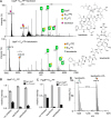
Inhibition of UppP and PgpB activities by antibiotics. (A) Spectra for a solution containing UppP (3.5 μM) and 20 μM C55-PP incubated in the presence of 100 μM bacitracin (top) and 100 μM teixobactin (lower panel) for 30 min. Only a small amount of enzyme–product complex is detected in the presence of both inhibitors, reflecting that both inhibitors sequester the lipid substrate C55-PP from the enzyme. The m/z values for antibiotics and their complexes with the substrate are labeled. Shown on the right are the chemical structures of bacitracin and teixobactin. (B,C) The relative intensities of enzyme–product complexes in the spectra for UppP- and PgpB-mediated formation of C55-PP. (D) The low m/z region of spectrum acquired for PgpB/LPA mixture in the negative electrospray ionization mode. Teixobactin form a stable complex with LPA.

Similar articles

Cited by

References

    1. Cortez K. J.; Roilides E.; Quiroz-Telles F.; Meletiadis J.; Antachopoulos C.; Knudsen T.; Buchanan W.; Milanovich J.; Sutton D. A.; Fothergill A.; et al. Infections caused by scedosporium spp. Clin. Microbiol. Rev. 2008, 21 (1), 157–197. 10.1128/cmr.00039-07. - DOI - PMC - PubMed
    1. Wang G.; Zhao Z.; Ke J.; Engel Y.; Shi Y.-M.; Robinson D.; Bingol K.; Zhang Z.; Bowen B.; Louie K.; et al. CRAGE enables rapid activation of biosynthetic gene clusters in undomesticated bacteria. Nat. Microbiol. 2019, 4 (12), 2498–2510. 10.1038/s41564-019-0573-8. - DOI - PubMed
    1. Mahler L.; Niehs S. P.; Martin K.; Weber T.; Scherlach K.; Hertweck C.; Roth M.; Rosenbaum M. A. Highly parallelized droplet cultivation and prioritization of antibiotic producers from natural microbial communities. eLife 2021, 10, e6477410.7554/eLife.64774. - DOI - PMC - PubMed
    1. Chu J.; Vila-Farres X.; Inoyama D.; Ternei M.; Cohen L. J.; Gordon E. A.; Reddy B. V. B.; Charlop-Powers Z.; Zebroski H. A.; Gallardo-Macias R.; et al. Discovery of MRSA active antibiotics using primary sequence from the human microbiome. Nat. Chem. Biol. 2016, 12 (12), 1004–1006. 10.1038/nchembio.2207. - DOI - PMC - PubMed
    1. Stokes J. M.; Yang K.; Swanson K.; Jin W.; Cubillos-Ruiz A.; Donghia N. M.; MacNair C. R.; French S.; Carfrae L. A.; Bloom-Ackermann Z.; et al. A deep learning approach to antibiotic discovery. Cell 2020, 181 (2), 475–483. 10.1016/j.cell.2020.04.001. - DOI - PubMed

Publication types