

G protein-coupled estrogen receptor (GPER) in the dorsal hippocampus regulates memory consolidation in gonadectomized male mice, likely via different signaling mechanisms than in female mice
- PMID: 38428223
- PMCID: PMC11065565
- DOI: 10.1016/j.yhbeh.2024.105516
G protein-coupled estrogen receptor (GPER) in the dorsal hippocampus regulates memory consolidation in gonadectomized male mice, likely via different signaling mechanisms than in female mice
Abstract
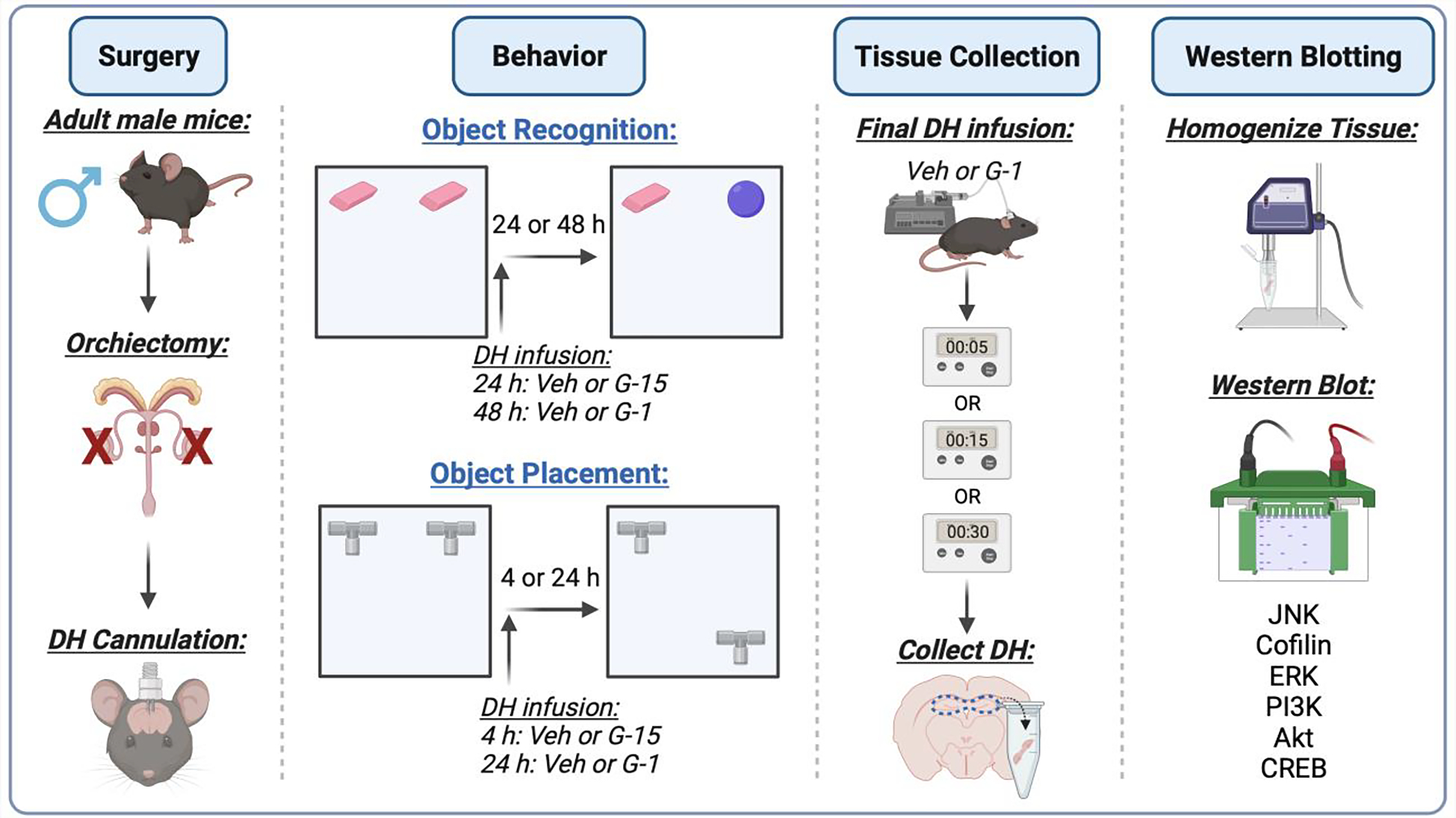
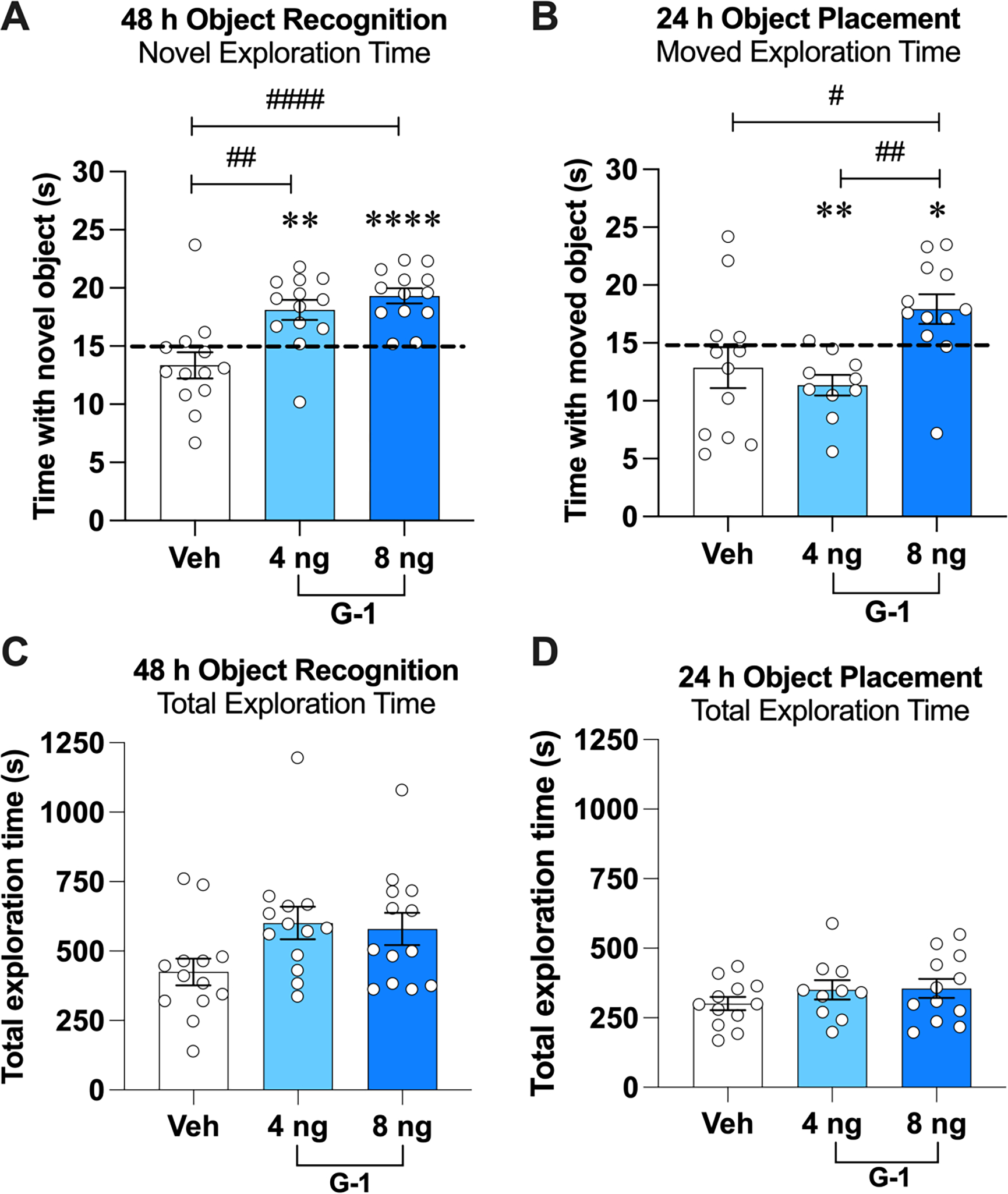
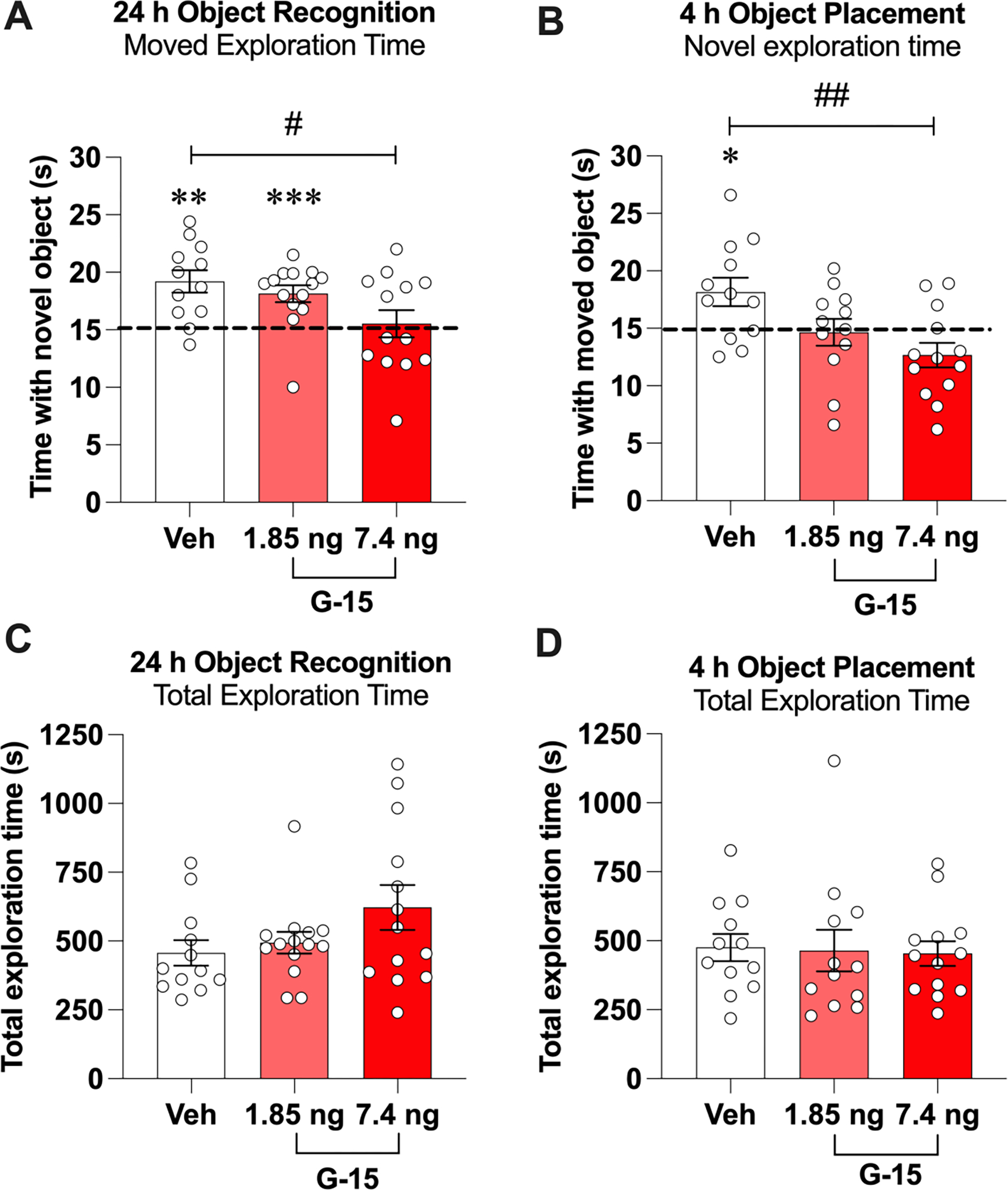
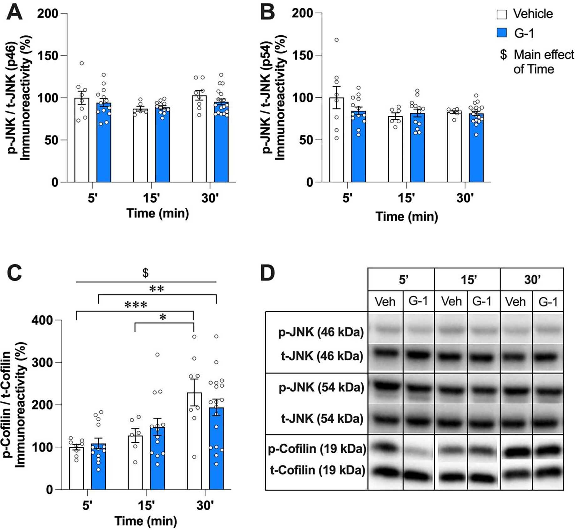
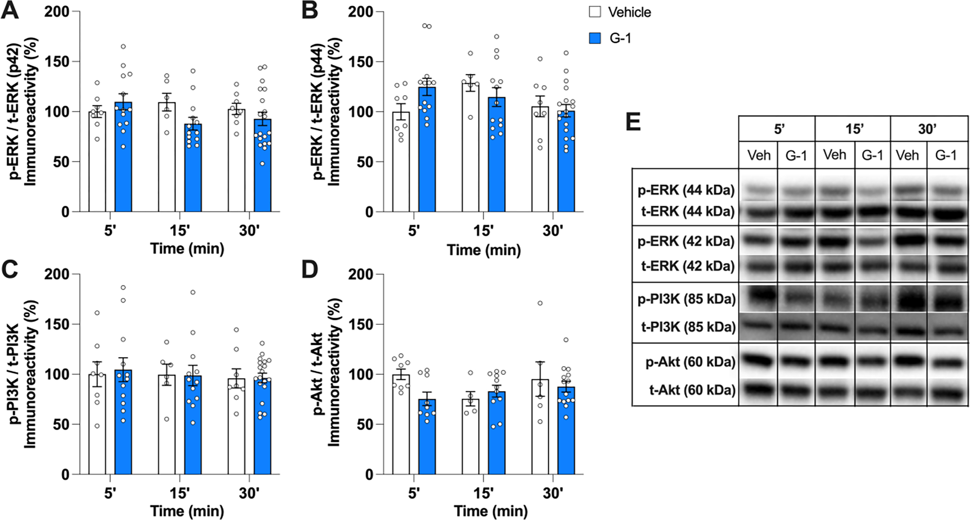
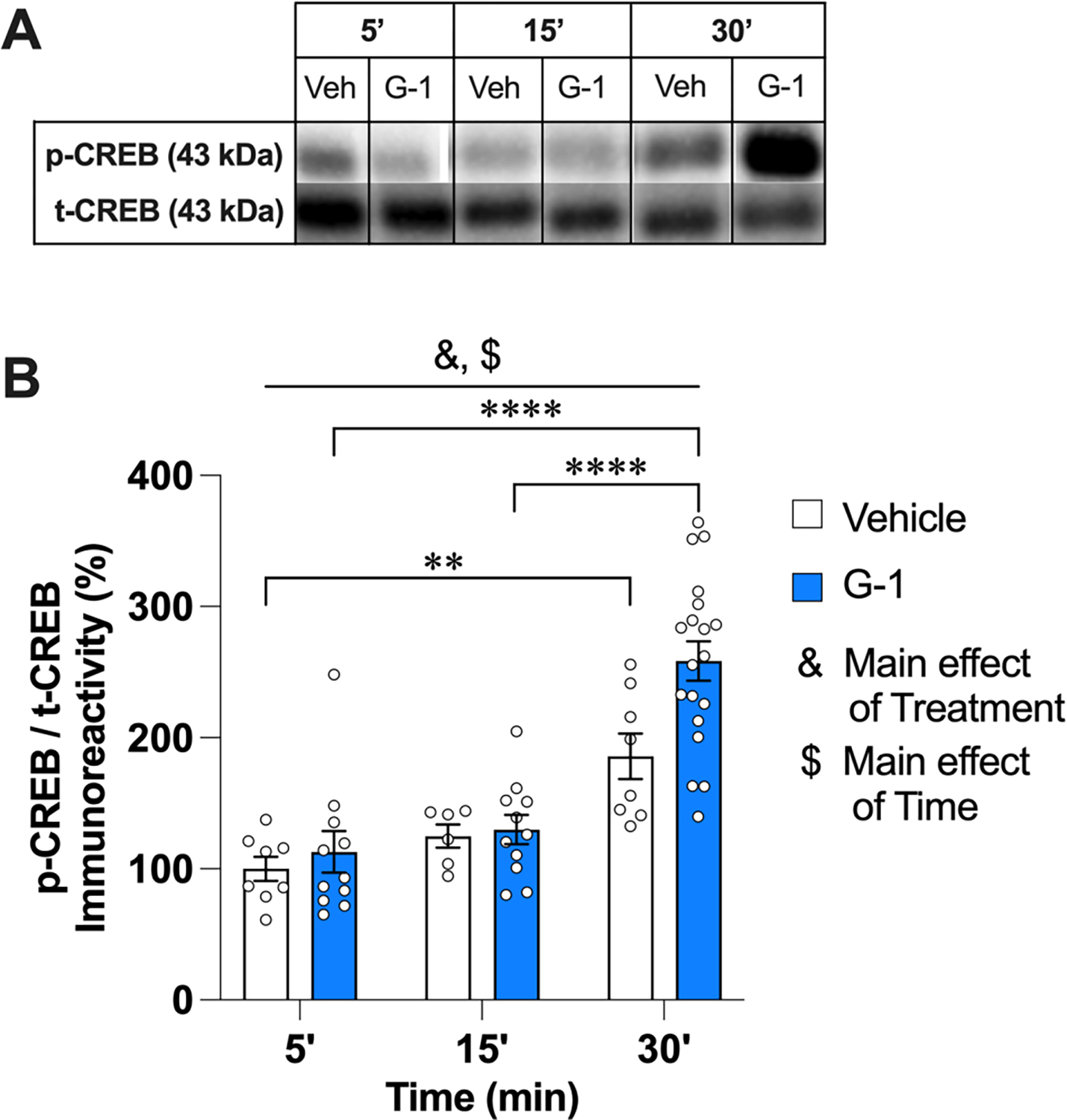
Studies in ovariectomized (OVX) female rodents suggest that G protein-coupled estrogen receptor (GPER) is a key regulator of memory, yet little is known about its importance to memory in males or the cellular mechanisms underlying its mnemonic effects in either sex. In OVX mice, bilateral infusion of the GPER agonist G-1 into the dorsal hippocampus (DH) enhances object recognition and spatial memory consolidation in a manner dependent on rapid activation of c-Jun N-terminal kinase (JNK) signaling, cofilin phosphorylation, and actin polymerization in the DH. However, the effects of GPER on memory consolidation and DH cell signaling in males are unknown. Thus, the present study first assessed effects of DH infusion of G-1 or the GPER antagonist G-15 on object recognition and spatial memory consolidation in gonadectomized (GDX) male mice. As in OVX mice, immediate post-training bilateral DH infusion of G-1 enhanced, whereas G-15 impaired, memory consolidation in the object recognition and object placement tasks. However, G-1 did not increase levels of phosphorylated JNK (p46, p54) or cofilin in the DH 5, 15, or 30 min after infusion, nor did it affect phosphorylation of ERK (p42, p44), PI3K, or Akt. Levels of phospho-cAMP-responsive element binding protein (CREB) were elevated in the DH 30 min following G-1 infusion, indicating that GPER in males activates a yet unknown signaling mechanism that triggers CREB-mediated gene transcription. Our findings show for the first time that GPER in the DH regulates memory consolidation in males and suggests sex differences in underlying signaling mechanisms.
Keywords: CREB; Cofilin; JNK; Mouse; Object placement; Object recognition; Spatial memory.
Copyright © 2024 The Authors. Published by Elsevier Inc. All rights reserved.
Conflict of interest statement
Declaration of competing interest Dr. Frick is a co-founder and the Chief Scientific Officer of Estrigenix Therapeutics, Inc., a company which aims to improve women's health by developing safe, clinically proven treatments for the mental and physical effects of menopause. The rest of the authors have no conflicts of interests to declare.
Figures
Similar articles
-
17β-Estradiol and Agonism of G-protein-Coupled Estrogen Receptor Enhance Hippocampal Memory via Different Cell-Signaling Mechanisms.J Neurosci. 2016 Mar 16;36(11):3309-21. doi: 10.1523/JNEUROSCI.0257-15.2016. J Neurosci. 2016. PMID: 26985039 Free PMC article.
-
Dorsal Hippocampal Actin Polymerization Is Necessary for Activation of G-Protein-Coupled Estrogen Receptor (GPER) to Increase CA1 Dendritic Spine Density and Enhance Memory Consolidation.J Neurosci. 2019 Nov 27;39(48):9598-9610. doi: 10.1523/JNEUROSCI.2687-18.2019. Epub 2019 Oct 18. J Neurosci. 2019. PMID: 31628182 Free PMC article.
-
Sex Differences in the Rapid Cell Signaling Mechanisms Underlying the Memory-Enhancing Effects of 17β-Estradiol.eNeuro. 2018 Oct 30;5(5):ENEURO.0267-18.2018. doi: 10.1523/ENEURO.0267-18.2018. eCollection 2018 Sep-Oct. eNeuro. 2018. PMID: 30406188 Free PMC article.
-
Molecular mechanisms underlying the memory-enhancing effects of estradiol.Horm Behav. 2015 Aug;74:4-18. doi: 10.1016/j.yhbeh.2015.05.001. Epub 2015 May 8. Horm Behav. 2015. PMID: 25960081 Free PMC article. Review.
-
Estrogenic regulation of memory consolidation: A look beyond the hippocampus, ovaries, and females.Physiol Behav. 2018 Apr 1;187:57-66. doi: 10.1016/j.physbeh.2017.07.028. Epub 2017 Jul 27. Physiol Behav. 2018. PMID: 28755863 Free PMC article. Review.
Cited by
-
Membrane progesterone and oestrogen receptors modulate GABAergic transmission in the prefrontal cortex of prepubertal male, but not female, mice.Exp Physiol. 2025 Jun;110(6):888-898. doi: 10.1113/EP092439. Epub 2025 May 1. Exp Physiol. 2025. PMID: 40309895 Free PMC article.
References
-
- Bai N, Zhang Q, Zhang W, Liu B, Yang F, Brann D, Wang R, 2020. G-protein-coupled estrogen receptor activation upregulates interleukin-1 receptor antagonist in the hippocampus after global cerebral ischemia: Implications for neuronal self-defense. J. Neuroinflammation 17, 45. 10.1186/s12974-020-1715-x - DOI - PMC - PubMed
-
- Bernabeu R, Bevilaqua L, Ardenghi P, Bromberg E, Schmitz P, Bianchin M, Izquierdo I, Medina JH, 1997. Involvement of hippocampal cAMP/cAMP-dependent protein kinase signaling pathways in a late memory consolidation phase of aversively motivated learning in rats. Proc. Natl. Acad. Sci. 94, 7041–7046. 10.1073/pnas.94.13.7041 - DOI - PMC - PubMed
Publication types
MeSH terms
Substances
Grants and funding
LinkOut - more resources
Full Text Sources
Molecular Biology Databases
Research Materials
Miscellaneous
