. 2024 Feb 29;187(5):1223-1237.e16.
doi: 10.1016/j.cell.2024.01.048.

The CARD8 inflammasome dictates HIV/SIV pathogenesis and disease progression

Affiliations

The CARD8 inflammasome dictates HIV/SIV pathogenesis and disease progression

Qiankun Wang et al. Cell. .

Abstract

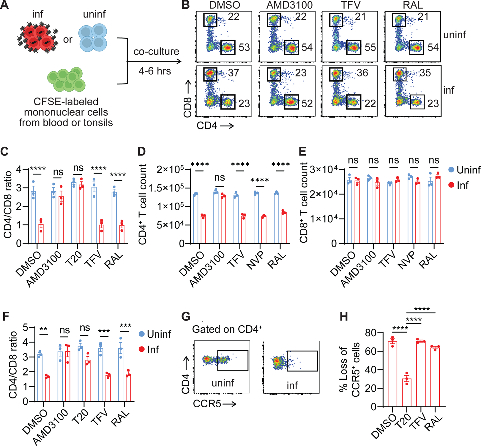
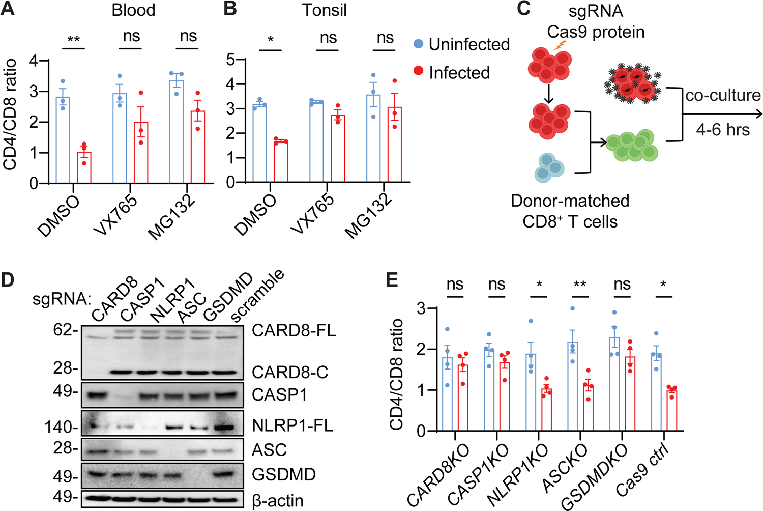
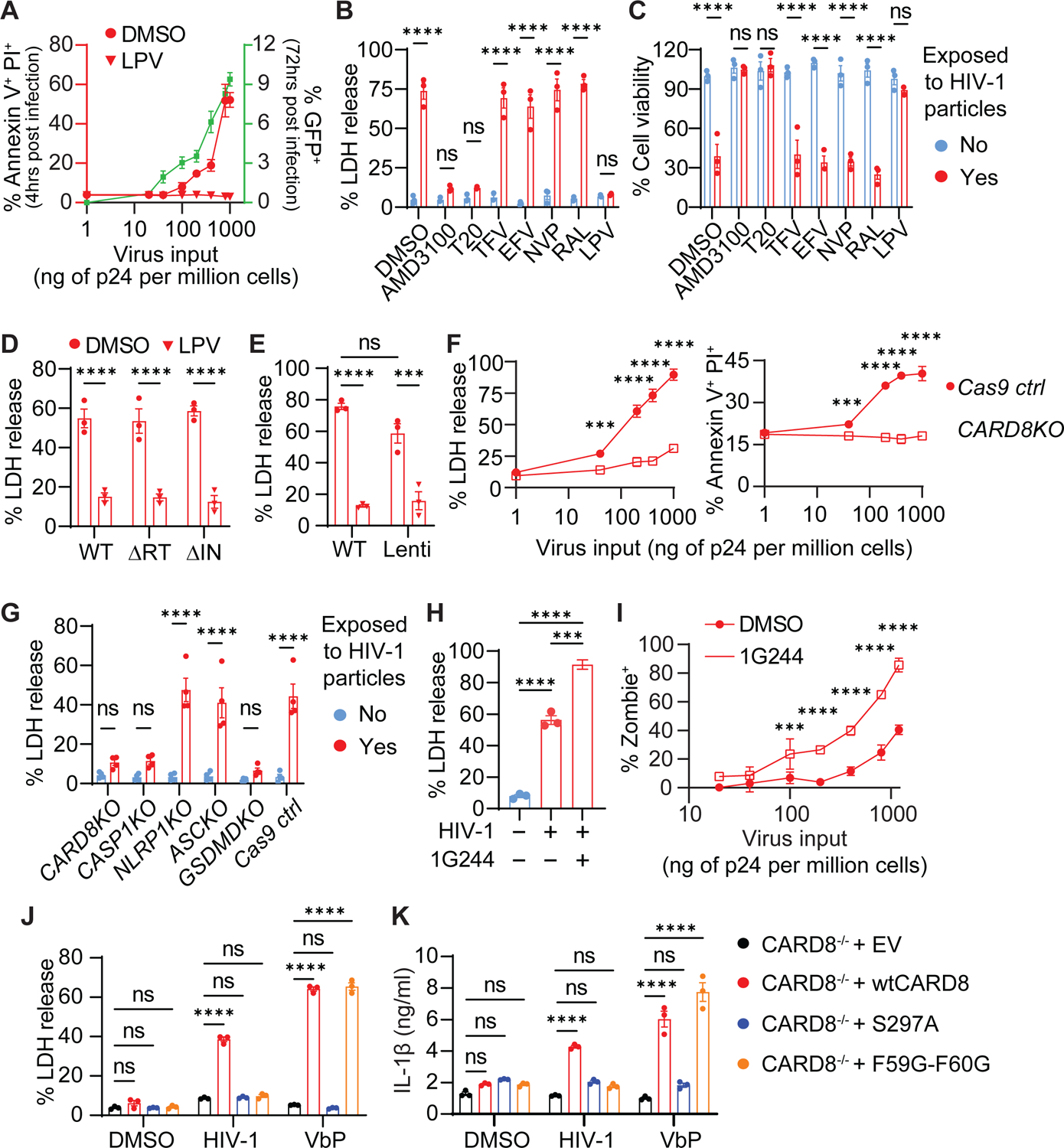
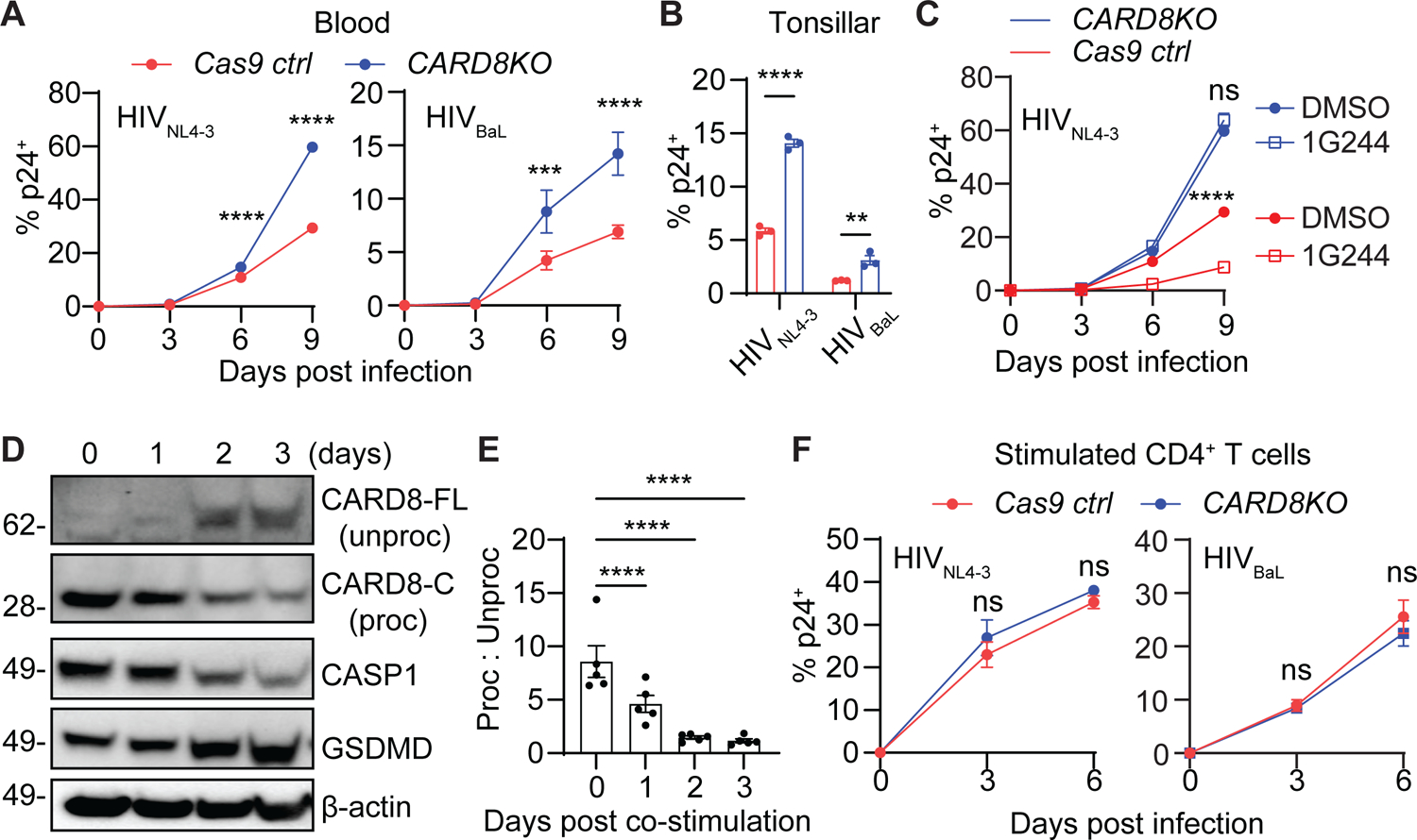
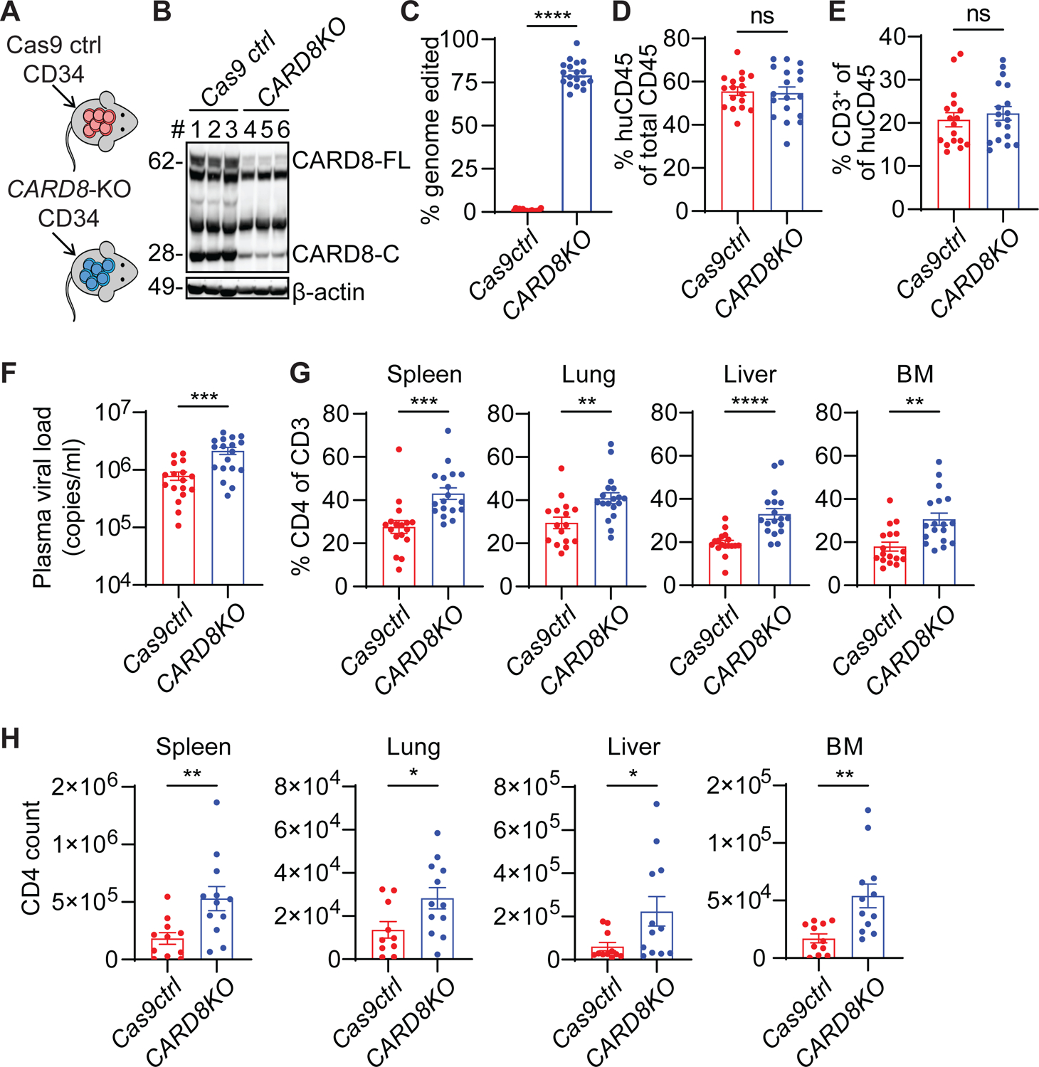
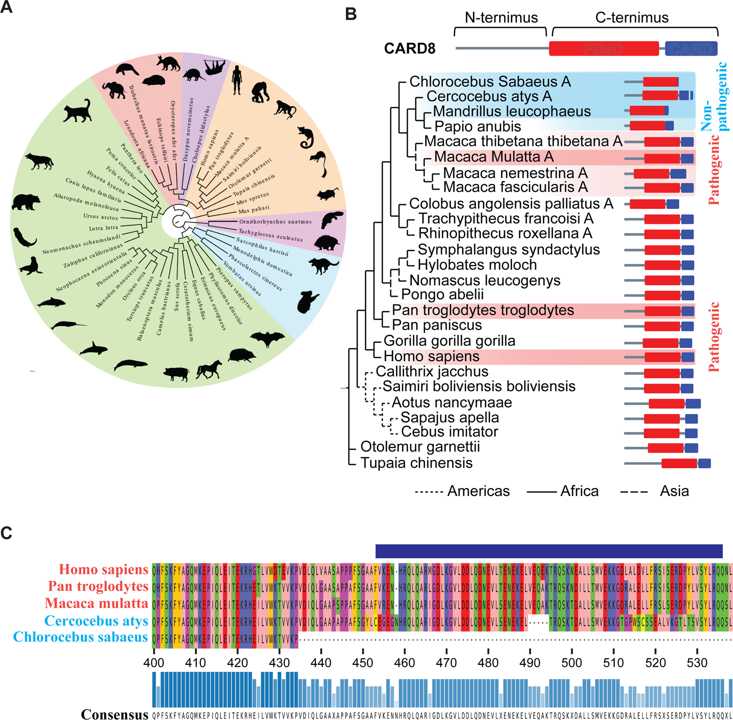
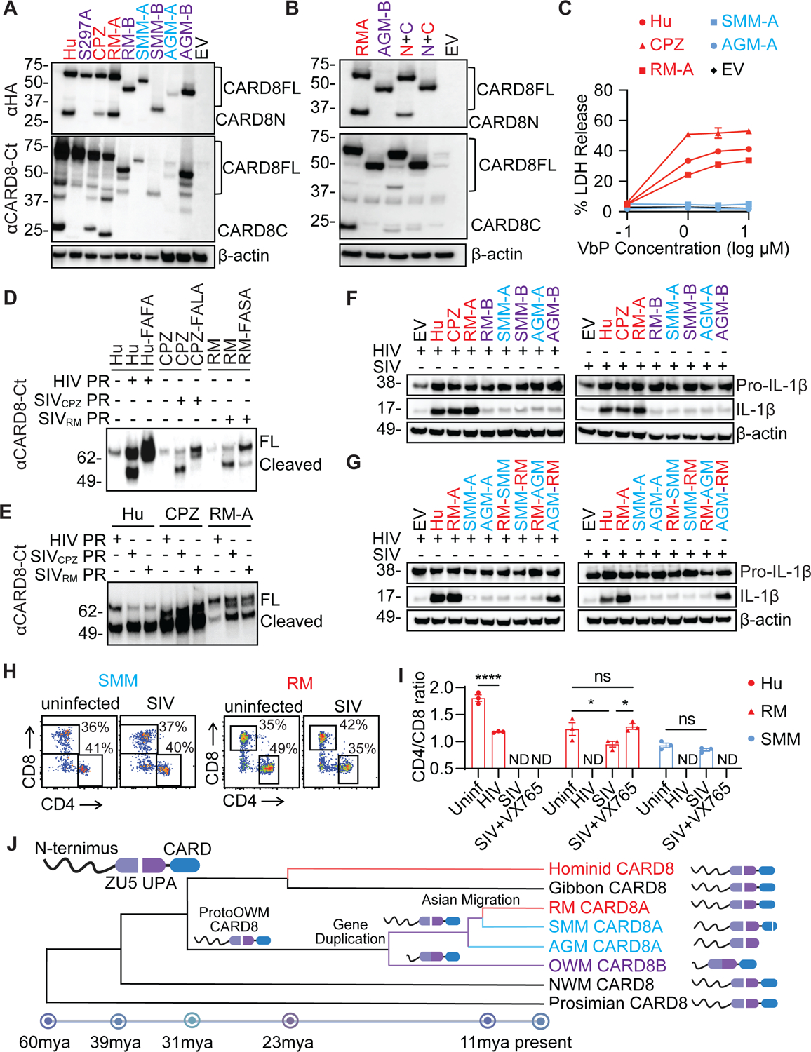
While CD4+ T cell depletion is key to disease progression in people living with HIV and SIV-infected macaques, the mechanisms underlying this depletion remain incompletely understood, with most cell death involving uninfected cells. In contrast, SIV infection of "natural" hosts such as sooty mangabeys does not cause CD4+ depletion and AIDS despite high-level viremia. Here, we report that the CARD8 inflammasome is activated immediately after HIV entry by the viral protease encapsulated in incoming virions. Sensing of HIV protease activity by CARD8 leads to rapid pyroptosis of quiescent cells without productive infection, while T cell activation abolishes CARD8 function and increases permissiveness to infection. In humanized mice reconstituted with CARD8-deficient cells, CD4+ depletion is delayed despite high viremia. Finally, we discovered loss-of-function mutations in CARD8 from "natural hosts," which may explain the peculiarly non-pathogenic nature of these infections. Our study suggests that CARD8 drives CD4+ T cell depletion during pathogenic HIV/SIV infections.

Keywords: CARD8; CD4(+) T cells; HIV; SIV; caspase 1; inflammasome; non-human primates; pyroptosis; viral entry; viral protease.


Conflict of interest statement

Declaration of interests The authors declare no competing interests.

Figures

Figure 1.
Figure 1.. HIV entry triggers rapid loss of CD4+ T cells.
(A) The co-culture schemes. Activated blood or tonsillar CD4+ T cells were infected with either HIVNL4-3 or HIVBaL for three days. Virus-producing cells were then co-cultured at a 1:1 ratio with CFSE-labeled donor-matched unstimulated mononuclear cells from blood or tonsils for four to six hours with the presence of indicated ARVs. (B) Representative plots were shown from blood cells infected with HIVNL4-3. (C-F) Rapid loss of blood and tonsillar CD4+ T cells. Virus-producing cells were infected with HIVNL4-3. Three blood samples (C-E) and three tonsillar samples (F) were used. (G and H) Rapid loss of CCR5+ CD4+ T cells. Virus-producing cells were infected with HIVBaL. Three blood samples were used. In C-F, p values were calculated using the two-way ANOVA with Šidák’s multiple comparison tests. In H, p values were calculated using the one-way ANOVA with Dunnett tests. ** p < 0.01. *** p < 0.001, **** p < 0.0001. Error bars show mean values with standard errors of the mean (SEM). n=3.
Figure 2.
Figure 2.. CD4+ T-cell depletion by HIV is mediated by the CARD8 inflammasome.
(A and B) Rapid CD4+ T-cell death is proteasome- and CASP1-dependent. Unstimulated PBMCs (A) or ToMCs (B) were pretreated with VX765 (50 μM) or MG132 (10 μM) for 30 minutes and then co-cultured with donor-matched uninfected or HIVNL4-3-producing CD4+ T cells for six hours before flow cytometry analyses. (C) The co-culture scheme of Cas9-edited unstimulated CD4+ T cells and HIV-infected autologous CD4+ T cells. Unstimulated CD4+ T cells electroporated with the indicated gene-specific sgRNA were cultured for three weeks and then mixed with donor-matched CD8+ T cells before co-culture with HIV-infected CD4+ T cells from the same donor. (D and E) The CARD8 inflammasome is required for the rapid loss of CD4+ T cells. Unstimulated CD4+ T cells with indicated knockouts were mixed with autologous CD8+ T cells at a 2:1 ratio before co-culture with HIVNL4-3-producing cells. Cas9 editing efficiency was confirmed in D. The immunoblots represent four independent experiments. Four blood samples were used. p values were calculated using two-way ANOVA with Šidák’s multiple comparison tests. * p < 0.05, ** p < 0.01, ns: not significant. Error bars show mean values with SEM from three or four independent blood donors.
Figure 3.
Figure 3.. HIV infection induces CARD8 inflammasome activation by virion-packaged protease.
(A-E) Rapid loss of CD4+ T cells induced by HIV protease encapsulated in the incoming viral particles. In (A), unstimulated CD4+ T cells were exposed to cell-free HIV reporter virus NL4-3-ΔEnv-EGFP pseudotyped with the NL4-3 envelope. Cell death and productive infection (GFP+) were measured four hours and three days post-infection, respectively. In (B and C), unstimulated CD4+ T cells were treated with indicated antiretroviral drugs for 30 minutes before being exposed to HIV reporter viral particles. In (D and E), unstimulated CD4+ T cells were pre-treated with LPV or DMSO for 30 minutes and then exposed to indicated enzyme inactive reporter HIV or lentiviral particles pseudotyped with the NL4-3 envelope. ΔRT: D110A and D185A. ΔIN: D116A. (F and G) Rapid loss of CD4+ T cells is mediated by the CARD8 inflammasome. Cell death was determined at indicated virus doses or in different knockout cells. (H and I) 1G244 enhances HIV entry-triggered pyroptosis of CD4+ T cells. Unstimulated CD4+ T cells were exposed to cell-free virions with or without 1G244 for four hours. (J and K) Activation of CARD8 inflammasome by HIV-1 in THP-1 cells. CARD8-KO THP-1 cells expressing wild-type or mutant CARD8 were exposed to HIV reporter virus NL4-3-ΔEnv-EGFP pseudotyped with the VSVG envelope for six hours before LDH or IL-1β measurement. VbP (5 μM) was used as positive controls. In (H), p values were calculated using the one-way ANOVA with Tukey’s multiple comparisons tests. Other p values were calculated using the two-way ANOVA with Šidák’s multiple comparison tests. * p < 0.05, **** p < 0.0001. *** p < 0.001, ** p < 0.01. The data points are means with SEM and represent three or more independent experiments.
Figure 4.
Figure 4.. The CARD8 inflammasome restricts HIV infection in unstimulated CD4+ T cells.
(A-C) Loss of CARD8 leads to increased susceptibility to HIV infection in unstimulated CD4+ T cells. CARD8-KO or Cas9 ctrl unstimulated CD4+ T cells from blood (A) or tonsil (B) were infected with HIVNL4-3 or HIVBaL at 40 ng HIVp24 per million cells. Infection was measured by intracellular p24 staining. Tonsillar samples were analyzed on day six post-infection. In (C), CARD8-KO or Cas9 ctrl unstimulated CD4+ T cells were infected with HIVNL4-3 or HIVBaL with or without 1G244 (500 nM). (D and E) Accumulation of unprocessed CARD8 in activated CD4+ T cells. Immunoblotting was performed to analyze CARD8 expression and autoprocessing following anti-CD3 and anti-CD28 stimulation of CD4+ T cells. (F) The CARD8 inflammasome does not restrict HIV infection in activated CD4+ T cells. CARD8-KO or Cas9 ctrl activated CD4+ T cells were infected with HIVNL4-3 or HIVBaL at 40 ng HIVp24 per million cells. Infection was measured by intracellular p24 staining. In (E), p values were calculated using the one-way ANOVA with Dunnett tests. Other p values were calculated using the two-way ANOVA with Šidák’s multiple comparison tests. * p < 0.05, **** p < 0.0001. *** p < 0.001, ** p < 0.01. Error bars show mean values with SEM. n=3.
Figure 5.
Figure 5.. The CARD8 inflammasome leads to the loss of CD4+ T cells in humanized mice with HIV infection.
(A) Strategy for generating CARD8-edited humanized mice. (B) Representative immunoblotting of total splenocytes from the uninfected CARD8-KO and Cas9 ctrl groups. Cas9 ctrl, n=3; CARD8-KO, n=3. (C) Blood samples were collected to determine the frequency of CARD8 editing by MiSeq. Cas9 ctrl, n=17; CARD8-KO, n=18. (D and E), Human immune cell reconstitution and T cell development in mice engrafted with CARD8-KO or Cas9 ctrl CD34+ cells. Blood samples were collected 10 weeks after transplantation. Cas9 ctrl, n=17; CARD8-KO, n=18. (F) Plasma viral loads was measured two weeks after infection. Cas9 ctrl, n=17; CARD8-KO, n=18. (G and H) The frequency and number of human CD4+ T cells in tissues. In G, Cas9 ctrl, n=17, except for lung (n=16); CARD8-KO, n=18. In H, Cas9 ctrl, n=11, except for lung (n=10); CARD8-KO, n=12. Data were pooled from three independent mouse cohorts. p values were calculated using an unpaired two-tailed t-test. * p < 0.05, ** p < 0.01, ***p < 0.001, and **** p < 0.0001. Error bars show mean values with SEM.
Figure 6.
Figure 6.. Genetic analysis of the NHP CARD8 inflammasome reveals functional defects in non-pathogenic hosts of SIV infection
(A) Phylogenetic analysis of mammalian CARD8. The protein sequences of the longest isoform for each CARD8 gene from each species were aligned and used to construct the maximum likelihood tree shown. CARD8 is present in every superorder (pink: Monotremata, blue: Marsupiala, purple: Xenartha, red: Afrotheria, green: Laurasiatheria, and orange: Euarchontoglires). Laurasiatheria is polyphyletic, Euarchontoglires is paraphyletic, and all other superorders are monophyletic. The sequences used to generate this tree may be found in Table S4. (B) Maximum likelihood gene tree of CARD8 or CARD8A from the selected NHP species. Dark red shading indicates known pathogenic HIV/SIV hosts and light red the suspected pathogenic hosts; dark blue depicts known non-pathogenic hosts, and light blue the suspected non-pathogenic hosts. The protein schemes on the right depict the predicted domain architecture based on the alignment to the known domain structure of the human protein. (C) Protein alignment of the CARD domains and part of the FIIND domains of the species of known pathogenicity status.
Figure 7.
Figure 7.. The CARD8 inflammasome is functional in pathogenic hosts and defective in non-pathogenic hosts of SIV infection
(A) Autoprocessing of NHP CARD8. HEK293T cells were transfected with plasmids expressing HA-tagged CARD8 from Hu and CPZ, and CARD8A and CARD8B from RM, SMM, and AGM. (B) CARD8 N-terminal truncation inhibits its C-terminal autoprocessing. The two chimeric CARD8 (N+C) were RMA N-terminus (red) plus AGMB C-terminus (cyan) and AGMB N-terminus (cyan) plus RMA C-terminus (red). (C) Validation of NHP CARD8 functions. CARD8-KO THP-1 cells replete with indicated NHP CARD8 were treated with VbP for six hours before cell death measurement by the LDH release assay. (D and E) In vitro cleavage of human and NHP CARD8 by HIV and SIV protease. Purified CARD8 proteins and SIV or HIV protease were incubated for one hour before immunoblotting. CARD8 cleavage site mutants included Hu-FAFA, CPZ-FALA, and RM-FASA. (F and G) Activation of human and NHP CARD8 inflammasome by HIV and SIV. CARD8-KO HEK293T cells were co-transfected with HIV or SIV molecular clones together with plasmids encoding CASP1, pro–IL-1β, and human or NHP CARD8. In (G), indicated chimeric CARD8 (N+C) proteins were used. (H and I) Rapid loss of CD4+ T cells. Co-culture of human, RM, and SMM PBMCs with SIVmac251-infected CEM-174 cells. CD4/CD8 ratio was measured 16 hours post co-culture. Two-way ANOVA with Šídák’s multiple comparisons test. * p < 0.05 and **** p < 0.0001. Error bars show mean values with SEM for PBMC from three separate primates. (J) The evolution of non-human primate CARD8 depicting key events in its evolution. CARD8 underwent a reverse tandem gene duplication in the ancestor to old world monkeys (OWM) giving rise to a non-functional CARD8B and a functional CARD8A. The CARD8A of non-pathogenic hosts in Africa underwent further selection leading to non-functionalization of the protein whereas the Asian migration of macaques allowed for escape from this selective pressure. Speciation dates are estimated based on recently published findings. NWM – new world monkeys.

Similar articles

Cited by

References

    1. Brinchmann JE, Albert J, and Vartdal F (1991). Few infected CD4+ T cells but a high proportion of replication-competent provirus copies in asymptomatic human immunodeficiency virus type 1 infection. J Virol 65, 2019–2023. 10.1128/JVI.65.4.2019-2023.1991. - DOI - PMC - PubMed
    1. Hsia K, and Spector SA (1991). Human immunodeficiency virus DNA is present in a high percentage of CD4+ lymphocytes of seropositive individuals. J Infect Dis 164, 470–475. 10.1093/infdis/164.3.470. - DOI - PubMed
    1. Psallidopoulos MC, Schnittman SM, Thompson LM 3rd, Baseler M, Fauci AS, Lane HC, and Salzman NP (1989). Integrated proviral human immunodeficiency virus type 1 is present in CD4+ peripheral blood lymphocytes in healthy seropositive individuals. J Virol 63, 4626–4631. 10.1128/JVI.63.11.4626-4631.1989. - DOI - PMC - PubMed
    1. Schnittman SM, Greenhouse JJ, Psallidopoulos MC, Baseler M, Salzman NP, Fauci AS, and Lane HC (1990). Increasing viral burden in CD4+ T cells from patients with human immunodeficiency virus (HIV) infection reflects rapidly progressive immunosuppression and clinical disease. Ann Intern Med 113, 438–443. 10.7326/0003-4819-113-6-438. - DOI - PubMed
    1. Simmonds P, Balfe P, Peutherer JF, Ludlam CA, Bishop JO, and Brown AJ (1990). Human immunodeficiency virus-infected individuals contain provirus in small numbers of peripheral mononuclear cells and at low copy numbers. J Virol 64, 864–872. 10.1128/JVI.64.2.864-872.1990. - DOI - PMC - PubMed

MeSH terms