. 2024 Feb 29;16(5):4609-4630.
doi: 10.18632/aging.205617. Epub 2024 Feb 29.

Myogenic exosome miR-140-5p modulates skeletal muscle regeneration and injury repair by regulating muscle satellite cells

Affiliations

Myogenic exosome miR-140-5p modulates skeletal muscle regeneration and injury repair by regulating muscle satellite cells

Xiaorui Cao et al. Aging (Albany NY). .

Abstract

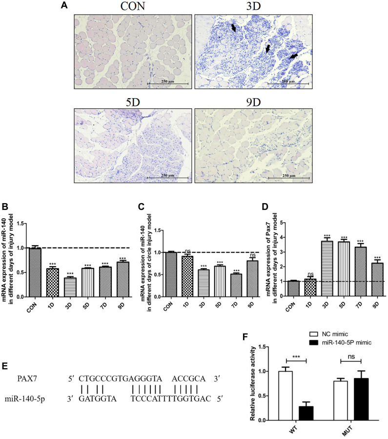
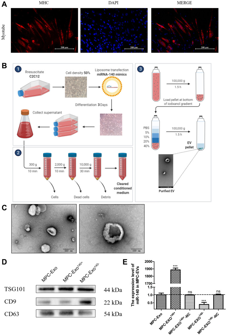
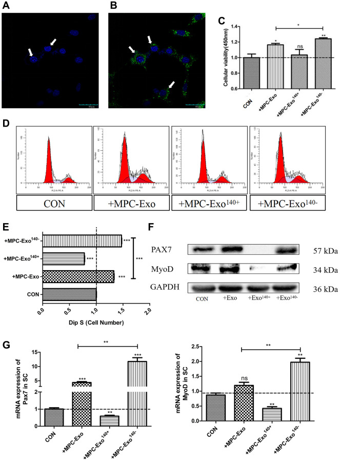
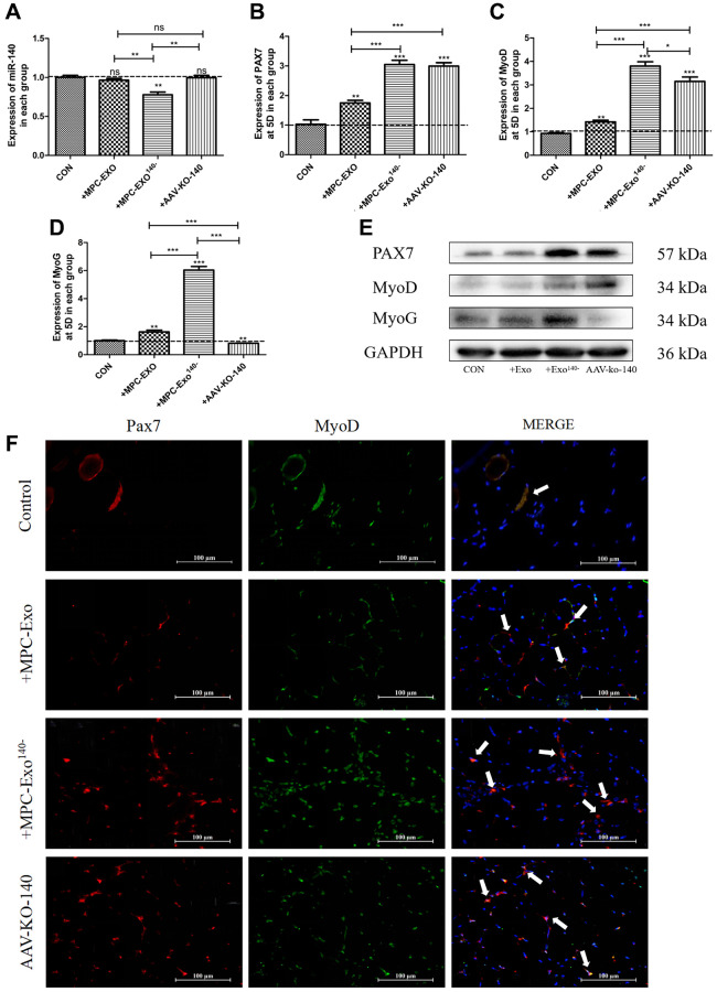
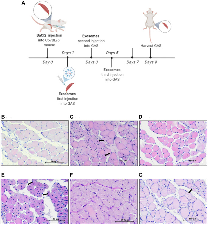
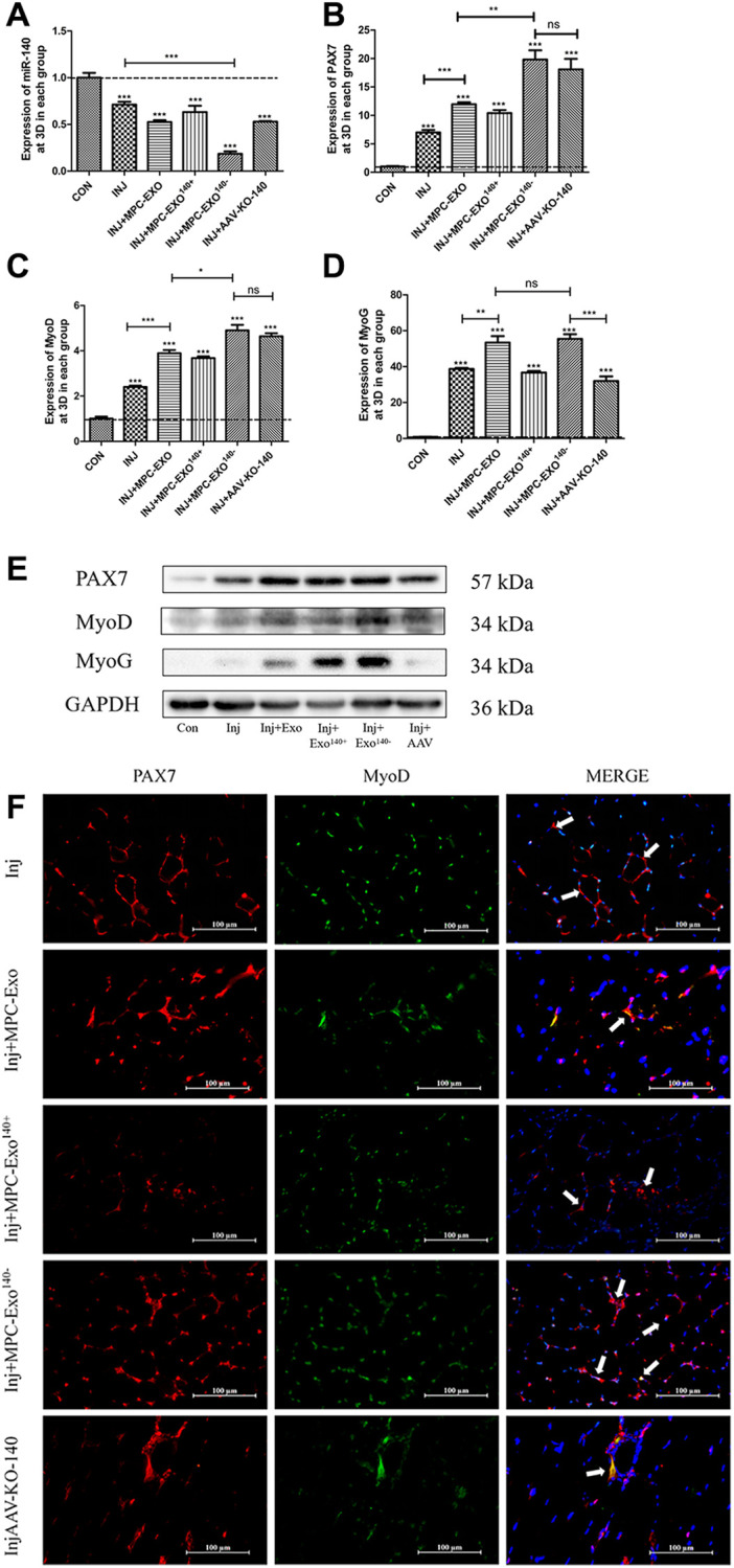
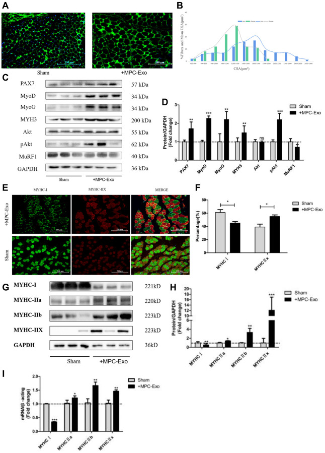
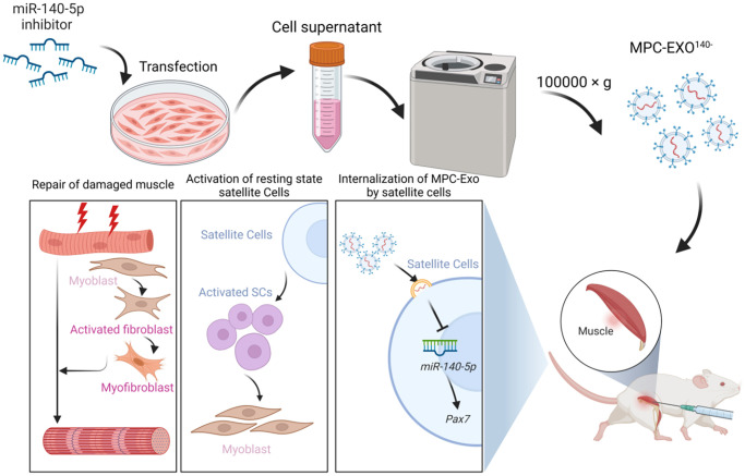
Muscle satellite cells (SCs) play a crucial role in the regeneration and repair of skeletal muscle injuries. Previous studies have shown that myogenic exosomes can enhance satellite cell proliferation, while the expression of miR-140-5p is significantly reduced during the repair process of mouse skeletal muscle injuries induced by BaCl2. This study aims to investigate the potential of myogenic exosomes carrying miR-140-5p inhibitors to activate SCs and influence the regeneration of injured muscles. Myogenic progenitor cell exosomes (MPC-Exo) and contained miR-140-5p mimics/inhibitors myogenic exosomes (MPC-Exo140+ and MPC-Exo140-) were employed to treat SCs and use the model. The results demonstrate that miR-140-5p regulates SC proliferation by targeting Pax7. Upon the addition of MPC-Exo and MPC-Exo140-, Pax7 expression in SCs significantly increased, leading to the transition of the cell cycle from G1 to S phase and an enhancement in cell proliferation. Furthermore, the therapeutic effect of MPC-Exo140- was validated in animal model, where the expression of muscle growth-related genes substantially increased in the gastrocnemius muscle. Our research demonstrates that MPC-Exo140- can effectively activate dormant muscle satellite cells, initiating their proliferation and differentiation processes, ultimately leading to the formation of new skeletal muscle cells and promoting skeletal muscle repair and remodeling.

Keywords: exosomes; miR-140-5p; muscle injury repair; muscle satellite cells; regeneration.


Conflict of interest statement

CONFLICTS OF INTEREST: The authors declare that the research was conducted in the absence of any commercial or financial relationships that could be construed as a potential conflict of interest.

Figures

Figure 1
Figure 1
Significant downregulation of miR-140-5p expression following injury, contrasting with the expression trend of Pax7. Male wildtype C57BL/6 mice established damage model by intramuscular injection of 1.2% BaCl2 into the gastrocnemius muscle. (A) displays the muscle repair and regeneration process post-acute damage to the gastrocnemius muscle caused by BaCl2, as shown through histological examination staining (Scale bar in 250 μm). (BD) present the RT-PCR results (fold change) illustrating the expression trends of miR-140-5p and Pax7 in the gastrocnemius muscle and circulation at different time points postinjury (n = 4 biological replicates). (E, F) demonstrate the binding pattern between miR-140-5p and Pax7, along with the dual luciferase assay results (n = 4), where WT refers to the wild type and MUT to the mutant type. The results are reported in the line/bar graph as mean ± SE; n = 4/group; different letters between bars mean P ≤ 0.05 analyses followed by non-paired Student’s t-test. *P < 0.05; **P < 0.01; ***P < 0.001 vs. CON Group.
Figure 2
Figure 2
Production and identification engineered exosomes containing miR-140-5p mimics or inhibitors. Preparation and identification of MPC-EVs. (A) illustrates the formation of myotubes post differentiation of C2C12 cells, shown via IF with red representing the heavy chain myosin (MYH) and blue for DAPI-stained nuclei (Scale bar in 200 μm). (B) depicts the preparation process of MPC-EVs. (C) presents the TEM results, showing the characteristic cup-shaped or semispherical morphology of the myogenic extracellular vesicles, in agreement with the expected bilayer membrane ultrastructure of extracellular vesicles. (D) shows WB results indicating positive reactions for the marker proteins TSG101, CD9, and CD63 in all types of MPC-EVs. (E) illustrates RT-PCR results (fold change), showing the miR-140-5p levels in MPC-Exo140+ and MPC-Exo140−, which are 1800 times and 1/3 of normal extracellular vesicles, respectively. MPC-Exo140+-NC and MPC-Exo140−-NC showed no difference from normal extracellular vesicles. Different letters between bars mean P ≤ 0.05 analyses followed by non-paired Student’s t-test. nsp > 0.05, *p < 0.05, **p < 0.01 and ***p < 0.001 vs. MPC-Exo.
Figure 3
Figure 3
MPC-EVs and MPC-Exo140− internalized by SCs induced Pax7 and MyoD significantly increased and promoted satellite cell proliferation. Activation of muscle satellite cells by MPC-EVs and MPC-Exo140−. The process of SCs taking up myogenic extracellular vesicles was displayed under a live-cell confocal microscopy workstation, with (A) SC without MPC-Exond (B) treated with MPC-Exo140- green fluorescence indicates uptake myogenic extracellular vesicles (Scale bar in 40 μm). (C) shows CCK8 results indicating a significant increase in cell proliferation after coincubation of SCs with MPC-EVs, especially in the MPC-Exo140− group. (D, E) present the results of cell cycle analysis by flow cytometry, revealing a significant increase in the number of cells in the S phase in the MPC-Exo140− group, while a significant decrease is seen in the MPC-Exo140+ group. (F, G) show WB and RT-PCR results (fold change), showing that the expression of Pax7 and MyoD significantly increased in the MPC-Exo140− group, while they were significantly inhibited in the MPC-Exo140+ group. Data: by one-way ANOVA analysis. nsp > 0.05, *p < 0.05, **p < 0.01 and ***p < 0.001 vs. SC.
Figure 4
Figure 4
Muscle satellite cells in the gastrocnemius muscle activate by MPC-Exo, MPC-Exo140- or AAV-KO-140. Eight-week-old male wildtype C57BL/6 mice were treated with MPC-Exo and MPC-Exo140− 3 times. Five-week-old mice were injected with adeno-associated virus carrying miR-140-5p inhibitors. RT-qPCR was used to detect the expression (fold change) of miR-140-5p (A), Pax7 (B), MyoD (C) and MyoG (D). The amount of proteins for Pax 7, MyoD and myoD was analyzed by Western blot (E). Data: mean ± SEM (N = 5). *P < 0.05 by one-way ANOVA analysis. (F) presents the IF results: activated SCs are characterized by the coexpression of Pax7+/MyoD+. The marked locations in the figure indicate that in the control group, these cells are expressed only in the cell pool to maintain stem cell numbers. In the injected MPC-Exo group, there was a significant increase in activated SCs around the muscle fibers, indicating that muscle satellite cells were in a proliferative state. This proliferation of activated SCs was further augmented in the injected MPC-Exo140− group and AAV-KO-140 group (Scale bar in 100 μm).
Figure 5
Figure 5
MPC-EXO and MPC-Exo140− advanced the repair of gastrocnemius muscle after acute injury. Eight-week-old male wildtype C57BL/6 mice were treated with MPC-Exo, MPC-Exo140+ and MPC-Exo140− 3 times after acute injury. (A) presents a schematic diagram of muscle injury modeling treated with MPC-EVs. (BG) depict the HE staining results after three treatment sessions, with arrows pointing to central nuclei. The control group (B) shows closely arranged muscle cells with nuclei at the cell periphery. The injury group (C) presents newly formed muscle cells with centrally located nuclei and a few inflammatory cells between cells. In the MPC-Exo intervention group (D), the nuclei of newly formed muscle cells migrated toward the cell membrane. The MPC-Exo140+ intervention group (E) shows numerous centrally located nuclei in newly formed muscle cells and a loose extracellular matrix structure. In the MPC-Exo140− intervention group (F), the muscle cells were closely arranged, and the nuclei of newly formed muscle cells moved to the cell periphery. The AAV-KO-140 injury group (G) shows newly formed muscle cells with nuclei relocated to the cell periphery, but the fusion process between cells and newly formed multinucleated myotubes is inhibited, resulting in a significant increase in the number of nuclei (Scale bar in 100 μm).
Figure 6
Figure 6
MPC-Exo and MPC-Exo140− activate muscle satellite cells to promote repair of gastrocnemius muscle injury. (AC) and (D) show the mRNA expression changes (fold change) of miR-140-5p and muscle regeneration-related factors Pax7, MyoD, and myogenin after two interventions with MPC-EVs on the injured muscle. (E) presents changes in protein expression. (F) displays the impact of MPC-EVs on muscle satellite cells postinjury: activated SCs are characterized by the coexpression of Pax7+/MyoD+ (Figure 4F). The marked locations in the figure show that in the injury group, SCs enter an activated state stimulated by stress signals and move from the cell pool to between damaged muscle fibers. In the injected myogenic extracellular vesicle group, there was a significant increase in activated SCs around the muscle fibers. In the injected MPC-Exo140+ group, miR-140-5p mimics antagonize the activation effect of MPC-Exo on SCs, resulting in a significant decrease in the number of Pax7+/MyoD+ cells. In the injected MPC-Exo140− group and the AAV-KO-140 group, there was a significant increase in activated SCs. This suggests that under the intervention of miR-140-5p inhibitors, Pax7 and its downstream gene MyoD are activated in SCs, promoting SC proliferation (Scale bar in 100 μm). Different letters between bars mean P ≤ 0.05 analyses followed by non-paired Student’s t-test. nsp > 0.05, *p < 0.05, **p < 0.01 and ***p < 0.001 vs. CON.
Figure 7
Figure 7
MPC-Exo promote muscle development by activating satellite cells and fiber type transformation. Eight-week-old male wildtype C57BL/6 mice were treated with MPC-Exo 3 times. (A) uses laminin immunofluorescence staining to mark the changes in the cross-sectional area of the gastrocnemius muscle after injection (Scale bar in 200 μm). (B) presents a statistical analysis of its fiber area frequency distribution. (C, D) show WB results, revealing increased expression of muscle regeneration-related proteins and muscle protein synthesis metabolic proteins, as well as decreased expression of muscle protein breakdown metabolic proteins after three injections. (E) and (F) show IF results, indicating a decrease in MyHC I (green fluorescence) and an increase in MyHC IIx (red fluorescence) after injection (Scale bar in 100 μm). (GI) show WB and RT-PCR results, indicating a transition of muscle fiber type from slow-twitch to fast-twitch. Different letters between bars mean P ≤ 0.05 analyses followed by non-paired Student’s t-test. nsp > 0.05, *p < 0.05, **p < 0.01 and ***p < 0.001 vs. Sham.
Figure 8
Figure 8
MPC-Exo140− can activate dormant muscle satellite cells, initiating their proliferation and differentiation processes, ultimately leading to the formation of new skeletal muscle cells and promoting skeletal muscle repair and remodeling.

Similar articles

Cited by

References

    1. Fukada SI, Higashimoto T, Kaneshige A. Differences in muscle satellite cell dynamics during muscle hypertrophy and regeneration. Skelet Muscle. 2022; 12:17. 10.1186/s13395-022-00300-0 - DOI - PMC - PubMed
    1. Relaix F, Zammit PS. Satellite cells are essential for skeletal muscle regeneration: the cell on the edge returns centre stage. Development. 2012; 139:2845–56. 10.1242/dev.069088 - DOI - PubMed
    1. Whitham M, Parker BL, Friedrichsen M, Hingst JR, Hjorth M, Hughes WE, Egan CL, Cron L, Watt KI, Kuchel RP, Jayasooriah N, Estevez E, Petzold T, et al.. Extracellular Vesicles Provide a Means for Tissue Crosstalk during Exercise. Cell Metab. 2018; 27:237–51.e4. 10.1016/j.cmet.2017.12.001 - DOI - PubMed
    1. Bazgir B, Fathi R, Rezazadeh Valojerdi M, Mozdziak P, Asgari A. Satellite Cells Contribution to Exercise Mediated Muscle Hypertrophy and Repair. Cell J. 2017; 18:473–84. 10.22074/cellj.2016.4714 - DOI - PMC - PubMed
    1. Mourikis P, Relaix F. Activated Muscle Satellite Cells Chase Ghosts. Cell Stem Cell. 2016; 18:160–2. 10.1016/j.stem.2016.01.008 - DOI - PubMed

Publication types

LinkOut - more resources