. 2024 Mar 1;15(3):182.
doi: 10.1038/s41419-024-06560-6.

Caspase-2 protects against ferroptotic cell death

Affiliations

Caspase-2 protects against ferroptotic cell death

Swati Dawar et al. Cell Death Dis. .

Abstract

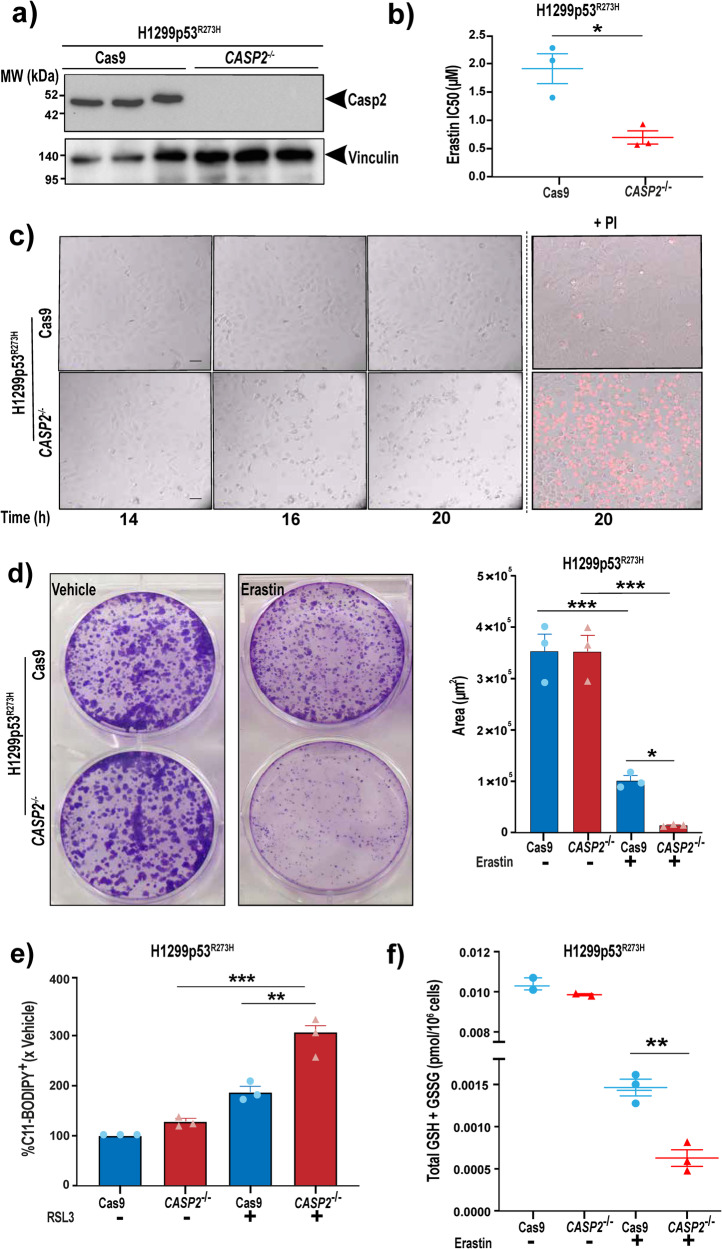
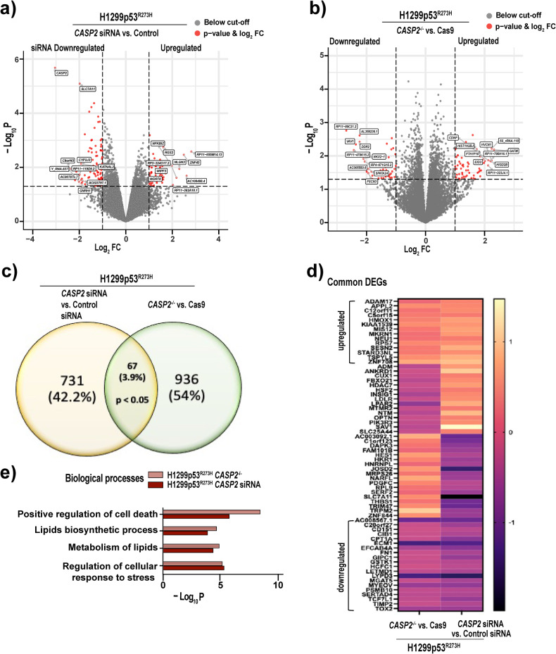
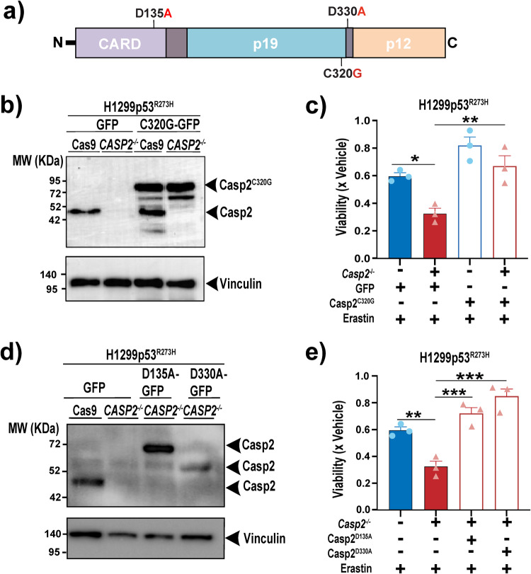
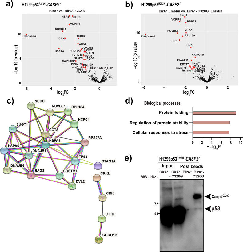
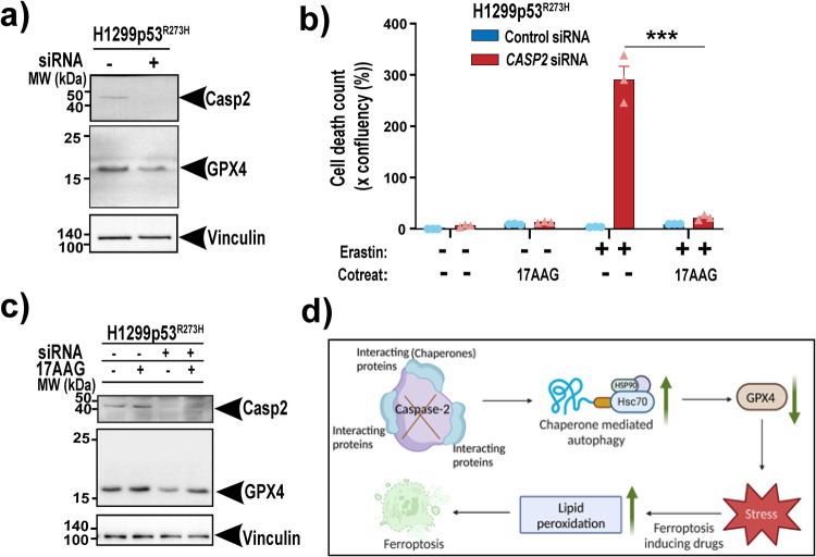
Caspase-2, one of the most evolutionarily conserved members of the caspase family, is an important regulator of the cellular response to oxidative stress. Given that ferroptosis is suppressed by antioxidant defense pathways, such as that involving selenoenzyme glutathione peroxidase 4 (GPX4), we hypothesized that caspase-2 may play a role in regulating ferroptosis. This study provides the first demonstration of an important and unprecedented function of caspase-2 in protecting cancer cells from undergoing ferroptotic cell death. Specifically, we show that depletion of caspase-2 leads to the downregulation of stress response genes including SESN2, HMOX1, SLC7A11, and sensitizes mutant-p53 cancer cells to cell death induced by various ferroptosis-inducing compounds. Importantly, the canonical catalytic activity of caspase-2 is not required for its role and suggests that caspase-2 regulates ferroptosis via non-proteolytic interaction with other proteins. Using an unbiased BioID proteomics screen, we identified novel caspase-2 interacting proteins (including heat shock proteins and co-chaperones) that regulate cellular responses to stress. Finally, we demonstrate that caspase-2 limits chaperone-mediated autophagic degradation of GPX4 to promote the survival of mutant-p53 cancer cells. In conclusion, we document a novel role for caspase-2 as a negative regulator of ferroptosis in cells with mutant p53. Our results provide evidence for a novel function of caspase-2 in cell death regulation and open potential new avenues to exploit ferroptosis in cancer therapy.


Conflict of interest statement

The authors declare no competing interests.

Figures

Fig. 1
Fig. 1. Acute silencing of caspase-2 enhances ferroptotic cell death in a mut-p53-dependent manner and can be rescued by ferroptosis inhibitors.
a Immunoblot analysis of caspase-2 and p53 expression in H1299 Parental p53null, H1299 p53R273H, and mut-p53 Flo-1 cells 48 h after transfection with control or CASP2 siRNA. Vinculin is shown as the loading control. Mean IC50 values after 72 h treatment with b erastin, c SAS, d RSL3 and e BSO in H1299 Parental p53null, H1299p53R273H and mut- p53 Flo-1 with control or CASP2 siRNA. f Viability of H1299p53R273H cells with control and CASP2 siRNA at 72 h post-treatment with 1 μM erastin, alone or in combination with ferrostatin-1 (Fer-1, 20 μM), deferoxamine (DFO, 100 μM), Trolox (Tro, 1 mM), β- mercaptoethanol (β-mer, 100 μM), N-acetylcysteine (NAC, 5 mM), glutathione-methylethyl ester (GSH-MEE, 5 mM) or QVD (25 μM). bf Data represented as mean ± s.e.m. from three or four independent experiments. Unpaired t-test and one-way ANOVA with Dunnett’s post hoc test were used to estimate significant differences in be and f, respectively. In f, comparisons were made between erastin only vs. cotreatment with ferroptosis inhibitors group in CASP2 siRNA cells. P-values are indicated with ns (not significant), *P < 0.05, **P < 0.01, ***P < 0.001.
Fig. 2
Fig. 2. Loss of caspase-2 leads to a similar increase in ferroptosis in mut-p53 cancer cells.
a Immunoblot analysis of caspase-2 expression in H1299p53R273H cells following CRISPR/Cas9 editing of CASP2 (CASP2−/−) vs. control cells (Cas9). Vinculin is shown as the loading control. b Mean IC50 values (μM) in H1299p53R273H Cas9 and H1299p53R273H -CASP2−/− cells after 72 h treatment with erastin. c Representative bright field images from live-cell imaging at the indicated time points of Cas9 and CASP2−/− H1299p53R273H cells treated with erastin (2 μM). Right-hand panels display dead cells stained red with propidium iodide (PI + ). Scale bar = 50 μm. d Representative images of crystal violet stained colonies of Cas9 or CASP2−/− H1299p53R273H cells treated with erastin or vehicle for 12 h, re-seeded and cultured over 10 days (left). Quantitation of crystal violet stained cell colonies using Cell Profiler software and represented as the area covered by the colonies (right). e Lipid peroxidation analysis by flow cytometry using C11-BODIPY post RSL3 (40 nM) or vehicle treatment for 18 h in H1299p53R273H Cas9 and H1299p53R273H -CASP2−/− cells. f Total intracellular glutathione (GSH + GSS pmol/106 cells), as determined by GR re-cycling assay after 12 h erastin (2 μM) treatment in H1299p53R273H Cas9 and H1299p53R273H -CASP2−/− cells. (b, d right, e, f) Data represented as mean ±s.e.m. from three independent experiments. Unpaired t-test and one-way ANOVA with Tukey’s post hoc test were used to estimate significant differences in b, f, and d, e respectively. P-values are indicated with *P < 0.05, **P < 0.01, and ***P < 0.001.
Fig. 3
Fig. 3. Loss of caspase-2 alters the expression of genes involved in the regulation of cellular stress pathways.
Volcano plots illustrating DEGs in a H1299p53R273H cells transfected with CASP2 siRNA vs. control siRNA and b CASP2−/− vs. Cas9 H1299p53R273H cells. Red dots indicate DEGs with nominal P-value < 0.05 and log2FC > or < –1. The top 10 (based on log2FC) upregulated and downregulated DEGs are labeled. c Venn diagram of unique and overlapping DEGs (P-value < 0.05) in CASP2 siRNA vs. control siRNA group and CASP2−/− vs. Cas9 group. d Heat-map displaying common up/downregulated genes with log2FC > 1 or < -1. e GO term analysis of the significantly enriched biological processes (nominal P-value < 0.01) in common DEGs in the absence of caspase-2 using Metascape [8].
Fig. 4
Fig. 4. The catalytic activity of caspase-2 is not required to execute its function in protecting mut-p53 cancer cells against ferroptosis.
a Diagram of caspase-2 consisting of prodomain or caspase recruitment domain (CARD), long subunit (p19), and small subunit (p12) along with the specific mutation sites introduced. b Immunoblot analysis of caspase-2 in H1299p53R273H Cas9 and H1299p53R273H-CASP2−/− cells transfected with a GFP-tagged catalytically inactive caspase-2-C320G (Casp2C320G/ C320G-GFP) expression plasmid or control plasmid (GFP). The higher MW of the ectopic Casp2C320G is because of the GFP tag. Vinculin is shown as the loading control. c Viability in H1299p53R273H Cas9 and H1299p53R273H-CASP2−/− cells ectopically expressing Casp2C320G or GFP 24 h post-treatment with erastin (2 μM) normalized to vehicle-treated cells. d Immunoblot analysis of caspase-2 in H1299p53R273H Cas9 cells transfected with GFP control plasmid, and H1299p53R273H-CASP2−/− cells transfected with GFP-tagged caspase-2-D135A (Casp2D135A/D135A-GFP) and caspase-2-D330A (Casp2D330A/D330A-GFP) expression plasmids or GFP control plasmid. Vinculin is shown as the loading control. The 60-kDa band in Casp2D330A mutant cells represents a partially processed form of caspase-2-GFP protein [5, 6]. e Viability in H1299p53R273H Cas9 cells and H1299p53R273H-CASP2−/− cells ectopically expressing Casp2D135A and Casp2D330A at 24 h post-treatment with erastin (2 μM) normalized to vehicle-treated cells. c, e Data represented as mean ±s.e.m. from three independent experiments. One-way ANOVA with Bonferroni’s post hoc test was used to estimate significant differences in c, e. P-values are indicated with *P < 0.05, **P < 0.01, and ***P < 0.001.
Fig. 5
Fig. 5. Identification of novel caspase-2 interacting proteins underpinning ferroptotic cell death in mut-p53 cancer cells.
Volcano plot illustrating the log2-fold-changes of biotinylated proteins for BirA* control vs. BirA*-Casp2C320G for a untreated samples and b erastin-treated samples. Five biological replicates per group were prepared for MS analysis. Proteins were deemed to exhibit differential expression if the log2-fold-change in protein expression was ≥1-fold and an adjusted P-value ≤ 0.05. c Protein–protein interaction network among the top 28 significant caspase-2 interacting proteins, based on STRING annotations. Edges indicate a range of protein–protein associations (physical and functional) such as experimentally determined (pink), curated databases (cyan), text mining (light green), co-expression (black), and protein homology (purple). Disconnected nodes (n = 8 proteins) in the network are not shown. d Functional enrichment for biological processes (P-value < 0.01) associated with caspase-2 interacting proteins as determined by Metascape. e Co-immunoprecipitation and immunoblotting for mut-p53 in H1299p53R273H-CASP2−/− cells expressing BirA* control and BirA*-Casp2C320G before and after pull-down with streptavidin agarose beads under basal conditions.
Fig. 6
Fig. 6. Caspase-2 limits chaperone-mediated autophagic degradation of GPX4.
a Immunoblot analysis of GPX4 and caspase-2 expression in H1299p53R273H control or CASP2 siRNA cells. Vinculin is shown as the loading control. b Viability of H1299p53R273H cells with control and CASP2 siRNA at 18 h post-treatment with 1 μM erastin, alone or in combination with 17AAG (500 nM). c Immunoblot analysis of GPX4 and caspase-2 expression in H1299p53R273H control or CASP2 siRNA cells following treatment with 17AAG. Vinculin is shown as the loading control. d Schematic diagram summarizing the role of caspase-2 in ferroptosis. Data represented as mean ± s.e.m. from three independent experiments. An unpaired t-test was used to estimate significant differences in b. P-value is indicated with ***P < 0.001.

References

    1. Kumar S. Caspase 2 in apoptosis, the DNA damage response and tumour suppression: enigma no more? Nat Rev Cancer. 2009;9:897–903. doi: 10.1038/nrc2745. - DOI - PubMed
    1. Terry MR, Arya R, Mukhopadhyay A, Berrett KC, Clair PM, Witt B, et al. Caspase-2 impacts lung tumorigenesis and chemotherapy response in vivo. Cell Death Differ. 2015;22:719–30. doi: 10.1038/cdd.2014.159. - DOI - PMC - PubMed
    1. Parsons MJ, McCormick L, Janke L, Howard A, Bouchier-Hayes L, Green DR. Genetic deletion of caspase-2 accelerates MMTV/c-neu-driven mammary carcinogenesis in mice. Cell Death Differ. 2013;20:1174–82. doi: 10.1038/cdd.2013.38. - DOI - PMC - PubMed
    1. Dorstyn L, Hackett-Jones E, Nikolic A, Norris MD, Lim Y, Toubia J, et al. Transcriptome profiling of caspase-2 deficient EmuMyc and Th-MYCN mouse tumors identifies distinct putative roles for caspase-2 in neuronal differentiation and immune signaling. Cell Death Dis. 2019;10:56. doi: 10.1038/s41419-018-1296-0. - DOI - PMC - PubMed
    1. Ho LH, Taylor R, Dorstyn L, Cakouros D, Bouillet P, Kumar S. A tumor suppressor function for caspase-2. Proc Natl Acad Sci USA. 2009;106:5336–41. doi: 10.1073/pnas.0811928106. - DOI - PMC - PubMed

Publication types

MeSH terms

Substances