. 2024 Mar 1;9(1):11.
doi: 10.1038/s41536-024-00355-1.

Inflammation-suppressing cornea-in-a-syringe with anti-viral GF19 peptide promotes regeneration in HSV-1 infected rabbit corneas

Affiliations

Inflammation-suppressing cornea-in-a-syringe with anti-viral GF19 peptide promotes regeneration in HSV-1 infected rabbit corneas

Egidijus Simoliunas et al. NPJ Regen Med. .

Abstract

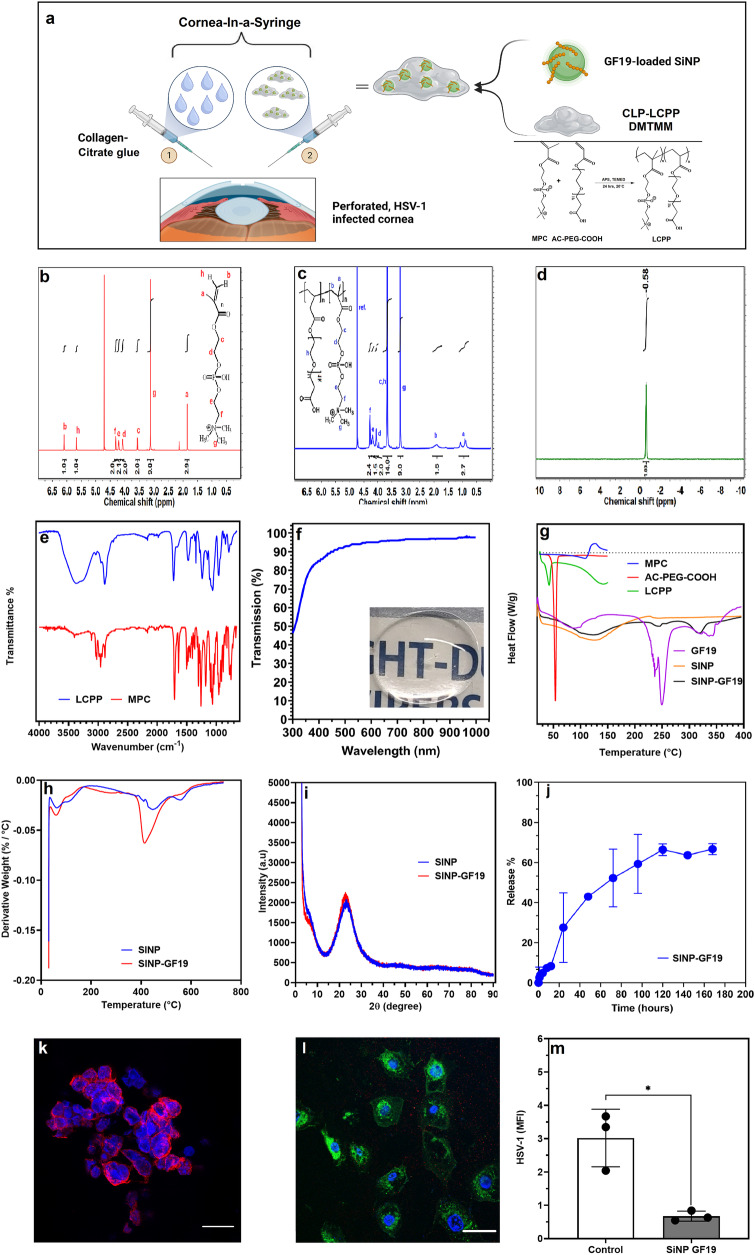
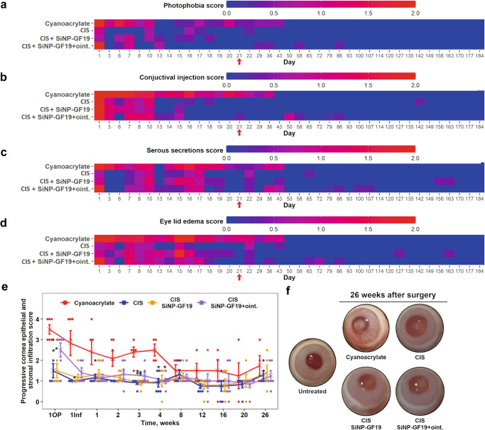
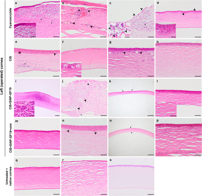
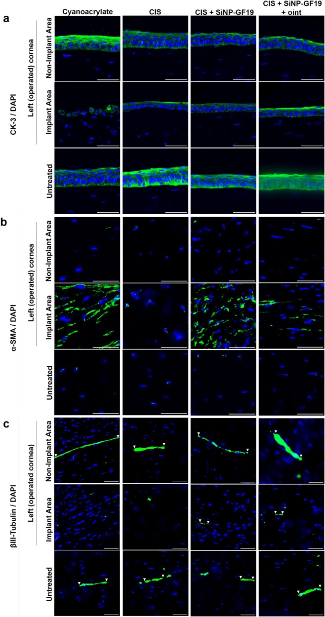
Pathophysiologic inflammation, e.g., from HSV-1 viral infection, can cause tissue destruction resulting in ulceration, perforation, and ultimately blindness. We developed an injectable Cornea-in-a-Syringe (CIS) sealant-filler to treat damaged corneas. CIS comprises linear carboxylated polymers of inflammation-suppressing 2-methacryloyloxyethyl phosphorylcholine, regeneration-promoting collagen-like peptide, and adhesive collagen-citrate glue. We also incorporated GF19, a modified anti-viral host defense peptide that blocked HSV-1 activity in vitro when released from silica nanoparticles (SiNP-GF19). CIS alone suppressed inflammation when tested in a surgically perforated and HSV-1-infected rabbit corneal model, allowing tissue and nerve regeneration. However, at six months post-operation, only regenerated neocorneas previously treated with CIS with SiNP-GF19 had structural and functional features approaching those of normal healthy corneas and were HSV-1 virus-free. We showed that composite injectable biomaterials can be designed to allow regeneration by modulating inflammation and blocking viral activity in an infected tissue. Future iterations could be optimized for clinical application.


Conflict of interest statement

M.Z.R. and M.G. have submitted a US provisional patent application on a related injectable MPC-based polymer formulation. The remaining authors declare no competing interests.

Figures

Fig. 1
Fig. 1. Hydrogel and SiNP-GF19 characterization.
a Preparation of SiNP-GF19 loaded CIS and LCPP. b 1H NMR spectrum of MPC. c 1H NMR spectrum of LCPP. d 31P NMR spectrum of LCPP. e FTIR spectra of MPC and LCPP. f Transparency of CLP-LCPP/citrate glue hydrogel shown by transmission of light through the hydrogel from the UV range through visible light (380 to 700 nm). n = 4 samples; average was reported. An image of a transparent CIS hydrogel is shown. g DSC curves of AC-PEG-COOH, MPC, LCPP, GF19, SiNP, and SiNP-GF19. h Derivative TGA curves of SiNP and SiNP-GF19. i XRD diffraction patterns of SiNP and SiNP-GF19. j GF19 release profile from SiNPs; n = 3 samples, mean ± SD. k HSV-1 infected and untreated human corneal epithelial cells (HCECs) at 48 h post-infection showing anti-HSV-1 antibody-stained virus (red) and cell nuclei were stained with DAPI (blue). Scale bar, 33 µm. l HCECs treated with SiNP loaded with FITC-labeled GF19 (green) for 1 h prior to HSV-1 infection. The cells were inoculated with virus for 1 h, after which both peptide and virus were removed. Cells are shown at 48 h post-infection. All cell nuclei were stained with DAPI (blue). Scale bars, 33 µm, m Mean fluorescence intensity (MFI) of HSV-1 staining in virus-infected untreated vs. SiNP-GF19-treated cells. *P = 0.098 by Student’s t-test, with statistical significance set at P ≤ 0.05. (n = 3 repeats per group, mean ± SD).
Fig. 2
Fig. 2. Timeline of the in vivo rabbit study.
The left eye of each animal received a surgically created perforation that was sealed and filled with one of cyanoacrylate, CIS, or CIS containing SiNP-GF19 (d0). On the second day after surgery (d2), the operated eyes were inoculated with 1×10⁴ PFU HSV-1 to create a peri-surgical infection. All operated eyes were treated daily with tobramycin antibiotic (Tobrex) ointment for 3 weeks after surgery. The rabbit eyes of the CIS+SiINP-GF19+oint. group were additionally treated with an ointment containing SiNP-GF19 for an additional three weeks after completion of the antibiotic course. Tear collection and healing of the left eyes were monitored as indicated over the 26-week post-operation follow-up.
Fig. 3
Fig. 3. Clinical follow-up of perforated and HSV-1 infected rabbit corneas after various treatments.
Heat maps show the clinical progression of corneal wound healing of the following parameters: a photophobia, b conjunctivitis, c tearing, and d periorbital edema. Blue corresponds to a healthy eye and red to a clinical worsening of the eye. The red arrows in the plots indicate when the CIS+SiNP-GF19+oint. group received additional ointment containing SiNP-GFP. e Infiltration of immune cells recorded at different time points: 1OP - the day after surgical perforation and filling, 1Inf − 1 day after infection with HSV-1, and 1, 2, 3, 4, 8, 12, 16, 20, and 26 weeks after surgery. Data are presented as a dot plot, where “x” represents the mean healing score of the group with standard error of the mean bars. A Kruskal–Wallis nonparametric test for ordinal data followed by a Dunn’s post hoc test for multiple comparisons was performed; statistical details are in Supplementary Table 3. *Indicates statistical significance (P ≤ 0.05) between the treatment and control (cyanoacrylate glue) groups, **when P ≤ 0.01. The graphs’ red arrows indicate the time at which the CIS+SiNP-GF19+oint. group was treated with extra SiNP-GF19 in an ointment. n = 6 animals, except for cyanoacrylate group where at days 21 and 29, the group number decreased to 5 and 4 respectively, and the CIS+SiNP-GF19 group at day 21, when the animal number decreased to 5. f Control untreated eye and treated corneas of rabbits at 26 weeks after surgery. The surgical perforations were made in the center of each cornea, in the area that is hazy. Different degrees of haze can be observed as in-growing corneal stromal cells will scatter light, thereby causing the haze. In the cyanoacrylate group, the central patched area is opaque and red blood vessels can be seen entering the patch (neovascularisation).
Fig. 4
Fig. 4. Hematoxylin & Eosin stained regenerated neocorneas at six months after treatment with various sealant-fillers, compared with untreated control corneas.
Representative sections from cyanoacrylate-treated corneas showing a moderate, diffuse, mixed inflammatory infiltrate (arrows) and lipid keratopathy (inset), and b diffuse, mixed perivascular inflammatory infiltrate (asterisk) with neovascularization (arrowheads). c Cyanoacrylate-treated cornea showing lipid keratopathy (arrow), swollen epithelial cells (arrowheads), neutrophils (inset, arrowheads), and presence of bacteria (inset, arrow). d Epithelial hyperplasia (arrowheads and inset) and mild stromal disarrangement in a cyanoacrylate-treated cornea. e Regenerated neocorneas from the CIS-treated group showing focal inflammatory infiltrate (arrow) and epithelial hyperplasia (arrowheads); f vacuolar degeneration of the epithelium; g swelling of epithelial cells (arrowheads) and a moderate degree of stromal disarrangement; and h mild degree of stroma disarrangement and epithelial hyperplasia. i A sample from the CIS+SiNP-GF19 group showing severe keratitis with loss of the epithelium and Bowman’s membrane, with a severe mixed inflammatory infiltrate with free erythrocytes (hemorrhage) (inset). Other representative sections from the CIS+SiNP-GF19 group showing j severe disarrangement of all stromal layers with loss of epithelium (asterisk), rupture of Descemet’s membrane (arrow), and diffuse mild mixed inflammatory infiltrate (arrowheads); k stromal thinning (arrows); l mild to moderate stromal disarrangement and epithelial hyperplasia. Regenerated neocornea sections from the CIS+SiNP-GF19+oint. group showing m mild stromal disarrangement; n epithelial hyperplasia; o slight stromal thinning (arrows); and p moderate stromal disarrangement. q Representative sections of untreated cornea without significant histological changes. r An untreated cornea with severely disarranged layers within the stroma. s Representative microphotograph of a healthy native cornea. Scale bars in (ac, g, h, j, n, p, r) represents 50 µm; (df, l, m, q, s) – 100 µm; (i) – 200 µm; (k, o) – 500 µm. Scale bars in inset from (a, c, d, f, i) represents 20 µm, and scale bar from (l) inset – 50 µm.
Fig. 5
Fig. 5. Regenerated rabbit neocorneas and adjacent areas in the different treatment groups, compared to untreated controls at six months post-operation.
Representative sections from regenerated neocorneas after treatment with different sealant-fillers, showing fluorescence immunohistochemistry with antibodies against a cytokeratin-3 (CK-3; green); b, α smooth muscle actin (α-SMA; green); and c βIII tubulin (green). The white arrowheads indicate sections of nerves that were stained by the βIII tubulin antibody. In all images, nuclei were stained with DAPI (blue). Scale bars, 50 µm.
Fig. 6
Fig. 6. HSV-1 infection in rabbits.
a Quantitative PCR analysis of HSV-1 DNA in rabbit tear samples. HSV-1 viral particles were detected in tear samples from the operated and virus-inoculated left eyes and untreated right eye at different time points: 1OP – 1 day after surgery; 1Inf – 1 day after infection (3 days after surgery); 1, 2, 3, 4, 8, 12, 16, 20 and 26 weeks after left eye surgery. Data are presented as group mean ± standard deviation. Points represent individual measurements. The red arrow in the graph indicates the time point at which the left eyes of the CIS+SiNP-GF19+oint. group animals started to receive anti-viral ointment. n = 3. b Representative images of regenerated neocorneas from HSV-1 infected and treated corneas at 6-month post-operation. Anti-HSV-1 staining (green) was performed to detect viruses within the tissues. The white arrowheads and arrows indicate HSV-1 staining in the anterior region of the stroma and the wing epithelial layer, respectively. In all images, nuclei were stained with DAPI (blue). Scale bars, 10 µm.

References

    1. Chen L, et al. Inflammatory responses and inflammation-associated diseases in organs. Oncotarget. 2018;9:7204–7218. doi: 10.18632/oncotarget.23208. - DOI - PMC - PubMed
    1. Meek KM, Dennis S, Khan S. Changes in the refractive index of the stroma and its extrafibrillar matrix when the cornea swells. Biophys. J. 2003;85:2205–2212. doi: 10.1016/S0006-3495(03)74646-3. - DOI - PMC - PubMed
    1. Cabrera-Aguas M, Khoo P, Watson SL. Infectious keratitis: a review. Clin. Exp. Ophthalmol. 2022;50:543–562. doi: 10.1111/ceo.14113. - DOI - PMC - PubMed
    1. Qazi Y, Hamrah P. Corneal allograft rejection: immunopathogenesis to therapeutics. J. Clin. Cell Immunol. 2013;2013:006. - PMC - PubMed
    1. Brunette I, et al. Alternatives to eye bank native tissue for corneal stromal replacement. Prog. Retin Eye Res. 2017;59:97–130. doi: 10.1016/j.preteyeres.2017.04.002. - DOI - PubMed