. 2024 Mar 1;10(1):49.
doi: 10.1038/s41531-024-00660-y.

G2019S selective LRRK2 kinase inhibitor abrogates mitochondrial DNA damage

Affiliations

G2019S selective LRRK2 kinase inhibitor abrogates mitochondrial DNA damage

Nicholas Pena et al. NPJ Parkinsons Dis. .

Abstract

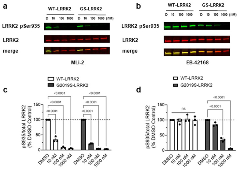
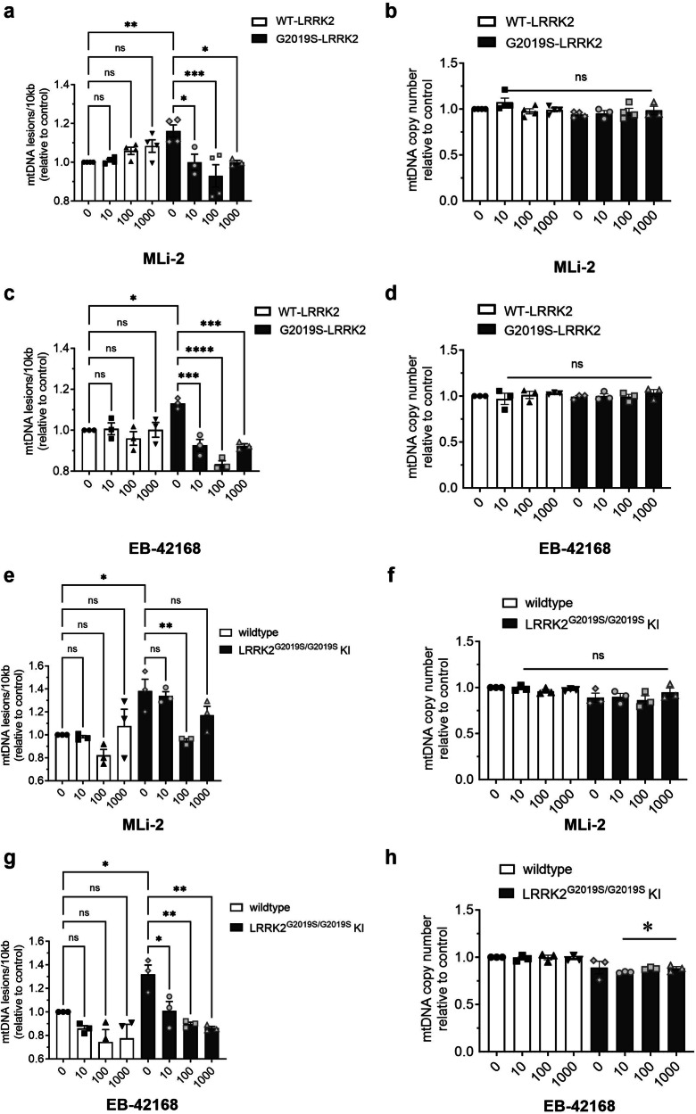
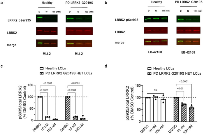
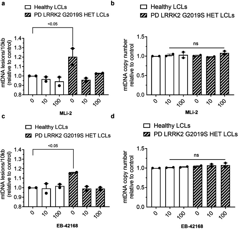
Pathogenic mutations in LRRK2 cause Parkinson's disease (PD). The G2019S variant is the most common, which results in abnormally high kinase activity. Compounds that target LRRK2 kinase activity are currently being developed and tested in clinical trials. We recently found that G2019S LRRK2 causes mitochondrial DNA (mtDNA) damage and treatment with multiple classes of LRRK2 kinase inhibitors at concentrations associated with dephosphorylation of LRRK2 reversed mtDNA damage to healthy control levels. Because maintaining the normal function of LRRK2 in heterozygous G2019S LRRK2 carriers while specifically targeting the G2019S LRRK2 activity could have an advantageous safety profile, we explored the efficacy of a G2019S mutant selective LRRK2 inhibitor to reverse mtDNA damage in G2019S LRRK2 models and patient cells relative to non-selective LRRK2 inhibitors. Potency of LRRK2 kinase inhibition by EB-42168, a G2019S mutant LRRK2 kinase inhibitor, and MLi-2, a non-selective inhibitor, was determined by measuring phosphorylation of LRRK2 at Ser935 and/or Ser1292 using quantitative western immunoblot analysis. The Mito DNADX assay, which allows for the accurate real-time quantification of mtDNA damage in a 96-well platform, was performed in parallel. We confirmed that EB-42168 selectively inhibits LRRK2 phosphorylation on G2019S LRRK2 relative to wild-type LRRK2. On the other hand, MLi-2 was equipotent for wild-type and G2019S LRRK2. Acute treatment with EB-42168 inhibited LRRK2 phosphorylation and also restored mtDNA damage to healthy control levels. We further investigated the relationship between LRRK2 kinase activity, mtDNA damage and mitophagy. Levels of mtDNA damage caused by G2019S LRRK2 were fully re-established within 2 h of a LRRK2 inhibitor wash out and recovery experiment, indicating the mtDNA damage phenotype is highly dynamic. G2019S LRRK2 mitophagy defects were not alleviated with LRRK2 kinase inhibition, suggesting that mitophagy is not mechanistically regulating LRRK2 kinase-mediated reversal of mtDNA damage in this acute timeframe. Abrogation of mtDNA damage with the mutant selective tool inhibitor EB-42168 demonstrates the potential of a precision medicine approach for LRRK2 G2019S PD. Levels of mtDNA damage may serve as a potential pharmacodynamic biomarker of altered kinase activity that could be useful for small molecule development and clinical trials.


Conflict of interest statement

The authors declare no competing interests. The former employees of Escape Bio do not have any financial ties to what is now a former company. They are all now employed by different companies and are committed to seeing the work made available to the scientific community and do not have any conflicts of interest as stated.

Figures

Fig. 1
Fig. 1. Analysis of LRRK2 and mtDNA damage in HEK293 cells lines stably overexpressing either WT-LRRK2 or G2019S-LRRK2.
a Representative western blot of WT-LRRK2 or G2019S-LRRK2 overexpressing cells show expression of LRRK2 pSer1292 and full-length LRRK2. β-actin was blotted as a loading control. b Quantification of western blots demonstrate ~6-fold increase of LRRK2 pSer1292 in G2019S-LRRK2 compared to WT-LRRK2 expressing cells. c Representative western blots of WT-LRRK2 or G2019S-LRRK2 overexpressing cells show expression of LRRK2 pSer935 and full-length LRRK2. β-actin was blotted as a loading control. d Quantification of western blots demonstrate no difference of LRRK2 pSer935 levels between G2019S-LRRK2 and WT-LRRK2 expressing cells. e Quantification of western blots demonstrate no difference of LRRK2 protein levels between the two cell lines. f Mitochondrial DNA damage was increased in G2019S-LRRK2 relative to WT-LRRK2 expressing cells. g The differences in mtDNA damage between the cell lines were not attributable to changes in steady state mtDNA levels. Data are presented as mean ± SEM. n = 3–4 replicates. Statistical significance was determined by unpaired t-test for all analyses. GS-LRRK2 G2019S-LRRK2, WT-LRRK2 wild-type LRRK2, ns non-significant.
Fig. 2
Fig. 2. EB-42168 demonstrates full inhibition of LRRK2 pSer935 in cells overexpressing G2019S-LRRK2 at concentrations that show no inhibition in WT-LRRK2 overexpressing cells.
a Representative western blot of WT-LRRK2 or G2019S-LRRK2 cells treated with DMSO, 10 nM, 100 nM or 1 µM MLi-2 for 2 h and assessed for LRRK2 pSer935 and full-length LRRK2. b Representative western blot of WT-LRRK2 or G2019S-LRRK2 cells treated with DMSO, 10 nM, 100 nM or 1 µM EB-42168 for 2 h and assessed for LRRK2 pSer935 and full-length LRRK2. c Quantification of western blots demonstrate a 70%, 90% and 95% decrease, respectively, in LRRK2 pSer935 levels with increasing concentration of MLi-2 in WT-LRRK2 expressing cells. Quantification of western blots demonstrate an 80%, 96% and 97% decrease respectively in LRRK2 pSer935 levels with increasing concentration of MLi-2 in G2019S-LRRK2 expressing cells. d Quantification of western blots demonstrate no change in LRRK2 pSer935 levels with EB-42168 treatment in WT-LRRK2 expressing cells. Quantification of western blots demonstrate a 65% and 95% decrease respectively in LRRK2 pSer935 levels with 100 nM and 1 µM of EB-42168 in G2019S-LRRK2 expressing cells. Data are presented as mean ± SEM. n = 3 replicates. (*p < 0.0001 determined by two-way ANOVA). GS-LRRK2 G2019S-LRRK2, WT-LRRK2 wild-type LRRK2, ns non-significant.
Fig. 3
Fig. 3. EB-42168 inhibits LRRK2 pSer1292 in G2019S-LRRK2 overexpressing cells at concentrations that also inhibited pSer935.
a Representative western blot of G2019S-LRRK2 cells treated with DMSO, 10 nM, 100 nM or 1 µM MLi-2 for 2 h and assessed for LRRK2 pSer1292 and full-length LRRK2. b Representative western blot of G2019S-LRRK2 cells treated with DMSO, 10 nM, 100 nM or 1 µM EB-42168 for 2 h and assessed for LRRK2 pSer1292 and full-length LRRK2. c Quantification of western blots demonstrate a 65%, 90% and 95% decrease in LRRK2 pSer1292 levels with increasing concentration of MLi-2 in G2019S-LRRK2 expressing cells. d Quantification of western blots demonstrate a 30%, 65% and 85% decrease in LRRK2 pSer1292 levels with 10 nM, 100 nM and 1 µM respectively of EB-42168 in G2019S-LRRK2 expressing cells. Data are presented as mean ± SEM. n = 3 replicates. (*p < 0.0001 determined by one-way ANOVA). GS-LRRK2 G2019S-LRRK2.
Fig. 4
Fig. 4. Mitochondrial DNA damage in G2019S-LRRK2 overexpression and LRRK2G2019S/G2019S KI cells was abrogated to normal levels with LRRK2 kinase inhibition.
a WT-LRRK2 and G2019S-LRRK2 cells were treated with DMSO, 10 nM, 100 nM or 1 µM MLi-2 for 2 h and analyzed for mtDNA damage. b mtDNA copy number was unaltered by the MLi-2 treatment. c WT-LRRK2 and G2019S-LRRK2 cells were treated with DMSO, 10 nM, 100 nM or 1 µM EB-42168 for 2 h and analyzed for mtDNA damage. d Treatment with EB-42168 did not change mtDNA copy number in either WT-LRRK2 or G2019S-LRRK2 cells. e LRRK2G2019S/G2019S KI cells and wild-type cells were treated with DMSO, 10 nM, 100 nM or 1 µM MLi-2 for 2 h and analyzed for mtDNA damage. f MLi-2 treatment did not change mtDNA copy number in either cell line. g LRRK2G2019S/G2019S KI cells and wild-type cells were treated with DMSO, 10 nM, 100 nM or 1 µM EB-42168 for 2 h and analyzed for mtDNA damage. h In contrast, while EB-42168 had no effect on mtDNA copy number in wild-type cells, mtDNA copy number was modestly decreased in EB-42618 treated LRRK2G2019S/G2019S KI cells. Data are presented as mean ± SEM. n = 3 replicates. (*p < 0.05, **p < 0.01, ***p = 0.0001, ****p < 0.0001 determined by one-way ANOVA). GS-LRRK2 G2019S-LRRK2, WT-LRRK2 wild-type LRRK2, ns non-significant.
Fig. 5
Fig. 5. G2019S LRRK2-dependent mtDNA damage is re-established within 2 h following washout of LRRK2 kinase inhibitor.
a Wild-type and LRRK2G2019S/G2019S KI cells were treated with vehicle or 100 nM MLi-2 for 2 h and analyzed for mtDNA damage. b mtDNA copy number was not affected by experimental conditions. c Wild-type and LRRK2G2019S/G2019S KI cells were treated with vehicle or 100 nM MLi-2 for 2 h, followed by washout and incubation in fresh media for 2 h and analyzed for mtDNA damage. d mtDNA copy number did not change with treatment condition. Data are presented as mean ± SEM. n = 2–3 replicates. (**p < 0.01, ***p < 0.001 determined by one-way ANOVA with a Bonferroni’s multiple comparisons test). ns non-significant.
Fig. 6
Fig. 6. Basal and depolarization-induced mitophagy in wild-type and LRRK2G2019S/G2019S KI cells pre-treated with vehicle or LRRK2 kinase inhibitor.
Representative images of (a) wild-type or (b) LRRK2G2019S/G2019S KI cells with the Lyso and Mitophagy dye with a 2 h pre-treatment with either 100 nM MLi-2, EB-42168, or vehicle, followed by a 3 h treatment with 20 μM FCCP or vehicle. c Quantification of pH-based Mitophagy dye fluorescence intensity within mitolysosomes at baseline and under FCCP-induced mitophagy. d Quantification of the number of lysosomes containing fluorescing mitochondria normalized by cell count. Data are presented as mean ± SEM. n = 2–4 replicates. (*p < 0.05 determined by one-way ANOVA).
Fig. 7
Fig. 7. EB-42168 demonstrates selective inhibition of pSer935 LRRK2 in heterozygous G2019S LRRK2 PD patient-derived LCLs relative to healthy controls.
a Representative western blot of PD patient-derived LRRK2 G2019S carriers and age-matched healthy controls LCLs treated with DMSO, 10 nM, or 100 nM MLi-2 for 2 h and assessed for LRRK2 pSer935 and full-length LRRK2. b Representative western blot of PD patient-derived LRRK2 G2019S carriers and age-matched healthy controls LCLs treated with DMSO, 10 nM, or 100 nM EB-42168 for 2 h and assessed for LRRK2 pSer935 and full-length LRRK2. c Quantification of western blots demonstrate an 85% and 95% decrease in LRRK2 pSer935 levels in MLi-2 treated LRRK2 G2019S PD patient-derived and age-matched healthy controls. d Quantification of western blots demonstrate no change in LRRK2 pSer935 levels with EB-42168 treatment in healthy controls. Quantification of western blots demonstrate a 25% and 40% decrease in LRRK2 pSer935 levels with 10 nM and 100 nM EB-42168 in PD patient-derived LRRK2 G2019S carriers. Data are presented as mean ± SEM. (**p < 0.01 determined by two-way ANOVA).
Fig. 8
Fig. 8. LRRK2 kinase inhibitor treatment restored LRRK2 G2019S induced mtDNA damage to basal healthy control levels.
a PD patient-derived heterozygous G2019S LRRK2 carriers and age-matched healthy controls LCLs treated with DMSO, 10 nM, or 100 nM MLi-2 or (c) EB-42168 for 2 h and analyzed for mtDNA damage. b, d mtDNA copy number was unaltered by these treatments. Data are presented as mean ± SEM. (*p < 0.05 determined by one-way ANOVA).

References

    1. Simon-Sanchez J, et al. Genome-wide association study reveals genetic risk underlying Parkinson’s disease. Nat. Genet. 2009;41:1308–1312. doi: 10.1038/ng.487. - DOI - PMC - PubMed
    1. Nalls MA, et al. Large-scale meta-analysis of genome-wide association data identifies six new risk loci for Parkinson’s disease. Nat. Genet. 2014;46:989–993. doi: 10.1038/ng.3043. - DOI - PMC - PubMed
    1. Nalls MA, et al. Identification of novel risk loci, causal insights, and heritable risk for Parkinson’s disease: a meta-analysis of genome-wide association studies. Lancet Neurol. 2019;18:1091–1102. doi: 10.1016/S1474-4422(19)30320-5. - DOI - PMC - PubMed
    1. Paisan-Ruiz C, et al. Cloning of the gene containing mutations that cause PARK8-linked Parkinson’s disease. Neuron. 2004;44:595–600. doi: 10.1016/j.neuron.2004.10.023. - DOI - PubMed
    1. Zimprich A, et al. Mutations in LRRK2 cause autosomal-dominant parkinsonism with pleomorphic pathology. Neuron. 2004;44:601–607. doi: 10.1016/j.neuron.2004.11.005. - DOI - PubMed

LinkOut - more resources