. 2024 Mar 1;81(1):110.
doi: 10.1007/s00018-024-05155-w.

MDSC-derived S100A8/9 contributes to lupus pathogenesis by promoting TLR7-mediated activation of macrophages and dendritic cells

Affiliations

MDSC-derived S100A8/9 contributes to lupus pathogenesis by promoting TLR7-mediated activation of macrophages and dendritic cells

Yonghong Yang et al. Cell Mol Life Sci. .

Abstract

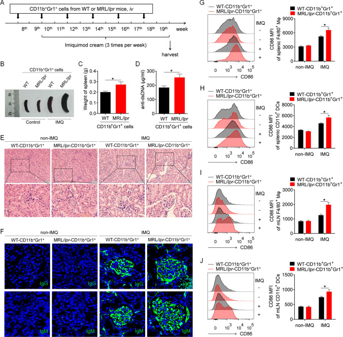
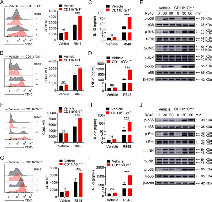
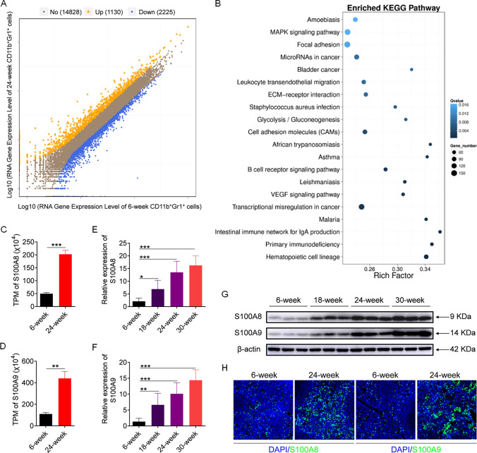
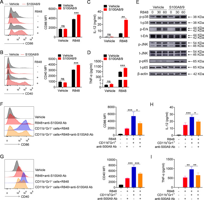
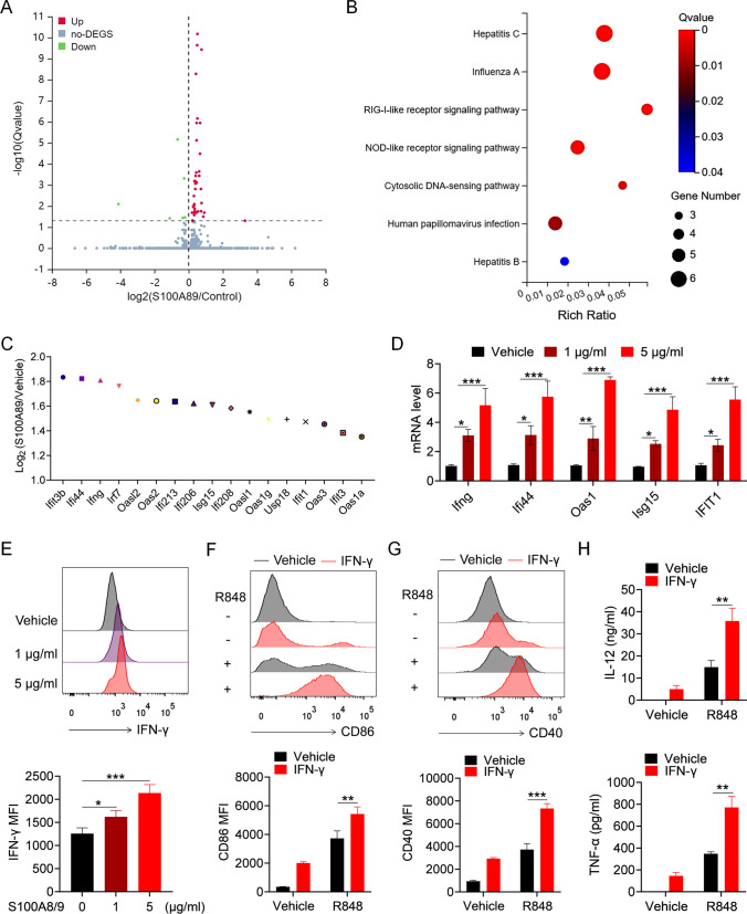
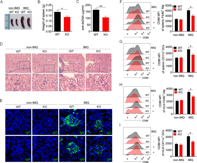
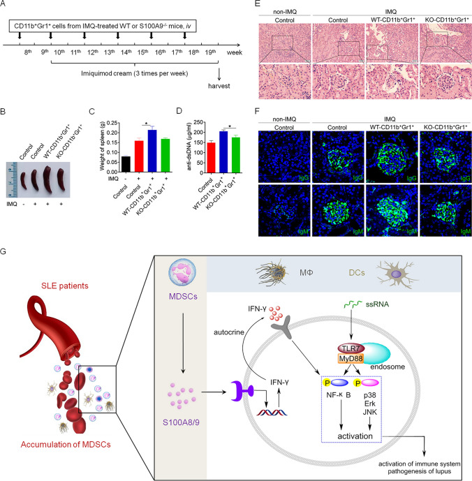
Toll-like receptors (TLRs), especially TLR7, play an important role in systemic lupus erythematosus (SLE) pathogenesis. However, the regulatory mechanism underlying the abnormal activation of TLR pathways in patients with SLE has not been elucidated. Notably, accumulating evidence indicates that myeloid-derived suppressor cells (MDSCs) are important regulators of inflammation and autoimmune diseases. Compared with healthy control subjects, patients with SLE have a greater proportion of MDSCs among peripheral blood mononuclear cells (PBMCs); however, the effect of MDSCs on TLR7 pathway activation has not been determined. In the present study, lupus MDSCs significantly promoted TLR7 pathway activation in macrophages and dendritic cells (DCs), exacerbating the imiquimod-induced lupus model. RNA-sequencing analysis revealed significant overexpression of S100 calcium-binding protein A8 (S100A8) and S100A9 in MDSCs from diseased MRL/lpr mice. In vitro and in vivo studies demonstrated that S100A8/9 effectively promoted TLR7 pathway activation and that S100A8/9 deficiency reversed the promoting effect of MDSCs on TLR7 pathway activation in lupus. Mechanistically, MDSC-derived S100A8/9 upregulated interferon gamma (IFN-γ) secretion by macrophages and IFN-γ subsequently promoted TLR7 pathway activation in an autocrine manner. Taken together, these findings suggest that lupus MDSCs promote TLR7 pathway activation and lupus pathogenesis through the S100A8/9-IFN-γ axis. Our study identified an important target for SLE therapy.

Keywords: MDSCs; Macrophage; S100A8/9; SLE; TLR7.


Conflict of interest statement

The authors have no relevant financial or non-financial interests to disclose.

Figures

Fig. 1
Fig. 1
Lupus MDSCs contribute to the disease progression of mice with lupus induced by imiquimide (IMQ). The splenic MDSCs isolated from 22- to 24-week-old C57BL/6 mice or MRL/lpr mice were transferred into 8-week-old C57BL/6 mice by caudal vein (n = 6), 2 × 10.6 cells/mice, once every 2 weeks. At the 10th week, mice were treated with IMQ to induce lupus model. After 10 weeks, the mice were killed for further study. A The schematic showing the experimental design. B Representative images of marked splenomegaly. C Weight of spleens. D ELISA analysis of the anti-dsDNA antibody in serum. E Representative images of kidney sections detected by H&E. F Immunofluorescence of IgG and IgM of kidney sections. GJ Flow cytometric analysis of the expression of CD86 on macrophages and dendritic cells in spleens (G, H) and mLN (I, J) of mice. *p < 0.05
Fig. 2
Fig. 2
Lupus MDSCs promote the TLR7-mediated activation of BMDMs and BMDCs in vitro. Splenic MDSCs, isolated from 22 to 24 weeks MRL/lpr mice, were placed in transwell chambers and co-cultured with BMDMs or BMDCs at a 1:1 ratio (1 × 106 cells). Twelve hours later, MDSCs were removed and R848 (1 μg/mL) was added into the culture system. A, B Flow cytometric analysis of the expression of CD86 and CD40 on BMDMs at 24 h. C, D ELISA analysis of the levels of IL-12/IL-23 p40 and TNF-α secreted by BMDMs at 24 h. E Western blot analysis of the phosphorylation of p38, Erk, JNK, and p65 in BMDMs at 30 and 60 min. F, G Flow cytometric analysis of the expression of CD86 and CD40 on BMDCs at 24 h. H, I ELISA analysis of the levels of IL-12/IL-23 p40 and TNF-α secreted by BMDCs at 24 h. J Western blot analysis of the phosphorylation of p38, Erk, JNK and p65 in BMDCs at 30 and 60 min. **p < 0.01, ***p < 0.001
Fig. 3
Fig. 3
S100A8/9 is overexpressed in lupus MDSCs. (A-D) Splenic MDSCs were isolated from 6-week-old and 24-week-old MRL/lpr mice and then RNA-seq analysis was performed. A Differential expression genes at FDR < 0.05 in MDSCs from 24-week-old MRL/lpr mice versus those from 6-week-old MRL/lpr mice. B KEGG signaling pathway enrichment analysis of differential gene expression in MDSCs from 24-week-old MRL/lpr mice versus those from 6-week-old MRL/lpr mice. C, D TPM values of S100A8 and S100A9 analyzed by RNA-seq. EG Splenic MDSCs were isolated from MRL/lpr mice of different ages (6, 12, 18 and 24 week), and the expression of S100A8 and S100A9 were analyzed by Q-PCR (E, F) and western blot (G). H Immunofluorescence staining of S100A8 and S100A9 in spleens of 6-week-old and 24-week-old MRL/lpr mice. *p < 0.05, **p < 0.01, ***p < 0.001
Fig. 4
Fig. 4
Lupus MDSCs promote the TLR7-mediated activation of BMDMs by secreting S100A8/9. A–E BMDMs were pretreated with mouse recombinant S100A8/9 protein (1 μg/mL) for 12 h, and then stimulated with R848 (1 μg/mL). Flow cytometric analysis of the expression of CD86 and CD40 at 24 h (A, B). ELISA analysis of the levels of IL-12/IL-23 p40 and TNF-α in culture supernatant at 24 h (C, D). Western blot analysis of the phosphorylation of p38, Erk, JNK and p65 at 30 and 60 min (E). FI Splenic MDSCs, isolated from 22 to 24-week-old MRL/lpr mice, were placed in transwell chambers and co-cultured with BMDMs or BMDCs at a 1:1 ratio (1 × 106 cells). Meanwhile, anti-S100A9 antibody or vehicle (1 μg/mL) was added. After 12 h, MDSCs were removed and R848 (1 μg/mL) was added into the culture system. Flow cytometric analysis of the expression of CD86 and CD40 at 24 h (F, G). ELISA analysis of the levels of IL-12/IL-23 p40 and TNF-α in culture supernatant at 24 h (H, I). *p < 0.05, **p < 0.01, ***p < 0.001
Fig. 5
Fig. 5
S100A8/9 augments the TLR7-mediated activation of BMDMs by promoting the secretion of IFN-γ. (A-C) BMDMs were treated with mouse recombinant S100A8/9 protein (1 μg/mL) or vehicle for 6 h, and then RNA-seq analysis was performed. A Differential expression genes at FDR < 0.05 in S100A8/9-treated BMDMs versus vehicle-treated BMDMs. B KEGG signaling pathway enrichment analysis of differential gene expression in S100A8/9-treated BMDMs versus vehicle-treated BMDMs. C The changes of the expression of IFN-γ and IFN-inducible genes in S100A8/9-treated BMDMs versus vehicle-treated BMDMs. D, E BMDMs were stimulated with mouse recombinant S100A8/9 protein (1 μg/mL and 5 μg/mL) for 6 or 12 h. The mRNA levels of IFN-γ and IFN-inducible genes were detected by Q-PCR (D). The protein level of IFN-γ was detected by FACS (E). F–H BMDMs were pretreated with mouse recombinant IFN-γ (20 ng/mL) for 12 h, followed by stimulation of R848 for 24 h. Flow cytometric analysis of the expression of CD86 and CD40 (F, G). ELISA analysis of the levels of IL-12/IL-23 p40 and TNF-α in culture supernatant (H). *p < 0.05, **p < 0.01, ***p < 0.001
Fig. 6
Fig. 6
S100A9 deficiency alleviates IMQ-induced lupus in mice. S100A9.−/− and wild-type mice (8-week-old) were treated with IMQ for 10 weeks, and killed for further experiment. A Representative images of marked splenomegaly. B Weight of spleens. C ELISA analysis of the anti-dsDNA antibody in serum. D Representative images of kidney sections detected by H&E. E Immunofluorescence of IgG and IgM of kidney sections. FI Flow cytometric analysis of the expression of CD86 on macrophages and dendritic cells in spleens (F, G) and mLN (H, I) of mice. *p < 0.05
Fig. 7
Fig. 7
Lupus MDSCs-derived S100A8/9 contributes to the disease progression of IMQ-induced lupus mice. The splenic MDSCs (S100A9−/−-MDSCs and WT-MDSCs) were isolated from S100A9−/− and wild-type mice which have been treated with IMQ for 10 weeks, and subsequently transferred into 8-week-old C57BL/6 mice by caudal vein (n = 6), 2 × 106 cells/mice, once every 2 weeks. At the 10th week, mice were treated with IMQ to induce lupus model. After 10 weeks, the mice were killed for further study. A The schematic showing the experimental design. B Representative images of marked splenomegaly. C Weight of spleens. D ELISA analysis of the anti-dsDNA antibody in serum. E Representative images of kidney sections detected by H&E. F Immunofluorescence of IgG and IgM of kidney sections. *p < 0.05

Similar articles

Cited by

References

    1. Connelly K, Eades LE, Koelmeyer R, Ayton D, Golder V, Kandane-Rathnayake R, et al. Towards a novel clinical outcome assessment for systemic lupus erythematosus: first outcomes of an international taskforce. Nat Rev Rheumatol. 2023 doi: 10.1038/s41584-023-00993-7. - DOI - PubMed
    1. Crow MK. Pathogenesis of systemic lupus erythematosus: risks, mechanisms and therapeutic targets. Ann Rheum Dis. 2023;82(8):999–1014. doi: 10.1136/ard-2022-223741. - DOI - PubMed
    1. Yoo EJ, Oh KH, Piao H, Kang HJ, Jeong GW, Park H, et al. Macrophage transcription factor TonEBP promotes systemic lupus erythematosus and kidney injury via damage-induced signaling pathways. Kidney Int. 2023;104(1):163–180. doi: 10.1016/j.kint.2023.03.030. - DOI - PubMed
    1. Soni C, Perez OA, Voss WN, Pucella JN, Serpas L, Mehl J, et al. Plasmacytoid dendritic cells and type I interferon promote extrafollicular B cell responses to extracellular self-DNA. Immunity. 2020;52(6):1022–1038. doi: 10.1016/j.immuni.2020.04.015. - DOI - PMC - PubMed
    1. Liu J, Zhang X, Cao X. Dendritic cells in systemic lupus erythematosus: from pathogenesis to therapeutic applications. J Autoimmun. 2022;132:102856. doi: 10.1016/j.jaut.2022.102856. - DOI - PubMed

MeSH terms