. 2024 Mar 1;14(1):5120.
doi: 10.1038/s41598-024-55058-w.

A tissue-silicone integrated simulator for right ventricular pulsatile circulation with severe functional tricuspid regurgitation

Affiliations

A tissue-silicone integrated simulator for right ventricular pulsatile circulation with severe functional tricuspid regurgitation

Jumpei Takada et al. Sci Rep. .

Abstract

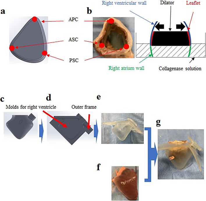
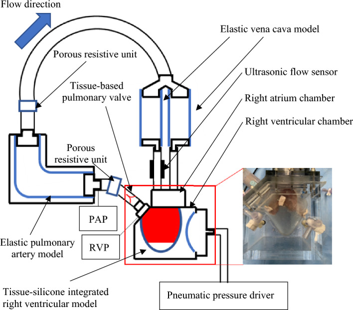
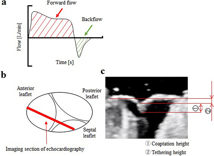
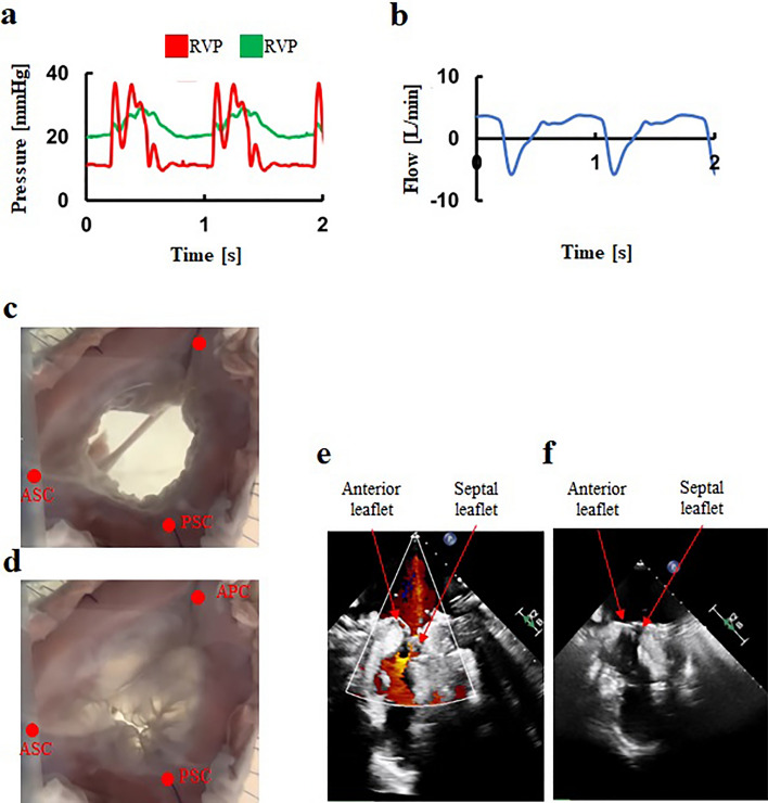
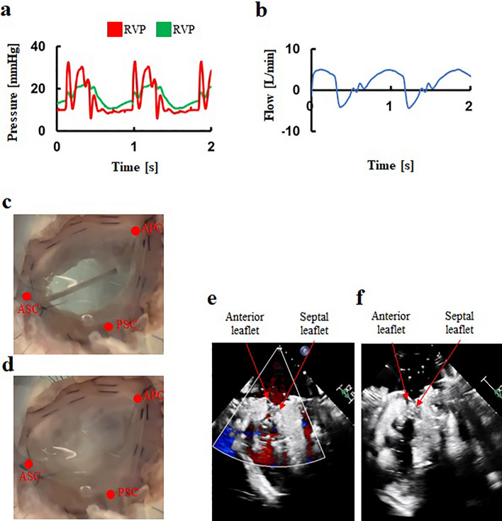
There is a great demand for development of a functional tricuspid regurgitation (FTR) model for accelerating development and preclinical study of tricuspid interventional repair devices. This study aimed to develop a severe FTR model by creating a tissue-silicone integrated right ventricular pulsatile circulatory simulator. The simulator incorporates the porcine tricuspid annulus, valve leaflets, chordae tendineae, papillary muscles, and right ventricular wall as one continuous piece of tissue, thereby preserving essential anatomical relationships of the tricuspid valve (TV) complex. We dilated the TV annulus with collagenolytic enzymes under applying stepwise dilation, and successfully achieved a severe FTR model with a regurgitant volume of 45 ± 9 mL/beat and a flow jet area of 15.8 ± 2.3 cm2 (n = 6). Compared to a normal model, the severe FTR model exhibited a larger annular circumference (133.1 ± 8.2 mm vs. 115.7 ± 5.5 mm; p = 0.009) and lower coaptation height (6.6 ± 1.0 mm vs. 17.7 ± 1.3 mm; p = 0.003). Following the De-Vega annular augmentation procedure to the severe FTR model, a significant reduction in regurgitant volume and flow jet area were observed. This severe FTR model may open new avenues for the development and evaluation of transcatheter TV devices.


Conflict of interest statement

The authors declare no competing interests.

Figures

Figure 1
Figure 1
A method for developing tissue-silicone integrated right ventricular model. (a) A dilator mimicking the human annulus morphology of FTR, (b) Method to insert a dilator into the tricuspid valve annulus. (c) Computer-aided design of a right ventricular model, (d) The gap between the right ventricular model and the outer frame, (e) A silicone right ventricular model, (f) The right ventricle tissue, (g) A tissue-silicone integrated model. FTR: Functional tricuspid regurgitation, APC: Anterior–posterior commissure, ASC: Anterior-septal commissure, PSC: Posterior-septal commissure.
Figure 2
Figure 2
A tissue-silicone integrated pulsatile right ventricular circulatory system incorporating a tricuspid valve. PAP; Pulmonary artery pressure, RVP; Right ventricular pressure.
Figure 3
Figure 3
Measurement of flow parameters and morphologies of the tricuspid valves. (a) Forward flow and backflow of a tricuspid valve, (b) Imaging section of echocardiography (c) Coaptation height and tethering height.
Figure 4
Figure 4
A representative hemodynamics and behaviors in the normal model. (a) Pressure waveforms, (b) Flow waveform, (c) Tricuspid valve at diastole, (d) Tricuspid valve at systole, (e) Transvalvular jet flow at systole, (f) Leaflet tethering at systole. RVP: Right ventricular pressure, PAV: Pulmonary artery pressure, APC: Anterior–posterior commissure, ASC: Anterior-septal commissure, PSC: Posterior-septal commissure.
Figure 5
Figure 5
An extreme hemodynamics and behaviors in the FTR model. (a) Pressure waveforms, (b) Flow waveform, (c) Tricuspid valve at diastole, (d) Tricuspid valve at systole, (e) Transvalvular jet flow at systole, (f) Leaflet tethering at systole. FTR: Functional tricuspid regurgitation, RVP: Right ventricular pressure, PAV: Pulmonary artery pressure, APC: Anterior–posterior commissure, ASC: Anterior-septal commissure, PSC: Posterior-septal commissure.
Figure 6
Figure 6
A representative hemodynamics and behaviors in the annular augmentation model using the De-Vega procedure. (a) Pressure waveforms, (b) Flow waveform, (c) Tricuspid valve at diastole, (d) Tricuspid valve at systole, (e) Transvalvular jet flow at systole, (f) Leaflet tethering at systole. RVP: Right ventricular pressure, PAV: Pulmonary artery pressure, APC: Anterior–posterior commissure, ASC: Anterior-septal commissure, PSC: Posterior-septal commissure.

References

    1. Dreyfus GD, Corbi PJ, Chan KM, Bahrami T. Secondary tricuspid regurgitation or dilatation: Which should be the criteria for surgical repair? Ann Thorac. Surg. 2005;79:127–132. doi: 10.1016/j.athoracsur.2004.06.057. - DOI - PubMed
    1. Gammie JS, et al. Concomitant tricuspid repair in patients with degenerative mitral regurgitation. N. Engl. J. Med. 2022;386:327–339. doi: 10.1056/NEJMoa2115961. - DOI - PMC - PubMed
    1. Mangieri A, et al. Mechanism and implications of the tricuspid regurgitation: From the pathophysiology to the current and future therapeutic options. Circ. Cardiovasc. Interv. 2017;10:e005043. doi: 10.1161/CIRCINTERVENTIONS.117.005043. - DOI - PubMed
    1. Kadri AN, et al. Outcomes of patients with severe tricuspid regurgitation and congestive heart failure. Heart. 2019;105:1813–1817. doi: 10.1136/heartjnl-2019-315004. - DOI - PubMed
    1. Vahanian A, et al. 2021 ESC/EACTS guidelines for the management of valvular heart disease. Eur. Heart J. 2022;43:561–632. doi: 10.1093/eurheartj/ehab395. - DOI - PubMed