

Deep Learning Imaging Reconstruction Algorithm for Carotid Dual Energy CT Angiography: Opportunistic Evaluation of Cervical Intervertebral Discs-A Preliminary Study
- PMID: 38429560
- PMCID: PMC11300736
- DOI: 10.1007/s10278-024-01016-x
Deep Learning Imaging Reconstruction Algorithm for Carotid Dual Energy CT Angiography: Opportunistic Evaluation of Cervical Intervertebral Discs-A Preliminary Study
Abstract
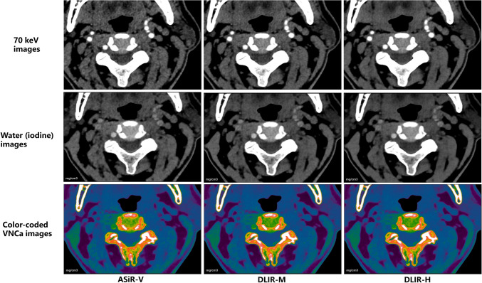
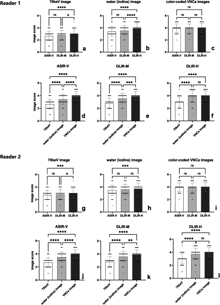
Thus, the aim of this study is to evaluate the performance of deep learning imaging reconstruction (DLIR) algorithm in different image sets derived from carotid dual-energy computed tomography angiography (DECTA) for evaluating cervical intervertebral discs (IVDs) and compare them with those reconstructed using adaptive statistical iterative reconstruction-Veo (ASiR-V). Forty-two patients who underwent carotid DECTA were included in this retrospective analysis. Three types of image sets (70 keV, water-iodine, and water-calcium) were reconstructed using 50% ASiR-V and DLIR at medium and high levels (DLIR-M and DLIR-H). The diagnostic acceptability and conspicuity of IVDs were assessed using a 5-point scale. Hounsfield Units (HU) and water concentration (WC) values of the IVDs; standard deviation (SD); and coefficient of variation (CV) were calculated. Measurement parameters of the 50% ASIR-V, DLIR-M, and DLIR-H groups were compared. The DLIR-H group showed higher scores for diagnostic acceptability and conspicuity, as well as lower SD values for HU and WC than the ASiR-V and DLIR-M groups for the 70 keV and water-iodine image sets (all p < .001). However, there was no significant difference in scores and SD among the three groups for the water-calcium image set (all p > .005). The water-calcium image set showed better diagnostic accuracy for evaluating IVDs compared to the other image sets. The inter-rater agreement using ASiR-V, DLIR-M, and DLIR-H was good for the 70 keV image set, excellent for the water-iodine and water-calcium image sets. DLIR improved the visualization of IVDs in the 70 keV and water-iodine image sets. However, its improvement on color-coded water-calcium image set was limited.
Keywords: Deep learning; Dual-energy computed tomography; Image reconstruction; Intervertebral disc.
© 2024. The Author(s).
Conflict of interest statement
The authors declare no competing interests.
Figures
Similar articles
-
Image quality improvement in head and neck angiography based on dual-energy CT and deep learning.BMC Med Imaging. 2025 Apr 10;25(1):115. doi: 10.1186/s12880-025-01659-4. BMC Med Imaging. 2025. PMID: 40211222 Free PMC article.
-
Multi-reader multiparametric DECT study evaluating different strengths of iterative and deep learning-based image reconstruction techniques.Eur Radiol. 2025 Feb;35(2):885-896. doi: 10.1007/s00330-024-10974-3. Epub 2024 Jul 24. Eur Radiol. 2025. PMID: 39046499
-
Deep learning image reconstruction algorithm for carotid dual-energy computed tomography angiography: evaluation of image quality and diagnostic performance.Insights Imaging. 2022 Nov 26;13(1):182. doi: 10.1186/s13244-022-01308-2. Insights Imaging. 2022. PMID: 36435892 Free PMC article.
-
A Deep Learning Image Reconstruction Algorithm for Improving Image Quality and Hepatic Lesion Detectability in Abdominal Dual-Energy Computed Tomography: Preliminary Results.J Digit Imaging. 2023 Dec;36(6):2347-2355. doi: 10.1007/s10278-023-00893-y. Epub 2023 Aug 14. J Digit Imaging. 2023. PMID: 37580484 Free PMC article.
-
Performance Evaluation of Image Segmentation Using Dual-Energy Spectral CT Images with Deep Learning Image Reconstruction: A Phantom Study.Tomography. 2025 Apr 27;11(5):51. doi: 10.3390/tomography11050051. Tomography. 2025. PMID: 40423253 Free PMC article.
Cited by
-
Metal artifact reduction combined with deep learning image reconstruction algorithm for CT image quality optimization: a phantom study.PeerJ. 2025 Jun 4;13:e19516. doi: 10.7717/peerj.19516. eCollection 2025. PeerJ. 2025. PMID: 40487060 Free PMC article.
-
Imaging of Spondylodiscitis: A Comprehensive Updated Review-Multimodality Imaging Findings, Differential Diagnosis, and Specific Microorganisms Detection.Microorganisms. 2024 Apr 29;12(5):893. doi: 10.3390/microorganisms12050893. Microorganisms. 2024. PMID: 38792723 Free PMC article. Review.
References
MeSH terms
Grants and funding
LinkOut - more resources
Full Text Sources
Medical