Review
. 2024 Mar 1;17(1):5.
doi: 10.1186/s13072-024-00530-0.

Intrafamily heterooligomerization as an emerging mechanism of methyltransferase regulation

Affiliations
Review

Intrafamily heterooligomerization as an emerging mechanism of methyltransferase regulation

Haley V Hobble et al. Epigenetics Chromatin. .

Abstract

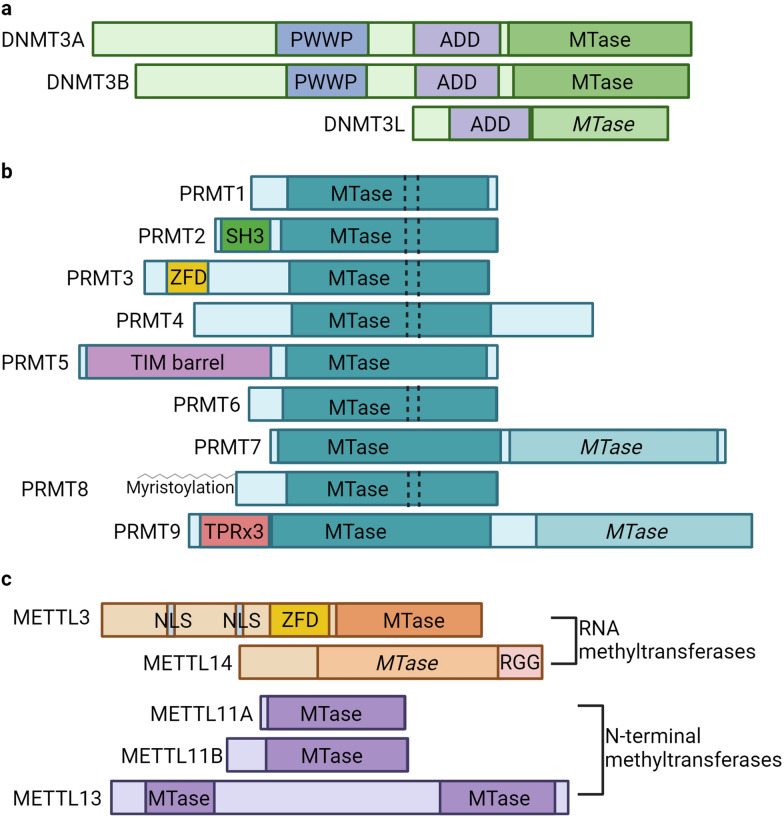
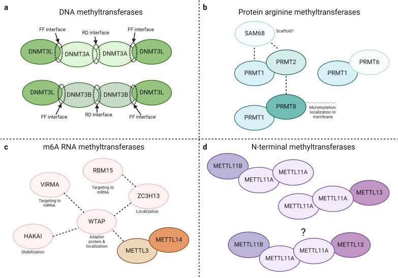
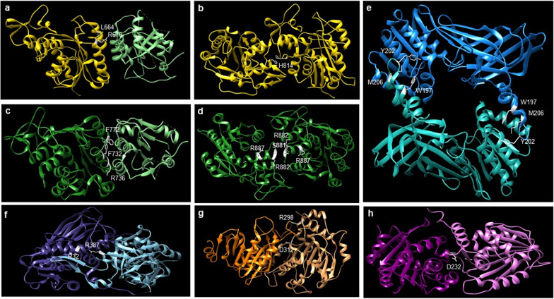
Protein and nucleic acid methylation are important biochemical modifications. In addition to their well-established roles in gene regulation, they also regulate cell signaling, metabolism, and translation. Despite this high biological relevance, little is known about the general regulation of methyltransferase function. Methyltransferases are divided into superfamilies based on structural similarities and further classified into smaller families based on sequence/domain/target similarity. While members within superfamilies differ in substrate specificity, their structurally similar active sites indicate a potential for shared modes of regulation. Growing evidence from one superfamily suggests a common regulatory mode may be through heterooligomerization with other family members. Here, we describe examples of methyltransferase regulation through intrafamily heterooligomerization and discuss how this can be exploited for therapeutic use.

Keywords: Methylation; Methyltransferase; Oligomerization; Regulation.


Conflict of interest statement

The authors declare that they have no competing interests.

Figures

Fig. 1
Fig. 1
Domain architecture of methyltransferase families. a The de novo DNA methyltransferases, DNMT3A, DNMT3B, and DNMT3L all share a similar ADD (Atrx-Dnmt3-Dnmt3l) domain. DNMT3A and DNMT3B share a PWWP (Pro-Trp-Trp-Pro) domain and a conserved catalytic methyltransferase (MTase) domain. DNMT3L possesses an inactive MTase domain variant, shown in a lighter color and italicized to distinguish it from the active MTase domains. b The PRMT family consists of 9 enzymes containing a conserved catalytic MTase core. Several notable features are found at the N-termini of some PRMTs: PRMT2 contains an SH3 domain, PRMT3 contains a zinc finger domain (ZFD), PRMT5 contains a TIM barrel, PRMT8 is N-terminally myristoylated, and PRMT9 contains three TRP (tetratricopeptide) motifs. PRMT7 and PRMT9 contain two tandem MTase domains, with the C-terminal MTase domains lacking some of the conserved motifs of the canonical PRMT core rendering them catalytically inactive. The inactive MTase domains are italicized and shown in a lighter color than the active. Type I PRMTs also have a conserved dimerization arm shown with dashed lines within the MTase domains. c The METTL family members discussed here include the RNA (m6A) methyltransferases METTL3 and METTL14, and the N-terminal methyltransferases METTL11A, METTL11B, and METTL13. The m6A methyltransferase METTL3 contains two nuclear localization signals (NLS), and a ZFD at its N-terminus, while METTL14 possesses a catalytically inactive MTase domain (italicized and shown in a lighter color than the active MTase domain) and RGG repeats (RGG) at its C-terminus. The N-terminal methyltransferases METTL11A and METTL11B each only contain one active MTase domain, while METTL13 contains two active MTase domains with distinct functions. Created with Biorender.com
Fig. 2
Fig. 2
Heterooligomeric methyltransferase complex formation. a DNMT3A–DNMT3L or DNMT3B–DNMT3L heterotetramers are depicted with the positions of the outer FF interfaces and inner RD interfaces shown. These interfaces would be in the same locations for both types of homotetramers as well. b The PRMT1–PRMT6, PRMT1–PRMT2, and PRMT1–PRMT8 heterodimers are shown along with dashed lines to indicate additional interactions between PRMT2 and PRMT8, and PRMT1, PRMT2, and SAM68 (KH domain-containing, RNA-binding, signal transduction-associated protein 1). c The METTL3–METTL14 heterodimer is shown along with dashed lines indicating interactions with other members of the m6A methyltransferase complex. WTAP Wilms Tumor 1-Associating Protein, VIRMA Vir Like m6A Methyltransferase Associated, HAKAI E3 ubiquitin-protein ligase, RBM15 RNA binding motif protein 15, ZC3H13 Zinc Finger CCCH-Type Containing 13. d The METTL11A dimer is shown as a complex with METTL11B or METTL13 individually and in combination as one possible larger complex consisting of all three N-terminal methyltransferases. Created with BioRender.com
Fig. 3
Fig. 3
Methyltransferase residues mutated in disease. a DNMT3B–DNMT3L (DNMT3B in yellow; DNMT3L in light green) heterodimer with ICF-associated mutated residues in the FF interface shown in white. PDB: 6KDA. b DNMT3B–DNMT3B (both subunits in yellow) homodimer with ICF-associated mutated residue in the RD interface shown in white. PDB: 6KDA. c DNMT3A–DNMT3L (DNMT3A in dark green; DNMT3L in light green) heterodimer with AML-associated mutated residues in the FF interface shown in white. PDB: 6F57. d DNMT3A–DNMT3A (both subunits in dark green) homodimer with AML-associated mutated residues in the RD interface shown in white. PDB: 6F57. e Rat PRMT1–PRMT1 (subunits in teal and light teal) homodimer with rat residues orthologous to human cancer-associated mutations shown in white. PDB: 3Q7E. f Mouse PRMT7 (NTD in dark blue; CTD in light blue) pseudodimer with mouse residues orthologous to human SBIDDS-associated mutated residues shown in white (amino acid numbering is the same for mouse and human). PDB: 4C4A. g METTL3–METTL14 (METTL3 in dark orange; METTL14 in light orange) heterodimer with METTL14 cancer-associated mutated residues shown in white. PDB: 5K7M. h METTL11A–METTL11B (METTL11A in purple; METTL11B in pink) heterodimer with METTL11B cancer-associated mutated residue shown in white. PDB: 2EX4; 6DUB; modeled as described previously [80]. All modeled using Chimera UCSF [96]

Similar articles

Cited by

References

    1. Strahl BD, Allis CD. The language of covalent histone modifications. Nature. 2000;403:41–45. doi: 10.1038/47412. - DOI - PubMed
    1. Holliday R, Pugh JE. DNA modification mechanisms and gene activity during development. Science. 1975;187:226–232. doi: 10.1126/science.187.4173.226. - DOI - PubMed
    1. Biggar KK, Li SS. Non-histone protein methylation as a regulator of cellular signalling and function. Nat Rev Mol Cell Biol. 2015;16(1):5–17. doi: 10.1038/nrm3915. - DOI - PubMed
    1. Malecki JM, Davydova E, Falnes PO. Protein methylation in mitochondria. J Biol Chem. 2022;298(4):101791. doi: 10.1016/j.jbc.2022.101791. - DOI - PMC - PubMed
    1. Clarke SG. Protein methylation at the surface and buried deep: thinking outside the histone box. Trends Biochem Sci. 2013;38(5):243–252. doi: 10.1016/j.tibs.2013.02.004. - DOI - PMC - PubMed

LinkOut - more resources