. 2024 Apr;54(4):e2350580.
doi: 10.1002/eji.202350580. Epub 2024 Mar 2.

Interleukin-2-induced skin inflammation

Affiliations

Interleukin-2-induced skin inflammation

Charline Sommer et al. Eur J Immunol. 2024 Apr.

Abstract

Recombinant human IL-2 has been used to treat inflammatory diseases and cancer; however, side effects like skin rashes limit the use of this therapeutic. To identify key molecules and cells inducing this side effect, we characterized IL-2-induced cutaneous immune reactions and investigated the relevance of CD25 (IL-2 receptor α) in the process. We injected IL-2 intradermally into WT mice and observed increases in immune cell subsets in the skin with preferential increases in frequencies of IL-4- and IL-13-producing group 2 innate lymphoid cells and IL-17-producing dermal γδ T cells. This overall led to a shift toward type 2/type 17 immune responses. In addition, using a novel topical genetic deletion approach, we reduced CD25 on skin, specifically on all cutaneous cells, and found that IL-2-dependent effects were reduced, hinting that CD25 - at least partly - induces this skin inflammation. Reduction of CD25 specifically on skin Tregs further augmented IL-2-induced immune cell infiltration, hinting that CD25 on skin Tregs is crucial to restrain IL-2-induced inflammation. Overall, our data support that innate lymphoid immune cells are key cells inducing side effects during IL-2 therapy and underline the significance of CD25 in this process.

Keywords: Drug‐induced skin inflammation; IL‐2 therapy; Immunotherapy; Innate lymphoid cells; γδ T cells.


Conflict of interest statement

Conflict of interest

The authors declare that they have no competing interests.

Figures
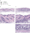
Figure 1:
Figure 1:. Intradermal IL-2 injections induce skin inflammation characterized by considerable immune cell infiltrates.
A Wild type mice were intradermally injected with IL-2 (aldesleukin) daily for 7 consecutive days before analysis of skin. Created with BioRender.com. Representative histological images of injections sides from control (B) and IL-2-treated skin (C). Overview pictures (upper images; scale bars 200 μm) and close-ups (middle and lower images; scale bares 40 μm) are shown.
Figure 2:
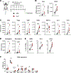
Figure 2:. IL-2-induced skin inflammation is characterized by increases in frequencies of innate immune cells.
A Wild type mice were intradermally injected with IL-2 (aldesleukin) daily for 7 consecutive days before analysis of skin and skin-draining lymph nodes (SDLN). Created with BioRender.com. B Absolute CD45+ immune cell numbers in skin and SDLN. C Absolute numbers of T cell subsets (Teffs: CD4+ T effector cells; dGDs: dermal γδ T cells; DETCs: dendritic epidermal T cells), innate lymphoid cells (ILCs), and CD3-NK1.1+ group 1 ILCs (ILC1s). D Absolute numbers of neutrophils and eosinophils in skin. E Frequencies of innate immune cells upon IL-2 injection. F CD25 expression as shown by CD25+ frequencies of the respective cells. cDC: conventional dendritic cell. n=5 mice from two independent experiments; * p<0.05, ** p<0.01, *** p<0.001 as determined by paired t-tests. All other conditions n.s.
Figure 3:
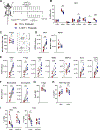
Figure 3:. Reduction of CD25 expression on skin cells reduces IL-2-induced increases of skin immune cell numbers.
A Cre recombinase of Rosa26CreERT2xCD25fl/fl mice was induced by topical 4-Hydroxytamoxifen (4-OHT) application prior to IL-2 injections on both sides to decrease CD25 on all skin cells. Acetone on the contralateral side of the back of the same mouse served as vehicle control. Created with BioRender.com. B CD25 expression as shown by CD25+ cell frequencies of the respective cell subsets. Teffs: CD4+ T effector cells; dGDs: dermal γδ T cells; DETCs: dendritic epidermal T cells; ILCs: innate lymphoid cells; ILC1s: CD3-NK1.1+ group 1 ILCs; cDC1: type 1 conventional dendritic cell. C Mean fluorescence intensity (MFI) of CD25 on respective cells. D Immune cell numbers on control and 4-OHT-treated sides in skin and skin-draining lymph nodes (SDLN). E Absolute cell numbers of lymphocyte immune cell subsets in skin tissue. F Absolute numbers of neutrophils and eosinophils in skin. G Expression of T-bet at GATA-3 transcription factors in Teffs and ILCs. n=4–8 mice from three independent experiments; * p<0.05, ** p<0.01, **** p<0.0001 as determined by paired t-tests. All other conditions n.s.
Figure 4:
Figure 4:. Reduction of CD25 on Tregs leads to significant increases in IL-2-induced skin inflammation.
A Cre recombinase of FoxP3CreERT2xCD25fl/fl mice was induced by topical 4-Hydroxytamoxifen (4-OHT) application to delete CD25 on skin Tregs prior to IL-2 injections on both sides. Acetone on the contralateral side of the back of the same mouse served as vehicle control. Created with BioRender.com. B CD25 expression as shown by CD25+ frequencies of the respective cells. Teffs: CD4+ T effector cells; dGDs: dermal γδ T cells; DETCs: dendritic epidermal T cells; ILCs: innate lymphoid cells; ILC1s: CD3-NK1.1+ group 1 ILCs; cDC1: type 1 conventional DC. C CD25 mean fluorescence intensity (MFI) of CD25+ Tregs (left) and representative CD25 gating on Tregs (right). D Immune cell numbers on control and 4-OHT-treated sides in skin and skin-draining lymph nodes (SDLN). E Absolute cell numbers of immune cell subsets in skin tissue. F Absolute numbers of neutrophils and eosinophils in skin. G MFI of FoxP3 in Tregs in skin. H Frequencies of skin Teffs and Tregs of CD4+ cells. I Expression of T-bet at GATA-3 transcription factors in Teffs and ILCs. n=7 mice; * p<0.05, ** p<0.01, *** p<0.001 as determined by paired t-tests. All other conditions n.s.
Figure 5:
Figure 5:. IL-2 injections induce a mixed type2/type 17 immune reaction in skin, associated with dermal γδ T cells and ILCs.
A Experimental set-up for injection of wild type mice with IL-2. Created with BioRender.com. B Expression of cytokines in CD45+ cells upon IL-2 injection. C Relative cytokine expression in immune cells in IL-2-treated vs. Ctrl skin. Frequencies of cytokine expression in CD45+ cells (type 1: IFN-γ + TNF-α; type 2: IL-4 + IL-13; type 17: IL-17) upon IL-2 injection was normalized to control conditions to yield fold changes. D Cytokine expression in different lymphocyte populations. Teffs: CD4+ T effector cells; dGDs: dermal γδ T cells; ILCs: innate lymphoid cells; ILC1s: CD3-NK1.1+ group 1 ILCs. n=4–5 mice representative for two independent experiments; * p<0.05, ** p<0.01 as determined by paired t-tests. All other conditions n.s.

References

    1. Wolkenstein P, Chosidow O, Wechsler J, Guillaume J-C, Lescs M-C, Brandely M, Avril M-F, Revuz J. Cutaneous side effects associated with interleukin 2 administration for metastatic melanoma. Journal of the American Academy of Dermatology 1993;28:66–70. - PubMed
    1. Raeber ME, Sahin D, Boyman O. Interleukin-2-based therapies in cancer. Sci Transl Med 2022;14:eabo5409. - PubMed
    1. Harris F, Arroyo Berdugo Y, Tree T. IL-2-based Approaches to Treg Enhancement. Clin Exp Immunol 2022. - PMC - PubMed
    1. Giri JG, Ahdieh M, Eisenman J, Shanebeck K, Grabstein K, Kumaki S, Namen A, Park LS, Cosman D, Anderson D. Utilization of the beta and gamma chains of the IL-2 receptor by the novel cytokine IL-15. EMBO J 1994;13:2822–30. - PMC - PubMed
    1. Yu A, Snowhite I, Vendrame F, Rosenzwajg M, Klatzmann D, Pugliese A, Malek TR. Selective IL-2 responsiveness of regulatory T cells through multiple intrinsic mechanisms supports the use of low-dose IL-2 therapy in type 1 diabetes. Diabetes 2015;64:2172–83. - PubMed

Substances

LinkOut - more resources