. 2024 Mar 2;108(1):248.
doi: 10.1007/s00253-023-12989-0.

Assessing immunogenicity of CRISPR-NCas9 engineered strain against porcine epidemic diarrhea virus

Affiliations

Assessing immunogenicity of CRISPR-NCas9 engineered strain against porcine epidemic diarrhea virus

Fengsai Li et al. Appl Microbiol Biotechnol. .

Erratum in

Abstract

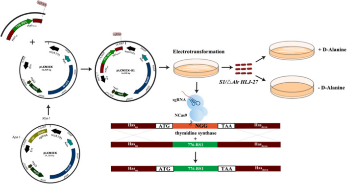
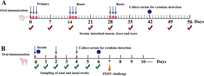
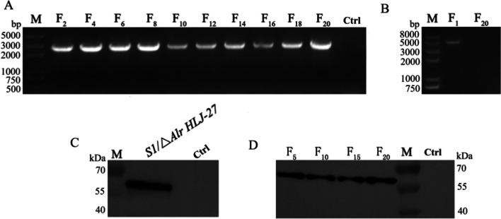
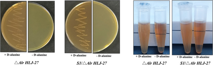
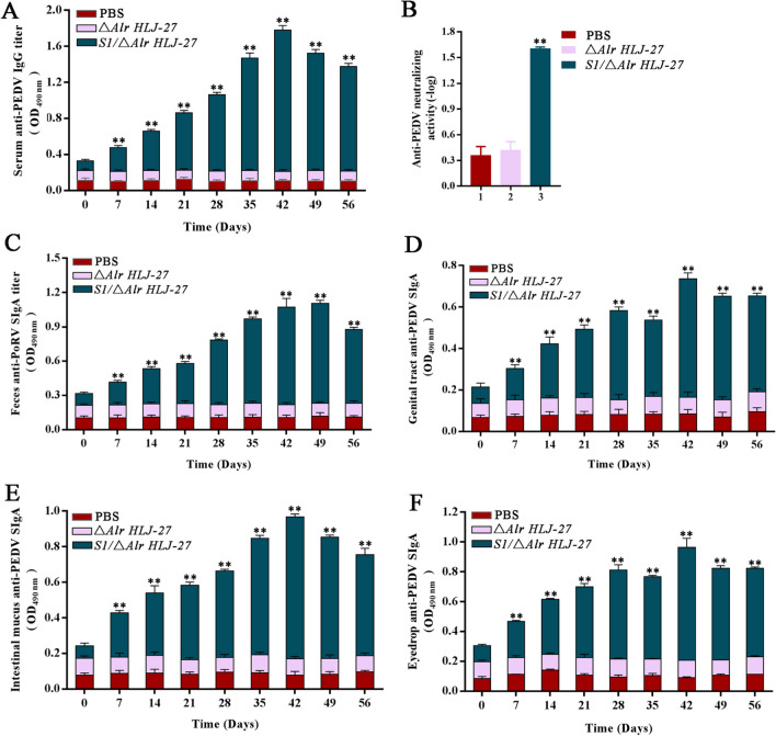
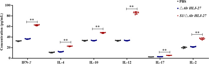
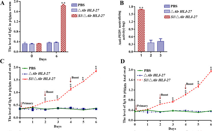
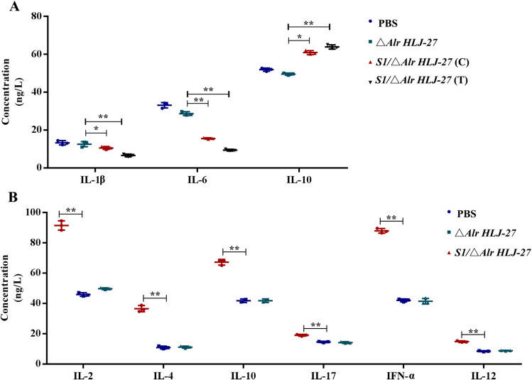
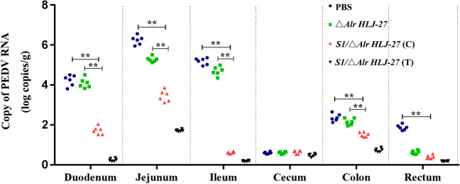
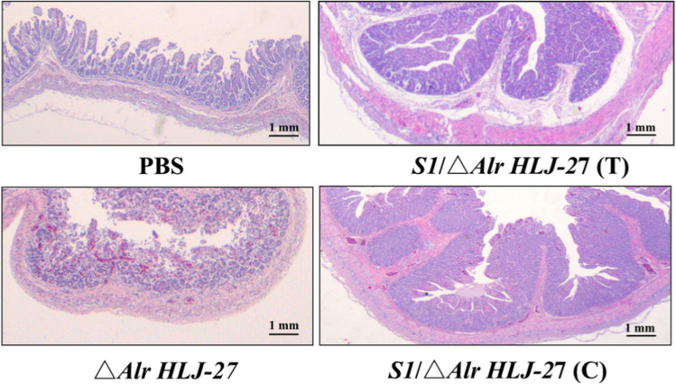
Porcine epidemic diarrhea (PED) caused by porcine epidemic diarrhea virus (PEDV), is an acute and highly infectious disease, resulting in substantial economic losses in the pig industry. Given that PEDV primarily infects the mucosal surfaces of the intestinal tract, it is crucial to improve the mucosal immunity to prevent viral invasion. Lactic acid bacteria (LAB) oral vaccines offer unique advantages and potential applications in combatting mucosal infectious diseases, making them an ideal approach for controlling PED outbreaks. However, traditional LAB oral vaccines use plasmids for exogenous protein expression and antibiotic genes as selection markers. Antibiotic genes can be diffused through transposition, transfer, or homologous recombination, resulting in the generation of drug-resistant strains. To overcome these issues, genome-editing technology has been developed to achieve gene expression in LAB genomes. In this study, we used the CRISPR-NCas9 system to integrate the PEDV S1 gene into the genome of alanine racemase-deficient Lactobacillus paracasei △Alr HLJ-27 (L. paracasei △Alr HLJ-27) at the thymidylate synthase (thyA) site, generating a strain, S1/△Alr HLJ-27. We conducted immunization assays in mice and piglets to evaluate the level of immune response and evaluated its protective effect against PEDV through challenge tests in piglets. Oral administration of the strain S1/△Alr HLJ-27 in mice and piglets elicited mucosal, humoral, and cellular immune responses. The strain also exhibited a certain level of resistance against PEDV infection in piglets. These results demonstrate the potential of S1/△Alr HLJ-27 as an oral vaccine candidate for PEDV control. KEY POINTS: • A strain S1/△Alr HLJ-27 was constructed as the candidate for an oral vaccine. • Immunogenicity response and challenge test was carried out to analyze the ability of the strain. • The strain S1/△Alr HLJ-27 could provide protection for piglets to a certain extent.

Keywords: CRISPR-Cas9; Genome expression; Lactic acid bacteria; Oral vaccine; PEDV S1 gene.


Conflict of interest statement

The authors declare no competing interests.

Figures

Fig. 1
Fig. 1
Schematic diagram of the construction of gene-editing plasmid pLCNICK-S1 and the gene-editing process CRISPR-Cas9D10A system. The gene-editing plasmid pLCNICK-S1 was constructed as showing as the steps. The fragment P776 promoter, RBS, and S1 were purified and fused into one fragment, generating the gene P776-RBS-S1 (P776-RS1); the fragment Hasup, P776-RS1, and Hasdown-sgRNA were amplified by the fusion PCR to yield the gene Hasup-P776-RS1-Hasdown-sgRNA, then ligated with the pMD19T-Simple, producing the cloning plasmid pMD19Ts-Hasup-P776-RS1-Hasdown-sgRNA (pMD19Ts-S1). The plasmid pMD19Ts-S1 was digested and then inserted into the temperature-sensitive vector pLCNICK at Apa I and Xba I sites, generating the gene-editing plasmid pLCNICK-S1. The pLCNICK-S1 was electrotransformed into competent cells △Alr HLJ-27 to initiate the gene-editing process, yielding the mutant strain S1/△Alr HLJ-27 with substituting the P776-RS1 gene for thymidine synthase (thyA) at the genome, and only growing with exogenous addition of d-alanine
Fig. 2
Fig. 2
The procedure of mice (A) or piglets (B) with oral immunization and sampling. A Serum, intestinal mucus, feces, and tears were collected at the day of 7, 14, 21, 28, 35, 42, 49, and 56. The mice were 90 in total, for 30 mice per group, and each sampling was repeated three times (three mice). Cytokine detection was carried out on 42 days post primary immunization with gathering the serum from the eyeball. The immunization procedure adopts continuous immunization for the interval time with 2 weeks between twice booster immunization and each immunization continuously for 3 days; B Large landrace piglets were 15 in total, including S1Alr HLJ-27 group (n = 6), ΔAlr HLJ-27 group (n = 3) and PBS group (n = 6). Piglet serum was collected on days 0 and 6 post immunization. Anal and nasal swabs were collected daily and soaked in PBS. The piglet immunization procedure was immunized for 2 days at a time, with a total of three immunizations. The cytokine levels in sera were measured on day 6 post immunization and challenged the piglets with PEDV to determine the immune protection of mutant S1/△Alr HLJ-27 with each group three piglets
Fig. 3
Fig. 3
The gene hereditary, the plasmid elimination, and the stability of protein expression were detected of mutant strain S1/Alr HLJ-27 (A, B, C). The genome of the strain was extracted every two generation within 20 generations to determine the stability of the gene inheritance by PCR amplification with thy-F/R as the primers and sequencing (A), M: Trans 2 k plus DNA marker; F2-F20: The F2-F20 generation strains of S1/Alr HLJ-27; Ctrl: the strain of △Alr HLJ-27. The plasmid elimination situation of F1 and F20 generation strains was evaluated by plasmid PCR amplification with XA-F/R as the primers and sequencing (B), M: Trans 2 k plus II DNA marker; F1/F20: The F1/F20 generation strain of S1/Alr HLJ-27. The expression of interest protein was detected by western blotting with mouse anti-S1 monoclonal antibody as a primary antibody (C). The stability of protein expression every five generation was determined (D)
Fig. 4
Fig. 4
The demand test of the mutant strain of S1/Alr HLJ-27. The growth results as the strain streaking on the MRS plate (A) and in the MRS liquid (B). “ + ” represent the MRS with the d-alanine; “ − ” represent the MRS without the d-alanine
Fig. 5
Fig. 5
Determination of anti-PEDV levels of immunoglobulin G (IgG) (A) and secreted immunoglobulin A (SIgA) (CF) in mice (n = 3 per group) with oral immunization the mutant strain S1/Alr HLJ-27. Meanwhile, the neutralizing antibody activity of serum IgG was measured by the method for immobilizing viral load and to dilute antibodies (B). The supernatant of fecal lysis and intestinal mucus by centrifugation, eyedrop, genital tract rinse, and serum diluting with 5% skim milk were used as the primary antibodies. Bars represent the mean ± standard error value of each group (**p < 0.01 compared to the control groups PBS and △Alr HLJ-27)
Fig. 6
Fig. 6
Detection of the levels of serum cytokines. The cytokines from the mice (n = 3 per group) were analyzed with oral immunization of the mutant strain S1/Alr HLJ-27. Bars represent the mean ± standard error value of each group (**p < 0.01 compared to the control groups PBS and △Alr HLJ-27)
Fig. 7
Fig. 7
Determination of anti-PEDV levels of immunoglobulin G (IgG) (A) and secreted immunoglobulin A (SIgA) (CD) in piglets (n = 3 per group) with oral immunization the mutant strain S1/Alr HLJ-27. Meanwhile, the neutralizing antibody activity of serum IgG was measured by the method for immobilizing viral load and to dilute antibodies (B). The supernatant of anal and nasal swabs by centrifugation and serum diluting with 5% skim milk was used as the primary antibodies to detect the levels of SIgA and IgG. Bars represent the mean ± standard error value of each group (**p < 0.01 compared to the control groups PBS and △Alr HLJ-27)
Fig. 8
Fig. 8
Detection of the levels of serum cytokines (piglets: n = 3 per group). The cytokines pre-challenge (B) and post-challenge (A) in piglets with oral immunization the mutant strain S1/Alr HLJ-27 were analyzed. C represents the challenge experiments groups, and T represents the piglets, housed in the same cage with the challenge groups. Bars represent the mean ± standard error value of each group (**p < 0.01 compared to the control groups PBS and △Alr HLJ-27)
Fig. 9
Fig. 9
Detection of viral load in different intestinal tissue in piglets (n = 3 per group) after challenge with PEDV. The piglets were administered infection with 1 mL of PEDV intestines crude extract at 6 days post primary immunization. C represents the challenge experiments groups, and T represents the piglets, housed in the same cage with the challenge groups. With the death of the piglets, necropsy was performed immediately to collect each intestinal fragment for virus detection. Bars represent the mean ± standard error value of each group (**p < 0.05, ** p < 0.01 compared to the control groups PBS and △Alr HLJ-27)
Fig. 10
Fig. 10
Histopathological analysis of the strain S1/△Alr HLJ-27 protective effect on immunized piglets. Histopathological examination of H&E-stained small intestinal tissues. The tissues were acquired from the groups administered PBS, △Alr HLJ-27 and S1/△Alr HLJ-27, post-challenge with 1 mL of PEDV intestines crude extract (RNA copy number of 1.0 × 106). “C” represents the S1/△Alr HLJ-27 challenge experiments groups, and “T” represents the S1/△Alr HLJ-27 piglets, housed in the same cage with the challenge groups. With the death of the piglets, necropsy was performed immediately to collect each intestinal for analysis. Original magnifications: 1 mm

Similar articles

Cited by

References

    1. Agarwal N, Gupta R (2021) History, evolution and classification of crispr-cas associated systems-sciencedirect. Prog Mol Biol Transl Sci 179:11–76. 10.1016/bs.pmbts.2020.12.012 - PubMed
    1. Biswas I, Gruss A, Ehrlich SD, Maguin E (1993) High-efficiency gene inactivation and replacement system for gram-positive bacteria. J Bacteriol 175(11):3628–3635. 10.1007/BF02186240 - PMC - PubMed
    1. Chen J, Cui Y, Wang Z, Liu G (2020) Identification and characterization of PEDV infection in rat crypt epithelial cells. Vet Microbiol 249:108848. 10.1016/j.vetmic.2020.108848 - PMC - PubMed
    1. Douglas GL, Klaenhammer TR (2011) Directed chromosomal integration and expression of the reporter gene gusA3 in Lactobacillus acidophilus NCFM. AEM 77(20):7365–7371. 10.1128/AEM.06028-11 - PMC - PubMed
    1. Egelkrout E, Hayden C, Fake G, Keener T, Arruda P, Saltzman R, Walker J, Howard J (2020) Oral delivery of maize-produced porcine epidemic diarrhea virus spike protein elicits neutralizing antibodies in pigs. Plant Cell Tiss Org 142(1):79–86. 10.1007/s11240-020-01835-0 - PMC - PubMed

LinkOut - more resources