. 2024 Mar 2;73(4):73.
doi: 10.1007/s00262-023-03596-z.

Oct4 activates IL-17A to orchestrate M2 macrophage polarization and cervical cancer metastasis

Affiliations

Oct4 activates IL-17A to orchestrate M2 macrophage polarization and cervical cancer metastasis

Zhuoqiong Bian et al. Cancer Immunol Immunother. .

Abstract

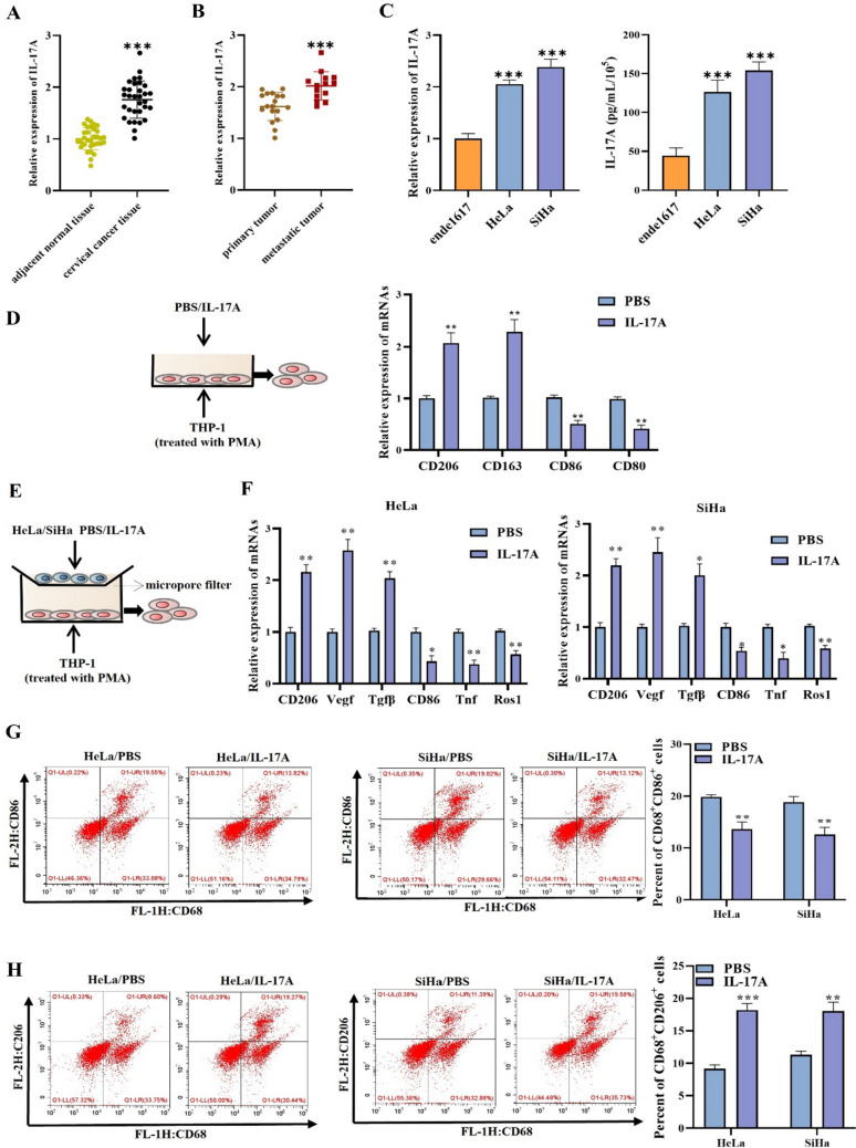
Background: Cervical cancer is a common malignant tumor in the female. Interleukin (IL)-17A is a proinflammatory factor and exerts a vital function in inflammatory diseases and cancers. M2 macrophage has been confirmed to promote tumor development. Nevertheless, it is not yet known whether IL-17A facilitates cervical cancer development by inducing M2 macrophage polarization. Therefore, this study was conducted to investigate the regulatory effect of IL-17A on M2 macrophage polarization and the underlying mechanism in cervical cancer development.

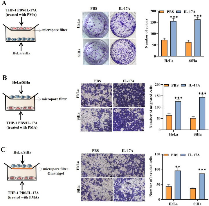
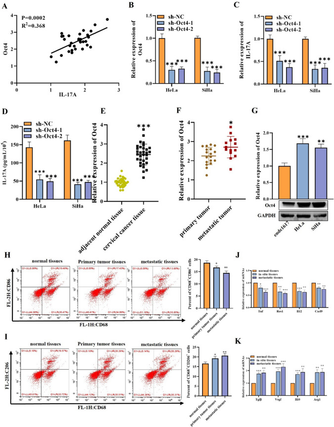
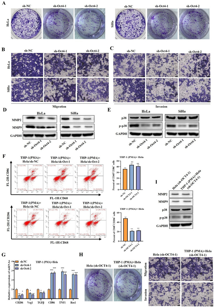
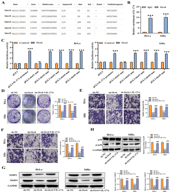
Methods: RT-qPCR was utilized for testing IL-17A expression in cancer tissues and cells. Flow cytometry was applied to evaluate the M1 or M2 macrophage polarization. Cell proliferative, migratory, and invasive capabilities were measured through colony formation and transwell assays. ChIP and luciferase reporter assays were applied to determine the interaction between IL-17A and octamer-binding transcription factor 4 (OCT4).

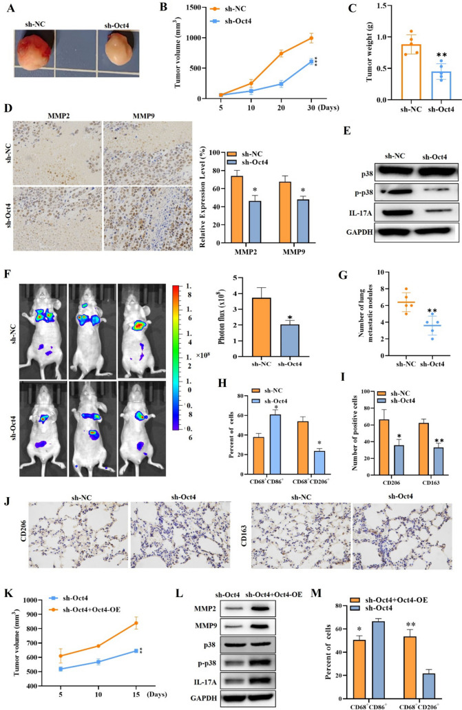
Results: IL-17A expression and concentration were high in metastatic tissues and cells of cervical cancer. IL-17A was found to facilitate M2 macrophage polarization in cervical cancer. Furthermore, IL-17A facilitated the macrophage-mediated promotion of cervical cancer cell proliferative, migratory, and invasive capabilities. Mechanistic assays manifested that Oct4 binds to and transcriptionally activated IL-17A in cervical cancer cells. Furthermore, Oct4 promoted cervical cancer cell malignant phenotype and M2 macrophage polarization by activating the p38 pathway that, in turn, upregulated IL-17A. Additionally, in vivo experiments confirmed that Oct4 knockdown reduced tumor growth and metastasis.

Conclusion: Oct4 triggers IL-17A to facilitate the polarization of M2 macrophages, which promotes cervical cancer cell metastasis.

Keywords: Cervical cancer; IL-17A; M2 macrophage; Oct4; p38 signaling.


Conflict of interest statement

The authors declare that they have no conflicts of interest.

Figures

Fig. 1
Fig. 1
IL-17A induces M2 macrophage polarization in cervical cancer. A, B RT-qPCR was used to measure IL-17A gene expression level in adjacent normal tissues, cervical cancer tissues, primary tumor tissues, and metastatic tumor tissues. C RT-qPCR and ELISA outcomes of IL-17A expression in ende1617, HeLa, and SiHa cells. D THP1 cells (treated with PMA) were treated with IL-17A (50 ng/mL) or PBS. RT-qPCR was utilized to measure the gene expression levels of CD206, CD163, CD86, and CD80 expression. E The diagrammatic sketch of cell co-culture system. F RT-qPCR was utilized to measure the gene expression levels of M1 macrophage markers (CD86, CD80, Tnf1, Ros1) and M2 macrophage markers (CD206, CD163, Veg1, Tgfβ). G, H Surface expression of CD86 and CD206 was detected in THP-1 (PMA) + Hela(sh-OCT4-1) cells using flow cytometry. The histograms represent the percent of CD86 or CD206 cells in THP-1 (PMA) + Hela(sh-OCT4-1) cells. *p < 0.05; **p < 0.01; ***p < 0.001. All experiments were repeated at least three times
Fig. 2
Fig. 2
IL-17A secreted by cervical cancer cells is conducive to macrophage-mediated facilitation of cell malignant phenotype. AC The proliferative, migratory, and invasive capabilities of HeLa or SiHa cells co-cultured with THP-1-derived TAM cells were evaluated by colony formation a and transwell assays bc in the PBS treatment group and IL-17A treatment group. **p < 0.01; ***p < 0.001. All experiments were repeated at least three times
Fig. 3
Fig. 3
Oct4 promotes the secretion of IL-17A. A The correlation between Oct4 and IL-17A in cervical cancer tissues was tested by Spearman correlation analysis. B, C RT-qPCR outcomes of Oct4 and IL-17A expression in cells transfected with sh-Oct4 or sh-NC. D ELISA was utilized to determine IL-17A content in cells transfected with sh-Oct4 or sh-NC. (E, F) RT-qPCR was used to measure the Oct4 gene expression level in adjacent normal tissues, cervical cancer tissues, primary tumor tissues, and metastatic tumor tissues. G RT-qPCR and western blot were used to evaluate Oct4 expression in indicated cells. H Surface expression of CD86 was detected in normal tissues, primary tumor tissues, and metastatic tumor tissues using flow cytometry. The histogram represents the percent of CD86 + cells in indicated groups. I Surface expression of CD206 was detected in normal tissues, primary tumor tissues, and metastatic tumor tissues using flow cytometry. The histogram represents the percent of CD206+ cells in indicated groups. J RT-qPCR was utilized to measure the gene expression levels of M1 macrophage markers (Tnf1, Ros1, Il12 and Cxcl9) in indicated groups. K RT-qPCR was utilized to measure the gene expression levels of M2 markers (Tgfβ, Vegf, Il10, and Arg1) in indicated groups. *p < 0.05; **p < 0.01; ***p < 0.001. All experiments were repeated at least three times
Fig. 4
Fig. 4
Oct4 promotes cell malignant phenotype and M2 macrophage polarization in cervical cancer by activating p38 pathway. (AC) Cell proliferative, migratory, and invasive capabilities were evaluated by colony formation a and transwell assays b–c when Oct4 was silenced. D, E Western blot was used to measure MMP2, MMP9, and p-p38 protein levels in cells when Oct4 was silenced. F Flow cytometry was utilized to analyze the frequency of M1 or M2 macrophages. G RT-qPCR outcomes of CD206, CD86, Tnf1, Ros1, Tgfβ and Vegf expression in cells when Oct4 was silenced. H Cell proliferative and migratory capabilities were evaluated by colony formation and transwell assays in groups of Hela (sh-OCT4-1) and THP-1(PMA) + sh-OCT4. I Western blot was used to measure MMP2, MMP9, and p-p38 protein levels in groups of Hela (sh-OCT4-1) and THP-1 (PMA) + sh-OCT4 cells. **p < 0.01; ***p < 0.001. All experiments were repeated at least three times
Fig. 5
Fig. 5
Oct4 activates IL-17A by binding to IL-17A promoter by activating p38 pathway. A JASPAR was utilized to predict the possible binding sites of Oct4 and IL-17A promoter. B, C ChIP assay and luciferase reporter assay were employed for verifying the interplay of Oct4 and IL-17A promoter. DF Cell proliferation, migration, and invasion were assessed by colony formation d and transwell assays ef when Oct4 was silenced, and IL-17A was added. G, H Western blot outcomes of MMP2, MMP9, and p-p38 protein levels in different cell groups. ***p < 0.001; ns means no significance. All experiments were repeated at least three times
Fig. 6
Fig. 6
Oct4 promotes tumor growth, metastasis, and M2 macrophage infiltration of cervical cancer. AC Tumor morphology, volume, and weight of mice injected with HeLa cells transfected with sh-Oct4 or sh-NC. D IHC staining of MMP2 and MMP9 in tumor tissues of different groups. E Western blot outcomes of IL-17A and p-p38 protein levels in mice tumor injected with HeLa cells transfected with sh-Oct4 or sh-NC. F Representative bioluminescence images and the histogram showing the lung metastatic lesions of mice. G Pulmonary metastatic nodules were calculated by manually counting individual lesions from metastatic tissue sections in different groups of mice. H The percent of CD86+ or CD206+ cells were assessed by flow cytometry. I The expression of CD206 and CD163 levels in mice was measured using IHC. J IHC staining was utilized for assessing the quantity of CD206-positive cells and CD163-positive cells in lung metastases. K Mice tumor volume of the sh-Oct4 + OCT4-OE group and the sh-Oct4 group. L Western blot results of MMP2, MMP9, and p-p38 expression in the tumor tissues of the sh-Oct4 + OCT4-OE group and the sh-Oct4 group. M Flow cytometry was utilized to analyze the frequency of M1 or M2 macrophages in different groups of mice. *p < 0.05; **p < 0.01; ***p < 0.001. All experiments were repeated at least three times

References

    1. Buskwofie A, David-West G, Clare CA. A review of cervical cancer: incidence and disparities. J Natl Med Assoc. 2020;112(2):229–232. - PubMed
    1. Sundström K, Elfström KM. Advances in cervical cancer prevention: efficacy, effectiveness, elimination? PLoS Med. 2020;17(1):e1003035. doi: 10.1371/journal.pmed.1003035. - DOI - PMC - PubMed
    1. Li H, Wu X, Cheng X. Advances in diagnosis and treatment of metastatic cervical cancer. J Gynecol Oncol. 2016;27(4):e43. doi: 10.3802/jgo.2016.27.e43. - DOI - PMC - PubMed
    1. Pan Y, Yu Y, Wang X, Zhang T. Tumor-associated macrophages in tumor immunity. Front Immunol. 2020;11:583084. doi: 10.3389/fimmu.2020.583084. - DOI - PMC - PubMed
    1. Nowak M, Klink M. The role of tumor-associated macrophages in the progression and chemoresistance of ovarian cancer. Cells. 2020;9(5):1299. doi: 10.3390/cells9051299. - DOI - PMC - PubMed

MeSH terms