. 2024 May;271(5):2285-2297.
doi: 10.1007/s00415-023-12171-6. Epub 2024 Mar 2.

Retinal imaging for the assessment of stroke risk: a systematic review

Affiliations

Retinal imaging for the assessment of stroke risk: a systematic review

Zain Girach et al. J Neurol. 2024 May.

Abstract

Background: Stroke is a leading cause of morbidity and mortality. Retinal imaging allows non-invasive assessment of the microvasculature. Consequently, retinal imaging is a technology which is garnering increasing attention as a means of assessing cardiovascular health and stroke risk.

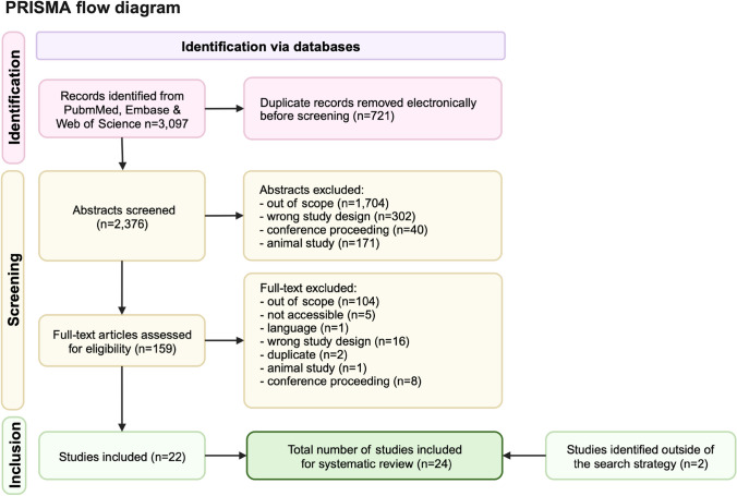
Methods: A biomedical literature search was performed to identify prospective studies that assess the role of retinal imaging derived biomarkers as indicators of stroke risk.

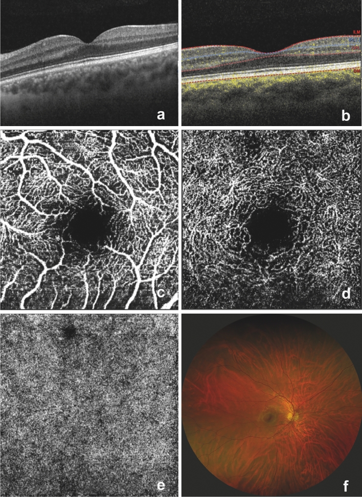
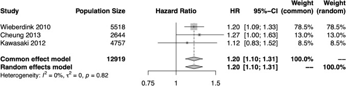
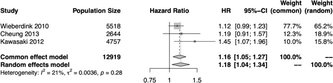
Results: Twenty-four studies were included in this systematic review. The available evidence suggests that wider retinal venules, lower fractal dimension, increased arteriolar tortuosity, presence of retinopathy, and presence of retinal emboli are associated with increased likelihood of stroke. There is weaker evidence to suggest that narrower arterioles and the presence of individual retinopathy traits such as microaneurysms and arteriovenous nicking indicate increased stroke risk. Our review identified three models utilizing artificial intelligence algorithms for the analysis of retinal images to predict stroke. Two of these focused on fundus photographs, whilst one also utilized optical coherence tomography (OCT) technology images. The constructed models performed similarly to conventional risk scores but did not significantly exceed their performance. Only two studies identified in this review used OCT imaging, despite the higher dimensionality of this data.

Conclusion: Whilst there is strong evidence that retinal imaging features can be used to indicate stroke risk, there is currently no predictive model which significantly outperforms conventional risk scores. To develop clinically useful tools, future research should focus on utilization of deep learning algorithms, validation in external cohorts, and analysis of OCT images.

Keywords: Artificial intelligence; Biomarkers; Deep learning; Prediction; Retina; Stroke.


Conflict of interest statement

The authors declare no conflicts of interest.

Figures

Fig. 1
Fig. 1
A selection of retinal images which illustrate normal anatomy in optical coherence tomography (OCT), OCT angiography (OCT-A), and scanning laser ophthalmoscopy: a a structural OCT image of the macula; b a structural OCT with OCT-A flow overlay (highlighted by yellow dots, which indicate blood flow) and with automated segmentation at the internal limiting membrane (ILM), inner plexiform layer (IPL), outer plexiform layer (OPL) and Bruch’s membrane (BM); c an en face macula OCT-A image (superficial vascular plexus slab); d an en face macula OCT-A image (deep vascular plexus slab); e an en face macula OCT-A image (choriocapillaris slab); f a wide-field retinal image acquired using scanning laser ophthalmoscopy
Fig. 2
Fig. 2
A PRISMA flow diagram outlining the exclusion/inclusion process
Fig. 3
Fig. 3
Hazard ratios (HR) for the central retinal vein equivalent (CRVE)–stroke relationship in each study eligible for meta-analysis, and the pooled HR and 95% confidence interval (CI) in both fixed effects and random effects models. There was no significant heterogeneity among these studies, and the results support a significant relationship between CRVE and stroke risk
Fig. 4
Fig. 4
Hazard ratios (HR) for the central retinal artery equivalent (CRAE)–stroke relationship in each study eligible for meta-analysis, and the pooled HR and 95% confidence interval (CI) in both fixed effects and random effects models. There was not significant heterogeneity among these studies, and the results support a significant relationship between narrow CRAE and stroke risk
Fig. 5
Fig. 5
Hazard ratios (HR) for the presence of retinopathy–stroke relationship in each study eligible for meta-analysis, and the pooled HR and 95% confidence interval (CI) in both fixed effects and random effects models. The HR of retinopathy is compared to a reference of no retinopathy. There was not significant heterogeneity among these studies, and the results support a significant relationship between retinopathy and stroke risk

References

    1. Donkor ES. Stroke in the 21 Century: A Snapshot of the Burden, Epidemiology, and Quality of Life. Stroke Res Treat. 2018;2018:3238165. - PMC - PubMed
    1. Kirshner HS, Biller J, Callahan AS., 3rd Long-term therapy to prevent stroke. J Am Board Fam Pract. 2005;18:528–540. - PubMed
    1. Shah A, Apte RS. Optical Coherence Tomography Angiography: A Window into Central Nervous System Neurodegeneration. Trends Mol Med. 2020;26:892–895. - PubMed
    1. Coronado I, Abdelkhaleq R, Yan J, et al. Towards Stroke Biomarkers on Fundus Retinal Imaging: A Comparison Between Vasculature Embeddings and General Purpose Convolutional Neural Networks. Conf Proc IEEE Eng Med Biol Soc. 2021;2021:3873–3876. - PMC - PubMed
    1. Rudnicka AR, Welikala R, Barman S, et al. Artificial intelligence-enabled retinal vasculometry for prediction of circulatory mortality, myocardial infarction and stroke. Br J Ophthalmol. 2022;106:1722–1729. - PMC - PubMed

Publication types

MeSH terms