. 2024 Summer;26(1):101031.
doi: 10.1016/j.jocmr.2024.101031. Epub 2024 Mar 1.

Impact of late gadolinium enhancement image acquisition resolution on neural network based automatic scar segmentation

Affiliations

Impact of late gadolinium enhancement image acquisition resolution on neural network based automatic scar segmentation

Tobias Hoh et al. J Cardiovasc Magn Reson. 2024 Summer.

Abstract

Background: Automatic myocardial scar segmentation from late gadolinium enhancement (LGE) images using neural networks promises an alternative to time-consuming and observer-dependent semi-automatic approaches. However, alterations in data acquisition, reconstruction as well as post-processing may compromise network performance. The objective of the present work was to systematically assess network performance degradation due to a mismatch of point-spread function between training and testing data.

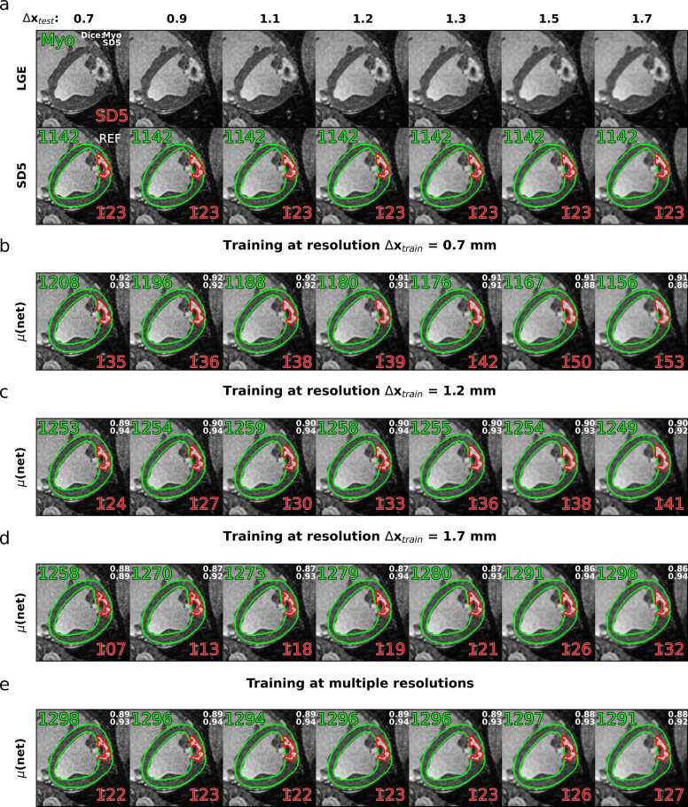
Methods: Thirty-six high-resolution (0.7×0.7×2.0 mm3) LGE k-space datasets were acquired post-mortem in porcine models of myocardial infarction. The in-plane point-spread function and hence in-plane resolution Δx was retrospectively degraded using k-space lowpass filtering, while field-of-view and matrix size were kept constant. Manual segmentation of the left ventricle (LV) and healthy remote myocardium was performed to quantify location and area (% of myocardium) of scar by thresholding (≥ SD5 above remote). Three standard U-Nets were trained on training resolutions Δxtrain = 0.7, 1.2 and 1.7 mm to predict endo- and epicardial borders of LV myocardium and scar. The scar prediction of the three networks for varying test resolutions (Δxtest = 0.7 to 1.7 mm) was compared against the reference SD5 thresholding at 0.7 mm. Finally, a fourth network trained on a combination of resolutions (Δxtrain = 0.7 to 1.7 mm) was tested.

Results: The prediction of relative scar areas showed the highest precision when the resolution of the test data was identical to or close to the resolution used during training. The median fractional scar errors and precisions (IQR) from networks trained and tested on the same resolution were 0.0 percentage points (p.p.) (1.24 - 1.45), and - 0.5 - 0.0 p.p. (2.00 - 3.25) for networks trained and tested on the most differing resolutions, respectively. Deploying the network trained on multiple resolutions resulted in reduced resolution dependency with median scar errors and IQRs of 0.0 p.p. (1.24 - 1.69) for all investigated test resolutions.

Conclusion: A mismatch of the imaging point-spread function between training and test data can lead to degradation of scar segmentation when using current U-Net architectures as demonstrated on LGE porcine myocardial infarction data. Training networks on multi-resolution data can alleviate the resolution dependency.

Keywords: Automatic segmentation; Cardiovascular magnetic resonance; Deep learning; LGE imaging; Neural networks; Scar quantification.


Conflict of interest statement

Declaration of Competing Interest The authors declare the following financial interests/personal relationships which may be considered as potential competing interests: Not applicable reports financial support was provided by Innosuisse Swiss Innovation Agency. If there are other authors, they declare that they have no known competing financial interests or personal relationships that could have appeared to influence the work reported in this paper.

Figures

Fig. 1
Fig. 1
Processing of images and reference (REF) segmentation. (a) Late gadolinium enhancement (LGE) image acquisition and SENSE reconstruction with manual expert observer segmentation of epi- and endo-contours as well as healthy remote myocardium. Semiautomatic reference scar segmentation using SD5 thresholding at acquisition resolution is followed by morphological denoising of scar masks. Healthy myocardium and dense scar (I > SD5) are shown in green and red, respectively. Resolution reduction by multiplication of k-space data with a low-pass filter, while keeping field-of-view and matrix size constant. (c) Examples of resulting images with in-plane resolutions Δxtrain = 0.7 mm, 1.2 mm and 1.7 mm, respectively. (d) Four segmentation networks are trained for the given resolutions. (e) Exemplary segmentation mask predictions for the network trained at Δxtrain = 0.7 mm (solid blue arrow) including identical (*) morphological denoising. SENSE, sensitivity encoding; SD: standard deviation
Fig. 2
Fig. 2
Example late-gadolinium-enhancement (LGE) dataset. Images with varying in-plane resolution Δx and reference SD5 thresholding segmentation masks are shown in (a). Corresponding predictions using networks trained on these resolutions are shown in (b-d), respectively. Predictions for a network trained on mixed resolutions from Δxtrain = 0.7 mm to 1.7 mm are shown in (e). Healthy myocardium is shown in green and scar (SD5) in red. Regional areas in mm2 for myocardium and scar are given as numbers in green and red, respectively. Dice scores relative to SD5 thresholding on REF are given in the top right corner of the shown frames. REF, reference; SD, standard deviation
Fig. 3
Fig. 3
Boxplot analysis of signed errors between network predictions and SD5 thresholding as a function of in-plane resolutions Δxtest from 0.7 mm to 1.7 mm is shown for networks trained on Δxtrain = 0.7 mm (a), 1.2 mm (b) and 1.7 mm (c), and multiple resolutions Δxtrain = 0.7 mm to 1.7 mm (d). Left and right columns show boxplots for myocardium (MYO) and scar (SCAR) predictions, respectively. Interquartile ranges (IQR), which indicate network precision, are given in the legend. SD, standard deviation
Fig. 4
Fig. 4
Signed errors and Dice score marginal distributions between network predictions and SD5 thresholding as a function of in-plane resolutions Δxtest = 0.7 mm to 1.7 mm are shown for networks trained on Δxtrain = 0.7 mm (a), 1.2 mm (b), 1.7 mm, and multiple resolutions Δxtrain = 0.7 mm to 1.7 mm (d). The two left panels show the signed errors as kernel density estimations of histograms for myocardium (MYO) and scar (SCAR), respectively. The two right panels show the Dice scores as kernel density estimations of histograms for MYO and SCAR, respectively. Actual histogram reflects distribution of signed errors and Dice scores at training data resolution.
Fig. 5
Fig. 5
Boxplot analysis of Dice scores between network predictions and SD5 thresholding as a function of in-plane resolutions Δxtest= 0.7 mm to 1.7 mm are shown for networks trained at Δxtrain = 0.7 mm (a), 1.2 mm (b), 1.7 mm (c) and mixed multiple resolutions Δxtrain = 0.7 mm to 1.7 mm (d). Left and right columns show boxplots for myocardium (MYO) and scar (SCAR) predictions, respectively. Interquartile ranges (IQR), which indicate network precision, are given in the legend. SD, standard deviation
Fig. 6
Fig. 6
Segmentation performance summary. (a) Interquartile ranges (IQR) of the signed scar errors over all investigated in-plane resolutions Δxtest = 0.7 mm to 1.7 mm are shown for networks trained on Δxtrain = 0.7 mm, 1.2 mm, 1.7 mm, and multiple resolutions. (b) Signed scar (SCAR) errors median and IQR, aggregated across all investigated in plane resolutions Δxtest = 0.7 mm to 1.7 mm, are shown for networks trained at Δxtrain = 0.7 mm, 1.2 mm, 1.7 mm, and multiple resolutions.

References

    1. Kellman P., Arai A.E. Cardiac imaging techniques for physicians: late enhancement. J Magn Reson Imaging. 2012;36:529–542. doi: 10.1002/JMRI.23605. - DOI - PMC - PubMed
    1. Puntmann V.O., Valbuena S., Hinojar R., et al. Society for Cardiovascular Magnetic Resonance (SCMR) expert consensus for CMR imaging endpoints in clinical research: part I - analytical validation and clinical qualification. J Cardiovasc Magn Reson. 2018;20:1–23. doi: 10.1186/S12968-018-0484-5. - DOI - PMC - PubMed
    1. Nijveldt R., Hofman M.B.M., Hirsch A., et al. Assessment of microvascular obstruction and prediction of short-term remodeling after acute mycoardial infarction: Cardiac MR imaging study. Radiology. 2009;250:363–370. doi: 10.1148/radiol.2502080739. - DOI - PubMed
    1. Klein C., Schmal T.R., Nekolla S.G., Schnackenburg B., Fleck E., Nagel E. Mechanism of late gadolinium enhancement in patients with acute myocardial infarction. J Cardiovasc Magn Reson. 2007;9:653–658. doi: 10.1080/10976640601105614. - DOI - PubMed
    1. Kim R.J., Wu E., Rafael A., et al. The use of contrast-enhanced magnetic resonance imaging to identify reversible myocardial dysfunction. N Engl J Med. 2000;343:1445–1453. doi: 10.1056/NEJM200011163432003. - DOI - PubMed

Publication types

MeSH terms

LinkOut - more resources