. 2024 Apr:368:413-429.
doi: 10.1016/j.jconrel.2024.02.037. Epub 2024 Mar 7.

Smart exosomes enhance PDAC targeted therapy

Affiliations

Smart exosomes enhance PDAC targeted therapy

Justin F Creeden et al. J Control Release. 2024 Apr.

Abstract

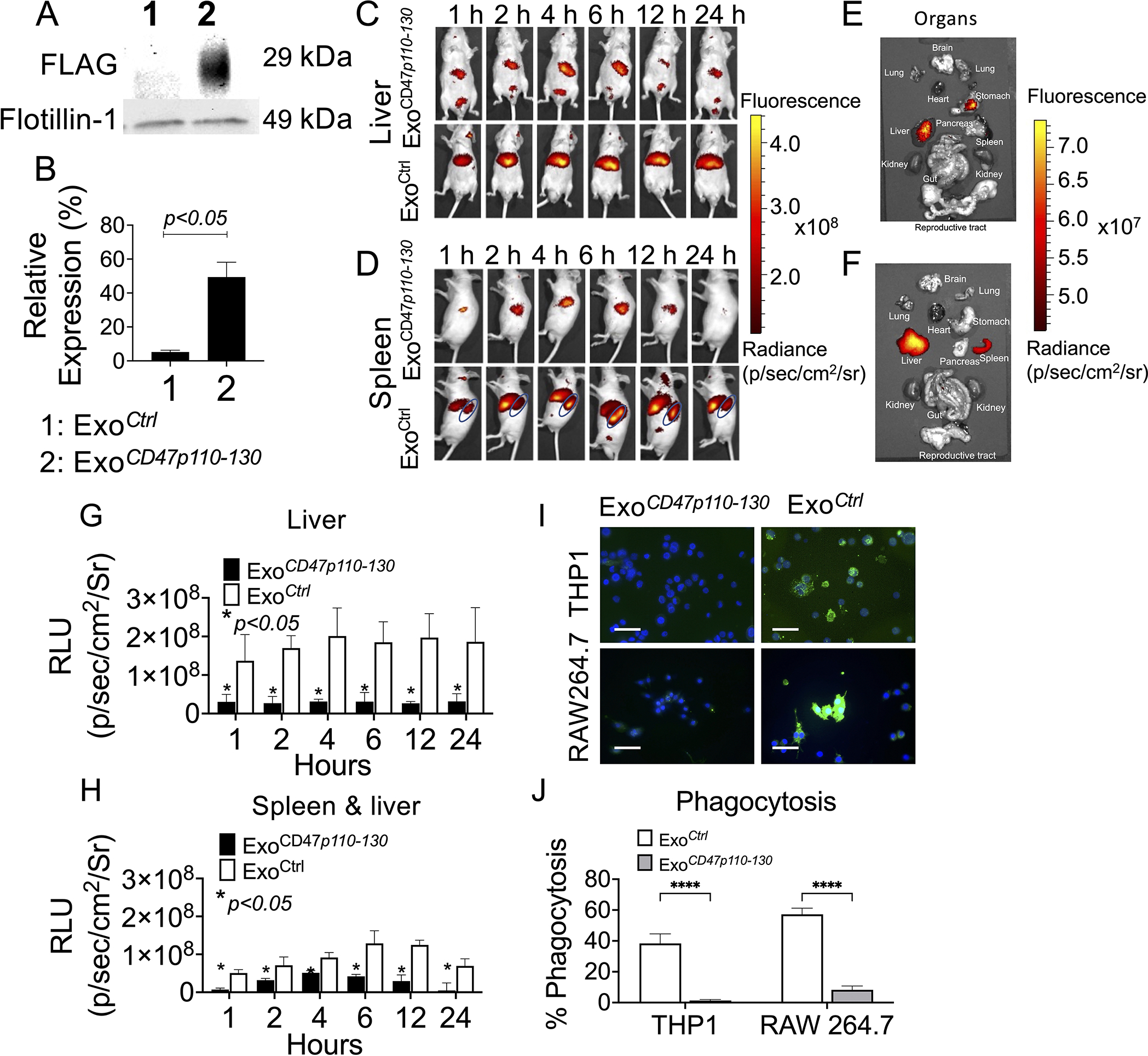
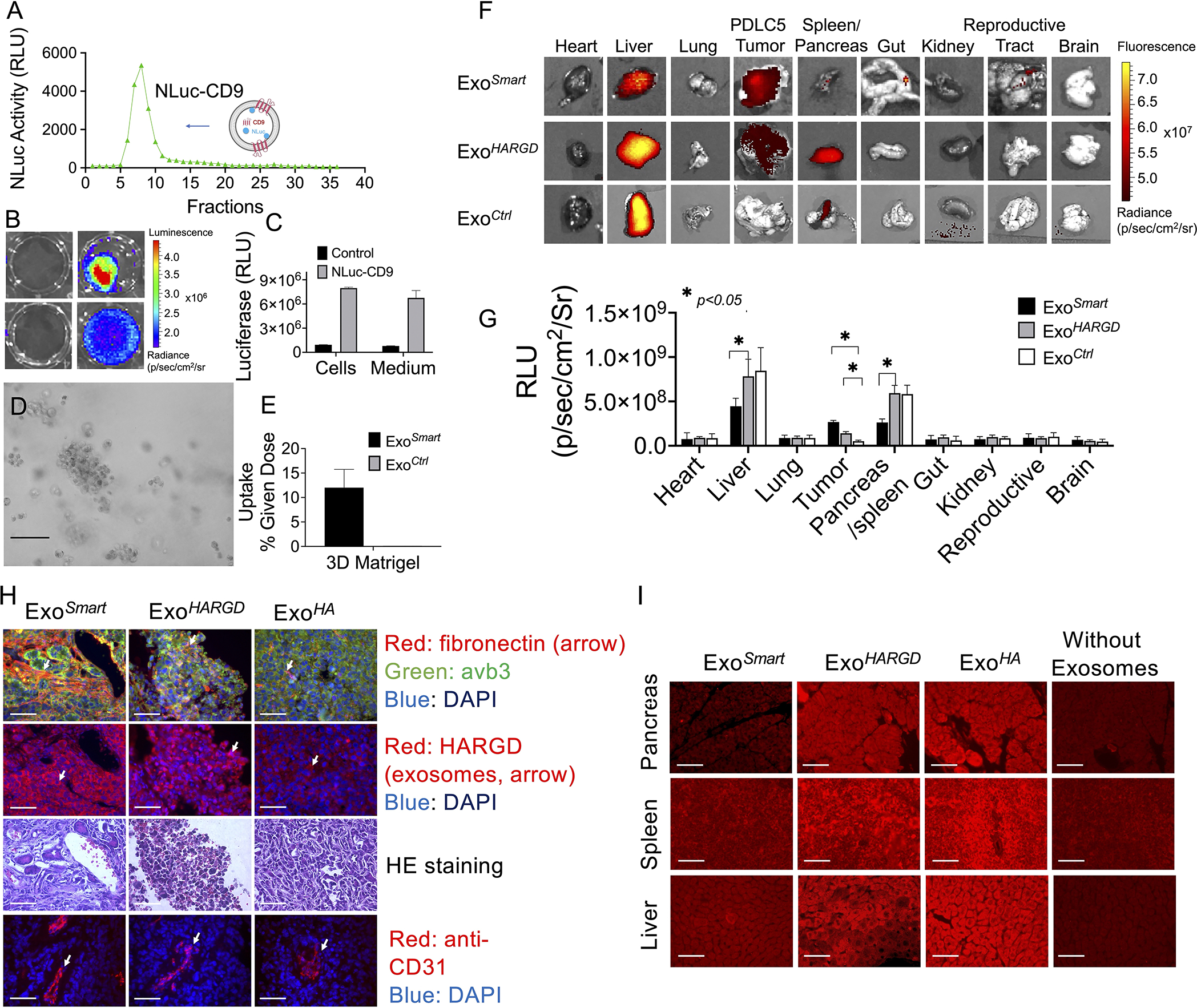
Exosomes continue to attract interest as a promising nanocarrier drug delivery technology. They are naturally derived nanoscale extracellular vesicles with innate properties well suited to shuttle proteins, lipids, and nucleic acids between cells. Nonetheless, their clinical utility is currently limited by several major challenges, such as their inability to target tumor cells and a high proportion of clearance by the mononuclear phagocyte system (MPS) of the liver and spleen. To overcome these limitations, we developed "Smart Exosomes" that co-display RGD and CD47p110-130 through CD9 engineering (ExoSmart). The resultant ExoSmart demonstrates enhanced binding capacity to αvβ3 on pancreatic ductal adenocarcinoma (PDAC) cells, resulting in amplified cellular uptake in in vitro and in vivo models and increased chemotherapeutic efficacies. Simultaneously, ExoSmart significantly reduced liver and spleen clearance of exosomes by inhibiting macrophage phagocytosis via CD47p110-130 interaction with signal regulatory proteins (SIRPα) on macrophages. These studies demonstrate that an engineered exosome drug delivery system increases PDAC therapeutic efficacy by enhancing active PDAC targeting and prolonging circulation times, and their findings hold tremendous translational potential for cancer therapy while providing a concrete foundation for future work utilizing novel peptide-engineered exosome strategies.

Keywords: Bioengineering; CD47; CD9; Drug delivery; Exosomes; Gemcitabine; Paclitaxel; Pancreatic cancer; RGD.


Conflict of interest statement

Declaration of competing interest The authors declare that there is no conflict of interest in this study.

Figures

Fig. 1.
Fig. 1.. Engineered CD9.
Insertion of RGD into CD9 LEL at E174 (A) did not affect expression of CD9 (B, red: mCherry). However, RGD insertion resulted in decreased binding of anti-CD9 antibody to overexpressed CD9 protein (C, red box) via WB. RGD insertion enhanced the relative binding activity of CD9 to αvβ3 protein (D) via ELISA assay. Scale bar = 50 μm.
Fig. 2.
Fig. 2.. Display of RGD on the Surface of Exosomes Changes Tropism Toward Cells Expressing αvβ3.
RGD peptide incorporation into CD9 resulted in overexpression of CD9-RGD on exosomes as determined by WB analysis (A). The engineered exosomes (B) did not demonstrate alterations in morphology shown by TEM (the control exosomes shown in Fig. S2A), hydrodynamic size (C), or zeta potential (D) measured by dynamic and electrophoretic light scattering. Flow cytometry confirmed the expression of HARGD on engineered exosomes (E) and control (F). RGD-exosomes demonstrated enhanced cellular uptake by PDCL5 and PDCL15 (G, H) but not PANC1 and Mia PaCa2 cells perhaps because of overexpression of αvβ3 in PDCL5 and PDLC15 cells (G, I). Control exosomes demonstrated low uptake by PDAC cells regardless of expression of αvβ3 (G, H). Scale bar in the fluorescence microscopy images = 50 μm. Scale bar in the electron microscopy images =100 nm.
Fig. 3.
Fig. 3.. Display of CD47p110–130 on Exosomes Reduced Exosome Accumulation in the Liver and Spleen Through Inhibition of Macrophage Phagocytosis.
Insertion of CD47p110–130-FLAG into CD9 at E174 resulted in overexpression of CD47p110–130-FLAG on exosomes (ExoCD47p110–130-FLAG) as determined by WB analysis of FLAG (A, B). 30μg of DiR labeled ExoCD47p110–130-FLAG and ExoCtrl were intravenously injected into NU/NU mice via tail vein followed by fluorescence imaging at designated schedule (C, D). The mice were sacrificed, and organs were imaged (E, F). Biodistribution studies demonstrated significantly reduced ExoCD47p110–130-FLAG accumulation in the liver (C, G) and spleen (D, H) in living animal imaging as well as in organ imaging (E, F). Differentiating liver and spleen signals in the living animal was difficult due to weak spleen imaging signals (D, H). DiO-labeled ExoCD47p110–130-FLAG significantly inhibited THP1 and RAW264.7 phagocytosis of exosomes (I, left column, green, J) as compared to ExoCtrl (I, right column, green, J).
Fig. 4.
Fig. 4.. Characterization of Smart Exosomes.
Exosomes were obtained from culture media of HEK 293 cells that were co-transfected with vectors CD9-HARGD and CD9-CD47p110–130-FLAG. The purified exosomes obtained through this procedure (ExoSmart) were characterized by nanoparticle tracking analysis for number-weighted diameter (A) and particle concentration (B); by DLS for intensity-weighted diameter (C) and zeta potential (D); by WB for exosome protein composition (E); by TEM for morphology of ExoSmart(F) and ExoCtrl (G); and by ELISA for binding capability of ExoSmart to HA and FLAG (H), as well as αvβ3 and SIRPa (I). Scale bar in the electron microscopy images =100 nm.
Fig. 5.
Fig. 5.. ExoSmart Enhances Cellular Uptake by PDAC cells.
Fluorescent imaging assays compare HARGD display by ExoSmart to HARGD display by LAMP2B—a commonly used exosome biomarker. Ten μg DiL-labeled ExoSmart and ExoLAMP2B-HARGD exosomes were incubated with pancreatic cancer cell lines PDCL5, PDCL15, PANC1, Mia PaCa2, Capan2, and AsPC1, as well as human pancreatic duct epithelial (HPDE) cells. ExoSmart exhibited greater uptake capability than ExoLAMP2B-HARGD in PDAC models (A, B), and both ExoSmart and ExoLAMP2B-HARGD exhibited significantly enhanced uptake by PDAC as compared to ExoCtrl (p<0.05, A, B) in PDAC models. We also measured relative expression of αvβ3 (C). The correlation between expression of β3 and uptake of ExoSmart was calculated (D). In a luminescence assay using NLuc labeled exosomes, ExoSmart resulted in 30% dose uptake by PDCL5 cells (E). A normalized specific binding curve was generated using NLuc-ExoSmart with and without RGD peptide overdose and KD values were calculated (F).
Fig. 6.
Fig. 6.. ExoSmart Targets PDCL5 Tumor Cells in 3D Model and in Orthotopic Xenograft Tumor Mouse Model.
NLuc-labeled ExoSmart and ExoCtrl (A) were used to evaluate matrix penetration (B, C), which was imaged in IVIS imager and quantificated by Live Image 4.2 software. NLuc-labeled ExoSmart and ExoCtrltargeting PDCL5 in 3D Matrigel model were imaged under phase contrast microscope (D) and measured by lysate luciferase activity (E). In vivo biodistribution signals were observed in liver, spleen, and tumor (F, G). Tumors from ExoSmart, ExoHARGD and ExoHA injected PDCL5/PSC mice were stained with anti-fibronectin, anti-αvβ3 (H, first row), anti-HA (H, second row) and HE (H, third row). The ExoSmart group demonstrated stronger HA signal (H, left column) than ExoHARGD (H, middle column) and ExoHA (H, right column) as indicated by white arrow. CD31 staining showed the vascularity in tumors (H, fourth row). ExoSmart demonstrated decreased accumulation in the liver and spleen as compared to ExoHARGD and ExoCtrl (F). Quantitative analysis showed significance (G, p<0.05). This was also confirmed by immunofluorescence analysis of tissues from the liver, spleen and pancreas using anti-HA (I, red color). Scale bars in (D, H and I) = 50 μm.
Fig. 7.
Fig. 7.. ExoSmart Demonstrated Decreased Susceptibility to Phagocytosis and Prolonged Circulation times.
Phagocytosis assays demonstrated decreased phagocytosis of ExoSmart in PMA-induced THP-1 (A, B) and RAW 264.7 cells (C, D); arrows point to phagocytosis of exosomes; scale bar = 50 μm. Pharmacokinetic studies of ExoSmart were performed in NU/NU mice with i.v.injection of 30 μg NLuc-ExoSmart or ExoCtrl followed by scheduled blood collection. Plasma exosome concentrations were determined by measuring NLuc activity (E). PK parameters (F) were calculated using PKsolver 2.0. NLuc-ExoSmart resulted in increased t1/2, AUC, and MRT, as well as decreased CL, compared to NLuc-ExoCtrl (F).
Fig. 8.
Fig. 8.. In Vitro and In Vivo Efficacy Evaluations of ExoSmart-encapsulated Chemotherapies.
When compared to ExoCtrl-PTX, ExoSmart-PTX resulted in significantly higher cytotoxicity in PDCL5 and PDCL15 (higher αvβ3 levels) (p<0.05), but not PANC1 cells (lower αvβ3 levels) (A). The ExoSmart-PTX/naked GEM combination demonstrated higher cytotoxicity than PTX+GEM without exosomes (C). Correlation between serum GLuc levels and tumor volume (D), tumor weight (E), and tumor imaging studies in living mice (F) were established using 5 μl of blood serum according to protocol timeline (G). Efficacy of ExoSmart-PTX+GEM therapy for PDAC compared to saline control, GEM+PTX, GEM+LNP-PTX, and GEM+ExoSmart-PTX was assessed via tumor weight (I). Tumor growth was assessed based on the serum GLuc levels (H). Therapeutic effect was further assessed via tumor cell apoptosis (J, K, red arrows indicate apoptosis, scale bar = 100 μm).

References

    1. Siegel RL, Miller KD, Fuchs HE, Jemal A, Cancer Statistics, 2021, CA Cancer J Clin, 71 (2021) 7–33. - PubMed
    1. Rawla P, Sunkara T, Gaduputi V, Epidemiology of Pancreatic Cancer: Global Trends, Etiology and Risk Factors, World J Oncol, 10 (2019) 10–27. - PMC - PubMed
    1. Krupa G, Jani RK, Active targeting of nanoparticles: An innovative technology for drug delivery in cancer therapeutics, Journal of Drug Delivery and Therapeutics, 9 (2019) 408–415.
    1. Li C, Wang J, Wang Y, Gao H, Wei G, Huang Y, Yu H, Gan Y, Wang Y, Mei L, Chen H, Hu H, Zhang Z, Jin Y, Recent progress in drug delivery, Acta Pharmaceutica Sinica B, (2019). - PMC - PubMed
    1. Kalaydina RV, Bajwa K, Qorri B, Decarlo A, Szewczuk MR, Recent advances in “smart” delivery systems for extended drug release in cancer therapy, Int J Nanomedicine, 13 (2018) 4727–4745. - PMC - PubMed