. 2024 Jul:222:109-122.
doi: 10.1016/j.biochi.2024.02.012. Epub 2024 Mar 1.

Biochemical and metabolic characterization of a G6PC2 inhibitor

Affiliations

Biochemical and metabolic characterization of a G6PC2 inhibitor

Emily M Hawes et al. Biochimie. 2024 Jul.

Abstract

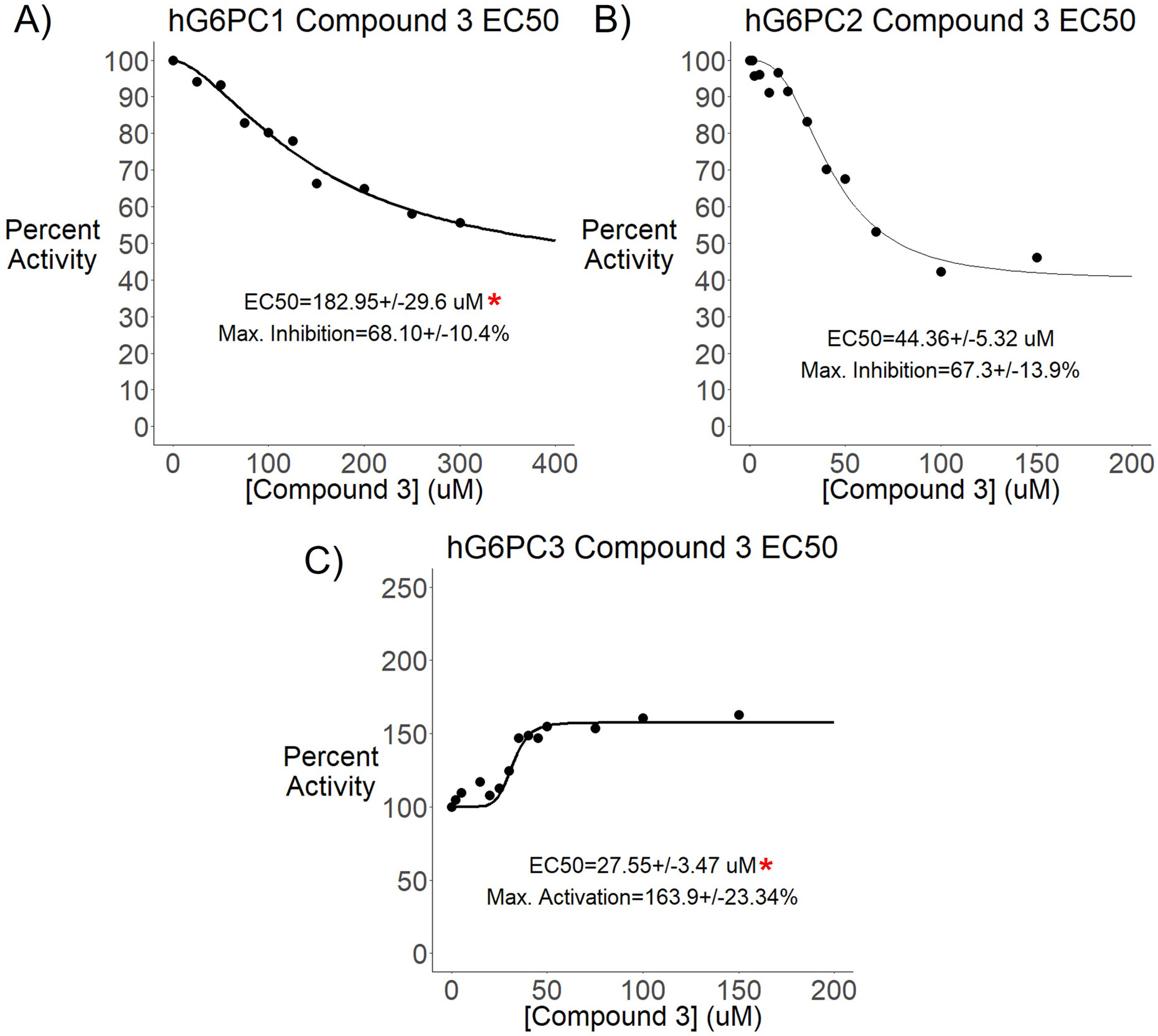
Three glucose-6-phosphatase catalytic subunits, that hydrolyze glucose-6-phosphate (G6P) to glucose and inorganic phosphate, have been identified, designated G6PC1-3, but only G6PC1 and G6PC2 have been implicated in the regulation of fasting blood glucose (FBG). Elevated FBG has been associated with multiple adverse clinical outcomes, including increased risk for type 2 diabetes and various cancers. Therefore, G6PC1 and G6PC2 inhibitors that lower FBG may be of prophylactic value for the prevention of multiple conditions. The studies described here characterize a G6PC2 inhibitor, designated VU0945627, previously identified as Compound 3. We show that VU0945627 preferentially inhibits human G6PC2 versus human G6PC1 but activates human G6PC3. VU0945627 is a mixed G6PC2 inhibitor, increasing the Km but reducing the Vmax for G6P hydrolysis. PyRx virtual docking to an AlphaFold2-derived G6PC2 structural model suggests VU0945627 binds two sites in human G6PC2. Mutation of residues in these sites reduces the inhibitory effect of VU0945627. VU0945627 does not inhibit mouse G6PC2 despite its 84% sequence identity with human G6PC2. Mutagenesis studies suggest this lack of inhibition of mouse G6PC2 is due, in part, to a change in residue 318 from histidine in human G6PC2 to proline in mouse G6PC2. Surprisingly, VU0945627 still inhibited glucose cycling in the mouse islet-derived βTC-3 cell line. Studies using intact mouse liver microsomes and PyRx docking suggest that this observation can be explained by an ability of VU0945627 to also inhibit the G6P transporter SLC37A4. These data will inform future computational modeling studies designed to identify G6PC isoform-specific inhibitors.

Keywords: Enzyme inhibition; Fasting blood glucose; Glucose-6-phosphatase; Islet.


Conflict of interest statement

Declaration of competing interest The authors declare that they have no known competing financial interests or personal relationships that could have appeared to influence the work reported in this paper.

Figures

Figure 1.
Figure 1.. Comparison of Human G6PC1, G6PC2 and G6PC3 Protein Expression Driven by the pJPA5 Vector.
832/13 cells were transiently transfected with pJPA5 expression vectors encoding either human G6PC1, G6PC2 or G6PC3 with C-terminal His-tags. Following transfection, cells were incubated for 18-20 hours in serum-containing media. Cells were subsequently harvested and protein expression assayed by western blotting as described in Experimental Procedures. Panel A: G6PC expression was assessed using an anti-His antibody and equal protein loading was confirmed using measurement of actin expression. A representative western blot is shown. Panel B: G6Pase activity was determined at pH 6.5 and 2 mM G6P in mock transfected (Control) and G6PC3 expression vector-transfected cells as described in Experimental Procedures. Reported values are the mean ± SEM (n=3). Statistical comparisons were made using a T test; *p<0.05.
Figure 2.
Figure 2.. G6P Hydrolysis by Human G6PC1, G6PC2 and G6PC3 Show Distinct Kinetics.
832/13 cells were transiently transfected with pJPA5 expression vectors encoding either human G6PC1, G6PC2 or G6PC3 with C-terminal His-tags. Following transfection, cells were incubated for 18-20 hours in serum-containing media. Cells were subsequently harvested, microsomal membranes were isolated and G6Pase activity determined at the pH shown as described in Experimental Procedures. Representative datasets for G6PC1 (Panel A), G6PC2 (Panel B) and G6PC3 (Panel C) are shown with the graph line indicating the average kinetic curve as calculated using R. Reported Km and Vmax values are the mean mM ± SEM or mean nmol/min/mg ± SEM, respectively (n=4-8). Data are graphed as relative activity with each dataset normalized to the activity at the maximum G6P concentration tested (80 mM for G6PC1 and G6PC3; 2 mM G6P for G6PC2). Statistical comparisons of Km and Vmax at pH 6.5 versus pH 8.0 were made using T tests; *p<0.05.
Figure 3.
Figure 3.. VU0945627 Inhibits Human G6PC1 and G6PC2 but Activates G6PC3.
832/13 cells were transiently transfected with pJPA5 expression vectors encoding either human G6PC1, human G6PC2 or human G6PC3 with C-terminal His-tags. Following transfection, cells were incubated for 18-20 hours in serum-containing media. Cells were subsequently harvested, microsomal membranes were isolated and G6Pase activity determined, as described in Experimental Procedures, at either pH 6.5 (for G6PC1 and G6PC3) or pH 8.0 (for G6PC2) in the presence or absence of the indicated concentrations of G6P and VU0945627. Representative datasets for G6PC1 (Panel A), G6PC2 (Panels B & C) and G6PC3 (Panel D) are shown with the graph line in Panels A, B & D indicating the average kinetic curve as calculated using R. Reported Km and Vmax values are the mean mM ± SEM or mean nmol/min/mg ± SEM, respectively (n=3-6). Data are graphed as relative activity with each dataset normalized to the activity at the maximum G6P concentration tested in the absence of VU0945627 (80 mM for G6PC1 and G6PC3; 2 mM G6P for G6PC2). For G6PC1 (Panel A) and G6PC3 (Panel D) statistical comparisons of Km and Vmax at pH 6.5, in the presence versus absence of VU0945627 were made using T tests; *p<0.05. For G6PC2 (Panel B) statistical comparisons of Km and Vmax at pH 8.0, in the presence versus absence of various concentrations of VU0945627 were made using one-way ANOVA with a Dunnett’s post-hoc test; *p<0.05.
Figure 4.
Figure 4.. VU0945627 exhibits a lower EC50 for Inhibition of Human G6PC2 than G6PC1.
832/13 cells were transiently transfected with pJPA5 expression vectors encoding either human G6PC1, human G6PC2 or human G6PC3 with C-terminal His-tags. Following transfection, cells were incubated for 18-20 hours in serum-containing media. Cells were subsequently harvested, microsomal membranes were isolated and G6Pase activity determined, as described in Experimental Procedures, at either pH 6.5 (for G6PC1 and G6PC3) or pH 8.0 (for G6PC2) in the presence or absence of the indicated concentrations of VU0945627 and either 10 mM G6P (for G6PC1), 2 mM G6P (for G6PC2) or 80 mM (for G6PC3) G6P. Representative datasets for G6PC1 (Panel A), G6PC2 (Panel B) and G6PC3 (Panel C) are shown with the graph line indicating the average EC50 curve as calculated using R. Reported EC50 values (mM) and maximal inhibition (%) values are means ± SEM (n=4-6). Data are graphed as percent activity with each dataset normalized to the activity detected in the absence of VU0945627. Statistical comparisons of EC50 values relative to G6PC2 were made using T tests; *p<0.05.
Figure 5.
Figure 5.. PyRx Software Predicts Two VU0945627 Binding Sites in G6PC2
PyRx (48) computational docking was performed for VU0945627 binding to the AlphaFold2-predicted structure for human G6PC2 (Uniprot Q9NQR9). VU0945627 was prepared for docking by energy minimization with 200 steps using a universal force field (UFF). The energy minimized VU0945627 was globally docked to human G6PC2 with AutoDock Vina (77) with 10 iterations of the 9 lowest energy conformations, resulting in 90 final ligand-protein conformations for each protein model were visualized in PyMOL (Panel A). Panel B and Panel C show an individual VU0945627 conformation that was selected to highlight G6PC2 residues that frequently interact with VU0945627 at both Site 1 and Site 2, respectively.
Figure 6.
Figure 6.. Multiple Amino Acids Influence the Inhibition of G6PC2 by VU0945627
832/13 cells were transiently transfected with pJPA5 expression vectors encoding either wild type (WT) or mutated human G6PC2 with a C-terminal His-tag. Following transfection, cells were incubated for 18-20 hours in serum-containing media. Cells were subsequently harvested, and G6PC2 expression in whole cell lysates was assessed by western blotting using an anti-6xHis antibody, with equal protein loading confirmed by measurement of actin expression, whereas G6Pase activity in the presence of 2 mM G6P and presence or absence of VU0945627 (150 mM) was measured at pH 8.0 in microsomal membranes, as described in Experimental Procedures. Panel A: Normalized expression was calculated as the ratio of 6x-His to actin expression obtained with the mutants shown and expressed relative to the ratio obtained with WT human G6PC2. Reported values are the means ± SEM (n=3-7). Panel B: G6Pase activity was determined as described in Experimental Procedures and expressed relative to WT G6PC2. Reported values are the mean ± SEM (n=3). Panel C: The effect of VU0945627 is graphed as percent inhibition, calculated as relative activity in the presence of inhibitor divided by the relative activity in the absence of inhibitor. Reported values are the mean ± SEM (n=3). Mutations outside Sites 1 and 2 are shown in gray; mutations in Site 1 are shown in red; mutations in Site 2 are shown in blue; combined mutations in Sites 1 are 2 are shown in purple. Statistical comparisons against WT G6PC2 were made using T tests; *p<0.05
Figure 7.
Figure 7.. VU0945627 Inhibits Human but not Mouse G6PC2
832/13 cells were transiently transfected with pJPA5 expression vectors encoding either wild type (WT) or mutated human or mouse G6PC1 or G6PC2 with C-terminal His-tags. Following transfection, cells were incubated for 18-20 hours in serum-containing media. Cells were subsequently harvested, and protein expression in whole cell lysates was assessed by western blotting using an anti-6xHis antibody, with equal protein loading confirmed by measurement of actin expression, whereas G6Pase activity in the presence of 2 mM G6P and presence or absence of VU0945627 (150 mM) was measured at either pH 6.5 (for G6PC1) or pH 8.0 (for G6PC2) in microsomal membranes, as described in Experimental Procedures. Panel A: The effect of VU0945627 is graphed as percent inhibition, calculated as relative activity in the presence of inhibitor divided by the relative activity in the absence of inhibitor. Reported values are the mean ± SEM (n=3-4). Statistical comparisons were made using one-way ANOVA with a Tukey’s post-hoc test; *p<0.05 as compared to all other samples. Panel B: Normalized expression was calculated as the ratio of 6x-His to actin expression obtained with the mutants shown and expressed relative to the ratio obtained with WT human G6PC2. Reported values are the means ± SEM (n=3-5). Panel C: G6Pase activity was determined as described in Experimental Procedures and expressed relative to WT G6PC2. Reported values are the mean ± SEM (n=3-5). Panels D & E: The effect of VU0945627 is graphed as percent inhibition, calculated as relative activity in the presence of inhibitor divided by the relative activity in the absence of inhibitor. Reported values are the mean ± SEM (n=3-5). For Panels B-E, statistical comparisons against WT G6PC2 were made using T tests; *p<0.05
Figure 8.
Figure 8.. VU0945627 Blocks G6P Hydrolysis in Intact Microsomes
Intact (Panel A) and disrupted (Panel B) microsomes were prepared from mouse liver and the hydrolysis of G6P (2 mM) and M6P (2 mM) at pH 6.5 in the absence or presence of the indicated concentration of VU0945627 was assessed as described in Experimental Procedures and expressed relative to the rate of G6P hydrolysis in the absence of VU0945627. Reported values are the mean ± SEM (n=3-4). Panel C: 832/13 cells were transiently transfected with pJPA5 expression vectors encoding wild type mouse G6PC1 with a C-terminal His-tag. Following transfection, cells were incubated for 18-20 hours in serum-containing media. Cells were subsequently harvested and G6Pase activity in the presence of 2 mM G6P at pH 6.5 and in the absence or presence of the indicated concentration of VU0945627 was assessed in microsomal membranes as described in Experimental Procedures. Reported values are the mean ± SEM (n=3). Statistical comparisons versus G6P hydrolysis in the absence of VU0945627 were made using one-way ANOVA with a Dunnett’s post-hoc test; *p<0.05.
Figure 9.
Figure 9.. VU0945627 Blocks Glucose Cycling in βTC-3 Cells
Comparison of glucose cycling rates (Panel A) and cell viability (Panel B) at 11 mM glucose in βTC-3 cells in the presence and absence of the indicated concentrations of VU0945627. Data represent means ± SEM (n=3-4).*p < 0.05 vs no VU0945627.
Figure 10.
Figure 10.. PyRx Software Predicts Two VU0945627 Binding Sites in SLC37A4
PyRx (48) computational docking was performed for VU0945627 binding to the AlphaFold2-predicted structural model for mouse SLC37A4 (Uniprot Q9D1F9). VU0945627 was prepared for docking by energy minimization with 200 steps using a universal force field (UFF). The energy minimized VU0945627 was globally docked to mouse SLC37A4 with AutoDock Vina (77) with 10 iterations of the 9 lowest energy conformations, resulting in 90 final ligand-protein conformations for each protein model. The putative interaction sites of the lowest energy confirmation for each run were visualized in PyMOL with lateral (Panel A) and vertical (Panel B) views shown.
Figure 11.
Figure 11.. VU0945627 Inhibits Purified G6PC2.
The human G6PC2 L219 or V219 variants with a C-terminal His-tag were expressed using baculovirus in HEK293S cells and purified as described in Experimental Procedures. G6Pase activity was then determined in the presence of 2 mM G6P at pH 6.5 in the presence or absence of VU0945627 (150 μM) and CHS (0.02 mM), as indicated. Reported values are the mean G6Pase activity (nmol/min/mg) (Panel A) or percent inhibition by VU0945627 (Panel B) ± SEM (n=3). Panel A: statistical comparisons versus the L219 or V219 G6PC2 variants in the absence of VU0945627 were made using T tests; *p<0.05. Panel B: no statistical differences were found using one-way ANOVA with a Tukey’s post-hoc test.

Similar articles

Cited by

References

    1. Hutton JC, and O'Brien RM (2009) The glucose-6-phosphatase catalytic subunit gene family. J Biol Chem 284, 29241–29245 - PMC - PubMed
    1. Van Schaftingen E, and Gerin I (2002) The glucose-6-phosphatase system. Biochem J 362, 513–532 - PMC - PubMed
    1. Arden SD, Zahn T, Steegers S, Webb S, Bergman B, O'Brien RM, and Hutton JC (1999) Molecular cloning of a pancreatic islet-specific glucose-6-phosphatase catalytic subunit-related protein. Diabetes 48, 531–542 - PubMed
    1. Martin CC, Bischof LJ, Bergman B, Hornbuckle LA, Hilliker C, Frigeri C, Wahl D, Svitek CA, Wong R, Goldman JK, Oeser JK, Lepretre F, Froguel P, O'Brien RM, and Hutton JC (2001) Cloning and Characterization of the Human and Rat Islet-Specific Glucose-6-Phosphatase Catalytic Subunit-Related Protein (IGRP) Genes. J Biol Chem 276, 25197–25207 - PubMed
    1. Xin Y, Kim J, Okamoto H, Ni M, Wei Y, Adler C, Murphy AJ, Yancopoulos GD, Lin C, and Gromada J (2016) RNA Sequencing of Single Human Islet Cells Reveals Type 2 Diabetes Genes. Cell Metab 24, 608–615 - PubMed

LinkOut - more resources