. 2024 Mar 2;22(1):32.
doi: 10.1186/s12969-024-00965-5.

Th17/1 and ex-Th17 cells are detected in patients with polyarticular juvenile arthritis and increase following treatment

Affiliations

Th17/1 and ex-Th17 cells are detected in patients with polyarticular juvenile arthritis and increase following treatment

Stephanie Wood et al. Pediatr Rheumatol Online J. .

Abstract

Background: A better understanding of the pathogenesis of polyarticular juvenile idiopathic arthritis (polyJIA) is needed to aide in the development of data-driven approaches to guide selection between therapeutic options. One inflammatory pathway of interest is JAK-STAT signaling. STAT3 is a transcription factor critical to the differentiation of inflammatory T helper 17 cells (Th17s). Previous studies have demonstrated increased STAT3 activation in adult patients with rheumatoid arthritis, but less is known about STAT3 activation in polyJIA. We hypothesized that Th17 cells and STAT3 activation would be increased in treatment-naïve polyJIA patients compared to pediatric controls.

Methods: Blood from 17 patients with polyJIA was collected at initial diagnosis and again if remission was achieved (post-treatment). Pediatric healthy controls were also collected. Peripheral blood mononuclear cells were isolated and CD4 + T cell subsets and STAT activation (phosphorylation) were evaluated using flow cytometry. Data were analyzed using Mann-Whitney U and Wilcoxon matched-pairs signed rank tests.

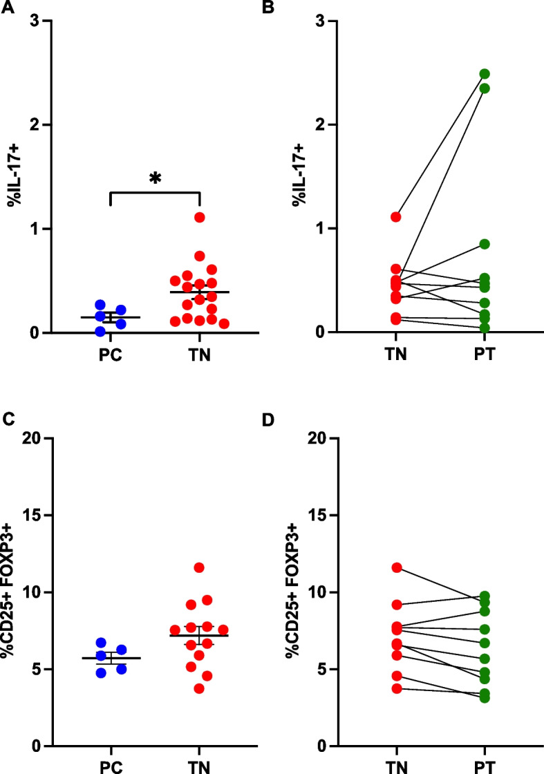
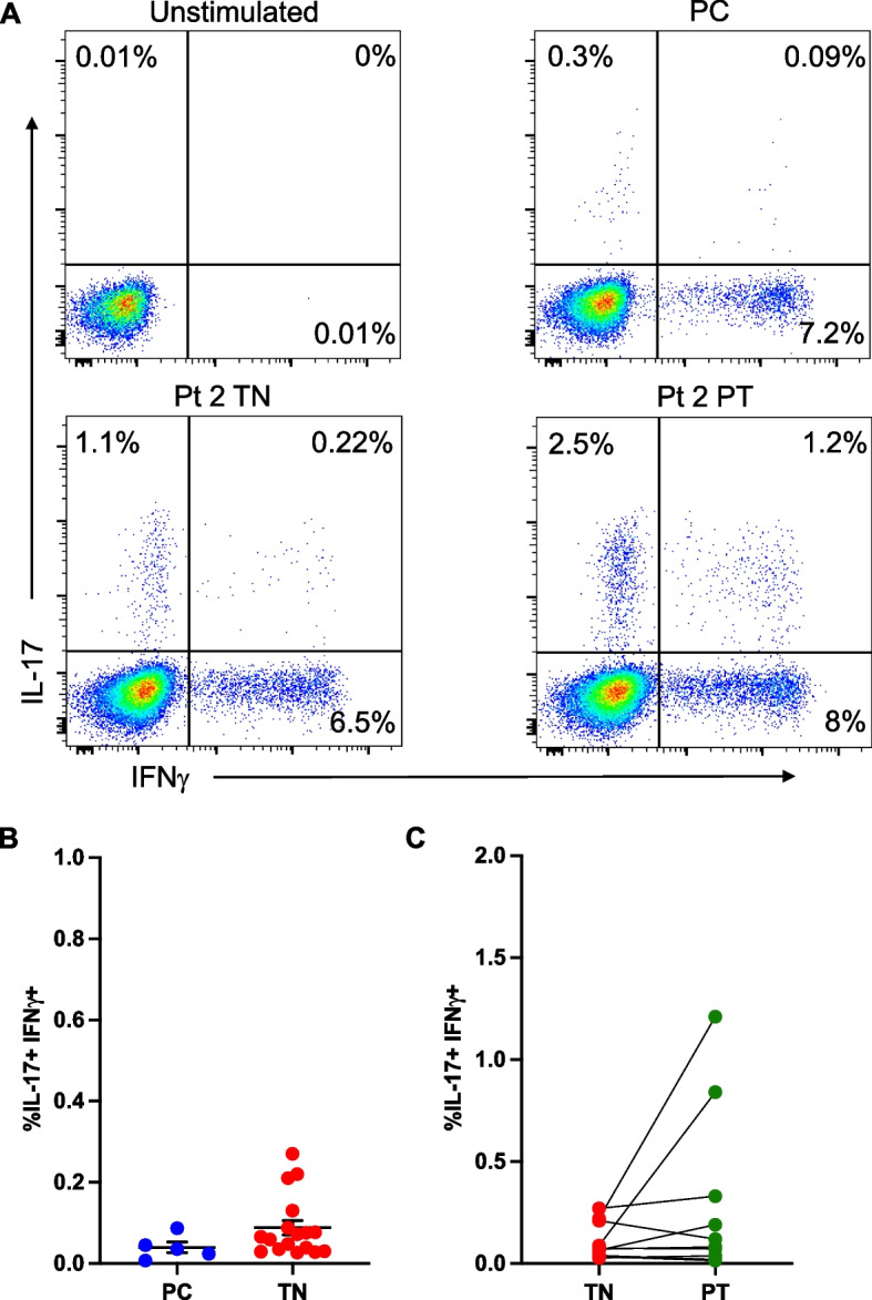
Results: Treatment-naïve polyJIA patients had increased Th17 cells (CD3 + CD4 + interleukin(IL)-17 +) compared to controls (0.15% v 0.44%, p < 0.05), but Tregs (CD3 + CD4 + CD25 + FOXP3 +) from patients did not differ from controls. Changes in STAT3 phosphorylation in CD4 + T cells following ex vivo stimulation were not significantly different in patients compared to controls. We identified dual IL-17 + and interferon (IFN)γ + expressing CD4 + T cells in patients, but not controls. Further, both Th17/1 s (CCR6 + CD161 + IFNγ + IL-17 +) and ex-Th17s (CCR6 + CD161 + IFNγ + IL-17neg) were increased in patients' post-treatment (Th17/1: 0.3% v 0.07%, p < 0.05 and ex-Th17s: 2.3% v 1.4%, p < 0.05). The patients with the highest IL-17 expressing cells post-treatment remained therapy-bound.

Conclusions: Patients with polyJIA have increased baseline Th17 cells, potentially reflecting higher tonic STAT3 activation in vivo. These quantifiable immune markers may identify patients that would benefit upfront from pathway-focused biologic therapies. Our data also suggest that inflammatory CD4 + T cell subsets not detected in controls but increased in post-treatment samples should be further evaluated as a tool to stratify patients in remission on medication. Future work will explore these proposed diagnostic and prognostic biomarkers.

Keywords: Ex-Th17; IL-6; Polyarticular juvenile idiopathic arthritis; STAT3; Th17; Th17/1; Treg.


Conflict of interest statement

TPV has consulted for Novartis, Pfizer, Moderna and SOBI and receives research support from AstraZeneca. All other authors report no disclosures.

Figures

Fig. 1
Fig. 1
Treatment-naïve polyJIA patients have altered CD4 + T cell subsets. A PBMCs from healthy pediatric controls (PC) and treatment-naïve polyJIA patients (TN) were analyzed for IL-17 producing CD3 + CD4 + (Th17) T cells after overnight stimulation with PMA + calcimycin. Treatment-naïve patients had more Th17 cells (PC 0.15 ± 0.05% v TN 0.4 ± 0.07%, *p < 0.05, Mann–Whitney U test). B Samples from polyJIA patients that had achieved remission on medication post-treatment (PT) were also analyzed for Th17 cells and compared to the treatment-naïve frequencies for that same patient without significant differences, but with two notable outliers (Wilcoxon matched-pairs signed rank test). C T regulatory cells (Tregs = CD3 + CD4 + CD25 + FOXP3 +) identified ex vivo from treatment-naïve patients were not different from healthy pediatric controls (PC 6.3 ± 0.8% v TN 7.1 ± 0.7%, Mann–Whitney U test). D Paired analysis of patients’ treatment-naïve and post-treatment samples did not reveal differences in Treg frequencies after treatment (Wilcoxon matched-pairs signed rank test). Black bars represent mean ± SEM. PBMCs, peripheral blood mononuclear cells; IL, interleukin; PMA, phorbol 12-myristate 13-acetate
Fig. 2
Fig. 2
STAT3 activation after ex vivo stimulation of CD4 + T cells from polyJIA patients. A PBMCs were left unstimulated (gray) or stimulated ex vivo for 30 min with IL-6 (purple), then activated/phosphorylated (p)STAT3 measured using flow cytometry. Histograms depict representative results from one experiment showing pSTAT3 in CD3 + CD4 + T cells from a healthy pediatric control (PC) and both treatment-naïve (TN) and post-treatment (PT) samples from 3 polyJIA patients (Pt 1–3). Black numbers represent the MFI of pSTAT3 in unstimulated cells and the purple numbers the MFI after IL-6 stimulation. B Samples were treated as in (A). Data represent the %pSTAT3 + cells in a stimulated sample distinct from when unstimulated, generated using Overton subtraction. Cells from treatment-naïve patients demonstrate less ex vivo response to cytokine stimuli than those from healthy pediatric controls, although not achieving significance (Mann–Whitney U test). Black bars represent mean ± SEM. IL-6: PC 85 ± 1.9% v TN 76 ± 2.4%, IFNα: PC 77 ± 1.5% v TN 69 ± 3.9%, and IL-27: PC 66 ± 2.1% v TN 64 ± 3.8%. PBMCs, peripheral blood mononuclear cells; IL, interleukin; MFI, mean florescence intensity; IFN, interferon
Fig. 3
Fig. 3
Dual-cytokine expressing CD4 + T cells are detected in a subset of polyJIA patients. A PBMCs were left unstimulated or stimulated overnight with PMA + calcimycin and CD3 + CD4 + T cells analyzed for cytokine production. Representative examples from an unstimulated sample and stimulated samples from a healthy pediatric control (PC) and a polyJIA patient (Pt 2), both treatment-naïve (TN) and post-treatment (PT), are shown. Percentages of CD3 + CD4 + T cells producing IL-17 or IFNγ or both are represented in the quadrants. B Samples were treated as in (A). Dual IL-17 and IFNγ expressing CD4 + T cells can be detected in a subset of treatment-naïve polyJIA patients, but not healthy pediatric controls (PC 0.04 ± 0.01% v TN 0.09 ± 0.02%, Mann–Whitney U test). C Paired analysis of patients’ treatment-naïve and post-treatment samples revealed that some of the outlying IL-17 expressing CD4 + T cells in post-treatment samples also co-express IFNγ (Wilcoxon matched-pairs signed rank test). Black bars represent mean ± SEM. PBMCs, peripheral blood mononuclear cells, PMA, phorbol 12-myristate 13-acetate; IL, interleukin; IFN, interferon
Fig. 4
Fig. 4
Evidence of altered CD4 + T cell plasticity in post-treatment samples from patients with polyJIA. A Naïve CD4 + T cells differentiate into numerous effector subsets and can become Th17 cells implicated in autoimmunity. One model of CD4 + T cell plasticity proposes transdifferentiation of homeostatic Th17 cells into pathogenic IFNγ-expressing Th17/1 cells or ex-Th17 cells. PBMCs were left unstimulated or stimulated overnight with PMA + calcimycin and CD3 + CD4 + T cells analyzed for cytokine production. Cells were additionally surface stained with CCR6 and CD161 to identify transitioned cells. B Treatment-naïve (TN) polyJIA patients had detectable Th17/1 cells, but Th17/1 cells were significantly higher in post-treatment (PT) samples (TN 0.07 ± 0.02% v PT 0.3 ± 0.1%, *p < 0.05, Wilcoxon matched-pairs signed rank test). C There were more previously IL-17 expressing ex-Th17 cells in samples from post-treatment polyJIA patients than in the same patients when treatment-naïve (TN 1.4 ± 0.4% v PT 2.3 ± 0.5%, *p < 0.05, Wilcoxon matched-pairs signed rank test). Black bars represent mean ± SEM. PBMCs, peripheral blood mononuclear cells, PMA, phorbol 12-myristate 13-acetate; IFN, interferon, IL, interleukin

References

    1. Prakken B, Albani S, Martini A. Juvenile idiopathic arthritis. The Lancet. 2011;377(9783):2138–2149. - PubMed
    1. Ravelli A, Consolaro A, Horneff G, Laxer RM, Lovell DJ, Wulffraat NM, et al. Treating juvenile idiopathic arthritis to target: recommendations of an international task force. Ann Rheum Dis. 2018;77:819. - PubMed
    1. Mannion ML, Cron RQ. Therapeutic strategies for treating juvenile idiopathic arthritis. Curr Opin Pharmacol. 2022;64:102226. - PubMed
    1. Ringold S, Angeles Han ST, Beukelman T, Lovell D, Cuello CA, Becker ML, et al. 2019 American College of Rheumatology/Arthritis Foundation guideline for the treatment of juvenile idiopathic arthritis: therapeutic approaches for non systemic Polyarthritis, Sacroiliitis, and Enthesitis. Arthritis Care Res. 2019;71(6):717–734. - PMC - PubMed
    1. Brunner HI, Ruperto N, Zuber Z, Keane C, Harari O, Kenwright A, et al. Efficacy and safety of tocilizumab in patients with polyarticular-course juvenile idiopathic arthritis: results from a phase 3, randomised, double-blind withdrawal trial. Ann Rheum Dis. 2015;74(6):1110–1117. - PMC - PubMed