Trim9 regulates the directional differentiation of retinal Müller cells to retinal ganglion cells
- PMID: 38432885
- PMCID: PMC10929896
- DOI: 10.11817/j.issn.1672-7347.2023.230108
Trim9 regulates the directional differentiation of retinal Müller cells to retinal ganglion cells
Abstract
Objectives: Glaucoma is a leading cause of irreversible blindness, and effective therapies to reverse the visual system damage caused by glaucoma are still lacking. Recently, the stem cell therapy enable the repair and regeneration of the damaged retinal neurons, but challenges regarding the source of stem cells remain. This study aims to investigate a protocol that allows the dedifferentiation of Müller cells into retinal stem cells, following by directed differentiation into retinal ganglion cells with high efficiency, and to provide a new method of cellular acquisition for retinal stem cells.
Methods: Epidermal cell growth factor and fibroblast growth factor 2 were used to induce the dedifferentiation of rat retinal Müller cells into retinal neural stem cells. Retinal stem cells derived from Müller cells were infected with a Trim9 overexpression lentiviral vector (PGC-FU-Trim9-GFP), and the efficiency of viral infection was assessed by fluorescence microscopy and flow cytometry. Retinoic acid and brain-derived neurotrophic factor treatments were used to induce the differentiation of the retinal stem cells into neurons and glial cells with or without the overexpression of Trim9. The expressions of each cellular marker (GLAST, GS, rhodopsin, PKC, HPC-1, Calbindin, Thy1.1, Brn-3b, Nestin, Pax6) were detected by immunofluorescence, PCR/real-time RT-PCR or Western blotting.
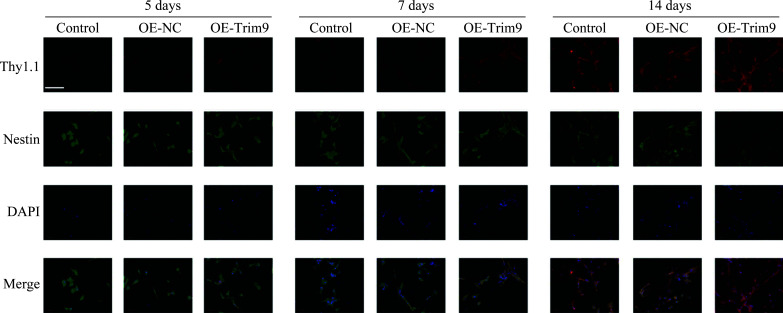
Results: Rat retinal Müller cells expressed neural stem cells markers (Nestin and Pax6) with the treatment of epidermal cell growth factor and fibroblast growth factor 2. The Thy1.1 positive cell rate of retinal stem cells overexpressing Trim9 was significantly increased, indicating their directional differentiation into retinal ganglion cells after treatment with retinoic acid and brain-derived neurotrophic factor.
Conclusions: In this study, rat retinal Müller cells are dedifferentiated into retinal stem cells successfully, and Trim9 promotes the directional differentiation from retinal stem cells to retinal ganglion cells effectively.
目的: 青光眼是不可逆性致盲性眼病的主要病因,目前尚无有效疗法逆转青光眼的视觉系统损害。最近发现干细胞疗法有望使受损的视网膜神经元修复和再生,但是在干细胞来源方面仍然存在较大挑战。本研究探寻一种将视网膜Müller细胞去分化为视网膜干细胞,进一步高效定向分化为视网膜神经节细胞的方案,以期为青光眼的干细胞治疗提供新的细胞获取途径。方法: 用表皮细胞生长因子和成纤维细胞生长因子2诱导大鼠视网膜Müller细胞去分化为视网膜干细胞。构建Trim9过表达慢病毒(PGC-FU-Trim9-GFP),感染由Müller细胞诱导去分化而来的视网膜干细胞,通过荧光显微镜观察和流式细胞术评估病毒感染效率。用视黄酸和脑源性神经营养因子处理过表达或未过表达Trim9的视网膜干细胞以诱导其分化为神经元和神经胶质细胞。采用免疫荧光、PCR/real-time RT-PCR和蛋白质印迹法检测各细胞标志物(GLAST、GS、rhodopsin、PKC、HPC-1、Calbindin、Thy1.1、Brn-3b、Nestin、Pax6)的表达。结果: 表皮细胞生长因子和成纤维细胞生长因子2处理后的大鼠视网膜Müller细胞表达视网膜干细胞标志物Nestin和Pax6。视黄酸和脑源性神经营养因子处理后的过表达Trim9的视网膜干细胞Thy1.1阳性细胞明显增多,表明其定向分化为视网膜神经节细胞。结论: 本研究成功将大鼠视网膜Müller细胞去分化为视网膜干细胞,并发现Trim9可有效促进由视网膜Müller细胞去分化而来的视网膜干细胞定向分化为视网膜神经节细胞。.
Keywords: Müller cells; Trim9; glaucoma; retinal ganglion cells; retinal stem cells.
Conflict of interest statement
作者声称无任何利益冲突。
Figures


Similar articles
-
TRIM9 promotes Müller cell-derived retinal stem cells to differentiate into retinal ganglion cells by regulating Atoh7.In Vitro Cell Dev Biol Anim. 2023 Sep;59(8):586-595. doi: 10.1007/s11626-023-00807-w. Epub 2023 Oct 4. In Vitro Cell Dev Biol Anim. 2023. PMID: 37792226
-
Promotion on the differentiation of retinal Müller cells into retinal ganglion cells by Brn-3b.Int J Ophthalmol. 2016 Jul 18;9(7):948-54. doi: 10.18240/ijo.2016.07.03. eCollection 2016. Int J Ophthalmol. 2016. PMID: 27500099 Free PMC article.
-
Atoh7 promotes the differentiation of Müller cells-derived retinal stem cells into retinal ganglion cells in a rat model of glaucoma.Exp Biol Med (Maywood). 2015 May;240(5):682-90. doi: 10.1177/1535370214560965. Epub 2015 Feb 20. Exp Biol Med (Maywood). 2015. PMID: 25710928 Free PMC article.
-
Müller glia: Stem cells for generation and regeneration of retinal neurons in teleost fish.Prog Retin Eye Res. 2014 May;40:94-123. doi: 10.1016/j.preteyeres.2013.12.007. Epub 2014 Jan 8. Prog Retin Eye Res. 2014. PMID: 24412518 Free PMC article. Review.
-
A Promising Tool in Retina Regeneration: Current Perspectives and Challenges When Using Mesenchymal Progenitor Stem Cells in Veterinary and Human Ophthalmological Applications.Stem Cell Rev Rep. 2017 Oct;13(5):598-602. doi: 10.1007/s12015-017-9750-4. Stem Cell Rev Rep. 2017. PMID: 28643176 Free PMC article. Review.
References
-
- 张青, 曹凯, 康梦田, 等. 青光眼临床诊疗若干问题问卷调查(2016年)[J]. 中华眼科杂志, 2017, 53(2): 115-120. 10.3760/cma.j.issn.0412-4081.2017.02.009. - DOI
- ZHANG Qing, CAO Kai, KANG Mengtian, et al. . The questionnaire survey on glaucoma diagnosis and treatment in China (2016)[J]. Chinese Journal of Ophthalmology, 2017, 53(2): 115-120. 10.3760/cma.j.issn.0412-4081.2017.02.009. - DOI - PubMed
-
- 韩冰, 吴志鸿. 青光眼数据库在临床的应用及现状[J]. 中国实用眼科杂志, 2016, 34(11): 1137-1139. 10.3760/cma.j.issn.1006-4443.2016.11.004. - DOI - PubMed
- HAN Bing, WU Zhihong. Clinical application and present situation of glaucoma database[J]. Chinese Journal of Practical Ophthalmology, 2016, 34(11): 1137-1139. 10.3760/cma.j.issn.1006-4443.2016.11.004. - DOI
MeSH terms
Substances
Grants and funding
LinkOut - more resources
Full Text Sources
Medical
Molecular Biology Databases
Miscellaneous