. 2024 Mar;30(3):e14654.
doi: 10.1111/cns.14654.

SPAK inhibitor ZT-1a attenuates reactive astrogliosis and oligodendrocyte degeneration in a mouse model of vascular dementia

Affiliations

SPAK inhibitor ZT-1a attenuates reactive astrogliosis and oligodendrocyte degeneration in a mouse model of vascular dementia

Mohammad Iqbal H Bhuiyan et al. CNS Neurosci Ther. 2024 Mar.

Abstract

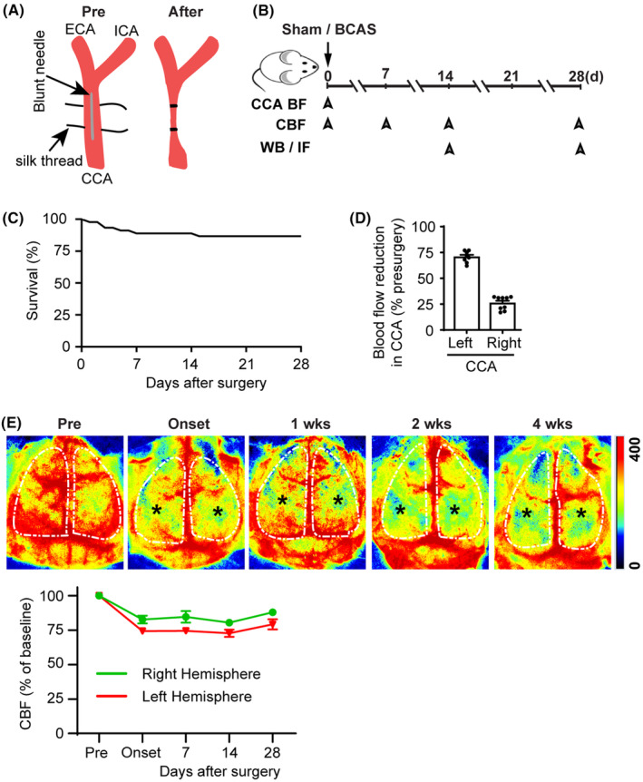
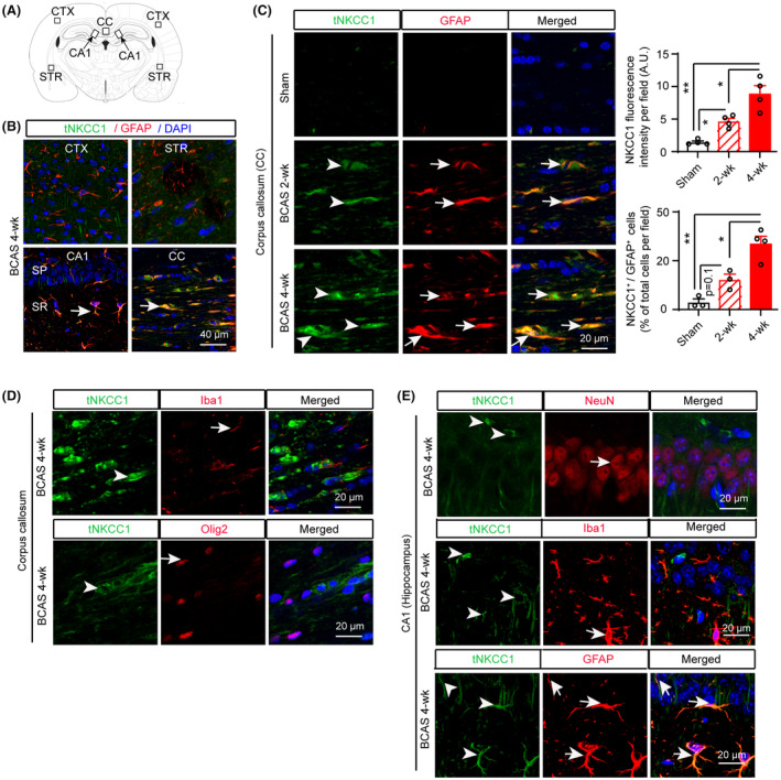
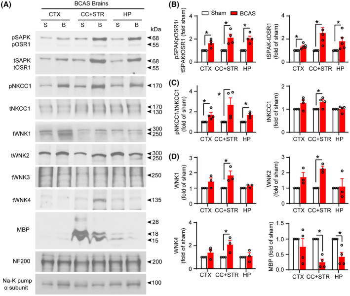
Background: Astrogliosis and white matter lesions (WML) are key characteristics of vascular contributions to cognitive impairment and dementia (VCID). However, the molecular mechanisms underlying VCID remain poorly understood. Stimulation of Na-K-Cl cotransport 1 (NKCC1) and its upstream kinases WNK (with no lysine) and SPAK (the STE20/SPS1-related proline/alanine-rich kinase) play a role in astrocytic intracellular Na+ overload, hypertrophy, and swelling. Therefore, in this study, we assessed the effect of SPAK inhibitor ZT-1a on pathogenesis and cognitive function in a mouse model of VCID induced by bilateral carotid artery stenosis (BCAS).

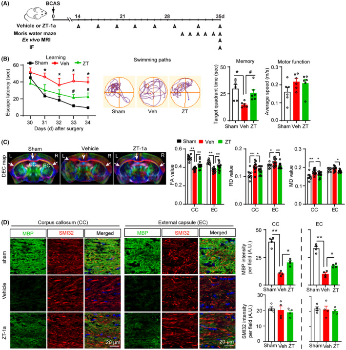
Methods: Following sham or BCAS surgery, mice were randomly assigned to receive either vehicle (DMSO) or SPAK inhibitor ZT-1a treatment regimen (days 14-35 post-surgery). Mice were then evaluated for cognitive functions by Morris water maze, WML by ex vivo MRI-DTI analysis, and astrogliosis/demyelination by immunofluorescence and immunoblotting.

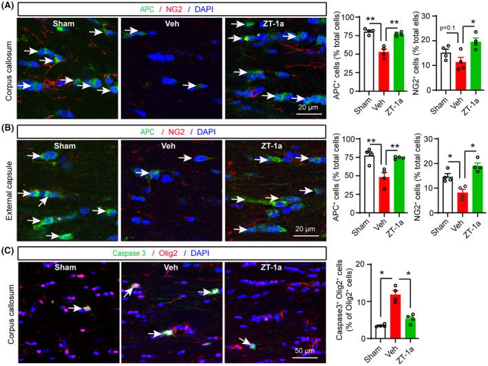
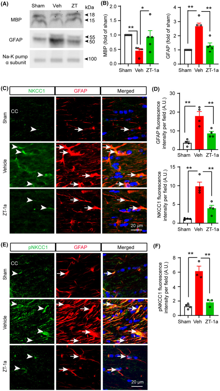
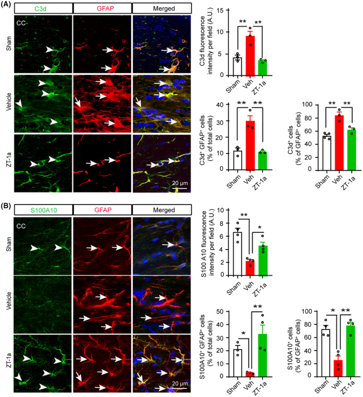
Results: Compared to sham control mice, BCAS-Veh mice exhibited chronic cerebral hypoperfusion and memory impairments, accompanied by significant MRI DTI-detected WML and oligodendrocyte (OL) death. Increased activation of WNK-SPAK-NKCC1-signaling proteins was detected in white matter tissues and in C3d+ GFAP+ cytotoxic astrocytes but not in S100A10+ GFAP+ homeostatic astrocytes in BCAS-Veh mice. In contrast, ZT-1a-treated BCAS mice displayed reduced expression and phosphorylation of NKCC1, decreased astrogliosis, OL death, and WML, along with improved memory functions.

Conclusion: BCAS-induced upregulation of WNK-SPAK-NKCC1 signaling contributes to white matter-reactive astrogliosis, OL death, and memory impairment. Pharmacological inhibition of the SPAK activity has therapeutic potential for alleviating pathogenesis and memory impairment in VCID.

Keywords: BCAS; NKCC1; VCID; ZT-1a; astrogliosis; vascular dementia.


Conflict of interest statement

The authors declare that they have no competing interests.

Figures

FIGURE 1
FIGURE 1
Chronic cerebral hypoperfusion and cognitive impairment in mice following bilateral carotid artery stenosis (BCAS). (A) Illustration of common carotid artery (CCA) stenosis. (B) Experimental protocol and outcome measurements. CCA—common carotid artery; BF—blood flow; CBF—cerebral blood flow; WB—Western blotting; IF—immunofluorescence. (C) Percent survival over 4 weeks after BCAS surgery. (D) Percent blood flow reduction in CCAs after BCAS compared to pre‐surgery baseline. Data are mean ± SEM; n = 9. (E) Representative images of cerebral blood flow (CBF) measured by two‐dimensional laser speckle imaging during 0–4 weeks post‐surgery. Black stars indicate brain regions with hypoperfusion. Quantification of CBF (lower panel), white dotted area from upper panel of E used for quantification. Data are mean ± SEM; n = 2.
FIGURE 2
FIGURE 2
Total NKCC1 protein expression in mouse brains after BCAS. (A) Brain section illustrates sample collection in the cortex (CTX), corpus callosum (CC), hippocampal CA1 subfield (stratum pyramidale [SP] and stratum radiatum [SR]), and striatum (STR) areas. (B) Compared to CTX, STR, and CA1 regions, a robust increase in tNKCC1 protein expression in GFAP+ astrocytes (arrow) was detected in the CC at 4 weeks after BCAS surgery. (C) Time‐dependent tNKCC1 protein expression (arrowhead) in GFAP+ astrocytes (arrow) at 2 weeks and 4 weeks after BCAS. Data are mean ± SEM; one‐way ANOVA, Tukey's post hoc test; n = 4, *p < 0.05, **p < 0.01. (D) In the CC, the increase in tNKCC1 protein expression (arrowhead) does not colocalize with Iba+ microglia/macrophages (arrow) or Olig2+ oligodendrocytes at 4 weeks after BCAS. (E) In the hippocampal CA1, an increase in tNKCC1 protein expression (arrowhead) does not colocalize with NeuN+ neurons (arrow) or Iba1+ microglia/ macrophages (arrow) but colocalizes with GFAP+ astrocytes (arrow) at 4 weeks after BCAS.
FIGURE 3
FIGURE 3
Stimulation of WNK‐SPAK‐NKCC1 complex in BCAS mouse brains. (A) Representative immunoblots of increased WNK‐SPAK/OSR1‐NKCC1 complex proteins and decreased myelin basic proteins (MBP) in BCAS‐induced hypoperfused mouse brains (membrane protein fractions) at 4 weeks after surgery. Na‐K pump (α subunit) was used as a loading control. (B–D) Quantitative analyses. Data are mean ± SEM; Student's t test; n = 3–4, *p < 0.05. B, BCAS; BCAS, bilateral carotid artery stenosis; CC, corpus callosum; CTX, cortex; HP, hippocampus; S, sham; STR, striatum.
FIGURE 4
FIGURE 4
SPAK inhibitor ZT‐1a prevents memory impairments and protects white matter microstructure integrity and MBP loss after BCAS. (A) Post‐BCAS administration of vehicle (Veh) or SPAK inhibitor ZT‐1a (5 mg/kg, i.p., during 14–35 days after BCAS, every 3 days), Morris water maze test, ex vivo MRI, and immunofluorescence (IF). (B) Compared to Veh‐treated BCAS mice, ZT‐a treatment reduces the escape latency times and swimming path, and increases target quadrant time. Data are mean ± SEM; two‐way repeated‐measures ANOVA, Tukey's post hoc test; n = 6, *p < 0.05 versus sham, # p < 0.05 versus Veh. (C) Representative images of DEC maps of ex vivo brain from sham, vehicle (Veh)‐treated, and ZT‐1a (ZT)‐treated mice at 5 weeks post‐BCAS detected by MRI‐DTI. Bar graphs show quantitative analyses of fractional anisotropy (FA), radial diffusivity (RD), and mean diffusivity (MD) of CC and EC. Data are mean ± SEM; one‐way ANOVA, Tukey's post hoc test; n = 7 (data points represent values from both hemispheres), *p < 0.05, **p < 0.01. (D) Changes in myelin basic protein (MBP) and nonphosphorylated neurofilament heavy chain (SMI32) in CC and EC of brains in B–C. Quantitative analyses of MBP and SMI32 immunofluorescence. Data are mean ± SEM; one‐way ANOVA, Tukey's post hoc test; n = 3–4, *p < 0.05, **p < 0.01.
FIGURE 5
FIGURE 5
SPAK inhibitor ZT‐1a prevents BCAS‐induced loss of both OPCs and OL in CC and EC. (A–C) Double‐immunofluorescence analysis of APC/NG2 in CC and EC, and caspase 3+/Olig2+ cells in CC of sham, Veh‐, and ZT‐1a‐treated brains at 5 weeks after BCAS surgery. The same cohort as Figure 4. Nuclei were counterstained with DAPI. Data are mean ± SEM; one‐way ANOVA, Tukey's post hoc test; n = 4, *p < 0.05, **p < 0.01.
FIGURE 6
FIGURE 6
SPAK inhibitor ZT‐1a inhibits expressions of tNKCC1, pNKCC1, and GFAP, and prevents MBP loss in the corpus callosum (CC) after BCAS. (A, B) ZT‐1a treatment prevents BCAS‐induced increased expression of GFAP and loss of MBP in mouse brain 5 weeks after BCAS. Data are mean ± SEM; one‐way ANOVA, Tukey's post hoc test; n = 3–4; *p < 0.05, **p < 0.01. (C–F) ZT‐1a‐treated mice exhibit decreased expressions of NKCC1 and GFAP proteins in GFAP+ astrocytes in CC compared to Veh‐treated mice at 5 weeks after BCAS. Data are mean ± SEM; one‐way ANOVA, Tukey's post hoc test; n = 3–4; **p < 0.01.
FIGURE 7
FIGURE 7
SPAK‐NKCC1 complex inhibition attenuates increment of cytotoxic astrocytes and increases homeostatic astrocytes in the BCAS mouse brains. (A) ZT‐1a‐treated mice exhibit decreased expressions of C3d protein in GFAP+ astrocytes in CC compared to Veh‐treated mice at 5 weeks after BCAS. Bar graphs show quantitative analyses of C3d fluorescence intensity, C3d+GFAP+ cells (% of total cells), and C3d+ cells (% of GFAP+ cells). Data are mean ± SEM; one‐way ANOVA, Tukey's post hoc test; n = 3–4; *p < 0.05, **p < 0.01. (B) ZT‐1a treatment increases S100A10+ homeostatic astrocytes in CC compared to vehicle‐treated mice at 5 weeks after BCAS. Bar graphs represent quantitative analyses of S100A10 fluorescence intensity, S100A10+GFAP+ cells (% of total cells), and S100A10+ cells (% of GFAP+ cells). Data are mean ± SEM; one‐way ANOVA, Tukey's post hoc test; n = 3–4; *p < 0.05, **p < 0.01.

References

    1. Snyder HM, Corriveau RA, Craft S, et al. Vascular contributions to cognitive impairment and dementia including Alzheimer's disease. Alzheimers Dement. 2015;11:710‐717. doi:10.1016/j.jalz.2014.10.008 - DOI - PMC - PubMed
    1. Corriveau RA, Bosetti F, Emr M, et al. The science of vascular contributions to cognitive impairment and dementia (VCID): a framework for advancing research priorities in the cerebrovascular biology of cognitive decline. Cell Mol Neurobiol. 2016;36:281‐288. doi:10.1007/s10571-016-0334-7 - DOI - PMC - PubMed
    1. Iadecola C. The pathobiology of vascular dementia. Neuron. 2013;80:844‐866. doi:10.1016/j.neuron.2013.10.008 - DOI - PMC - PubMed
    1. van der Flier WM, Skoog I, Schneider JA, et al. Vascular cognitive impairment. Nat Rev Dis Primers. 2018;4:18003. doi:10.1038/nrdp.2018.3 - DOI - PubMed
    1. Price BR, Norris CM, Sompol P, Wilcock DM. An emerging role of astrocytes in vascular contributions to cognitive impairment and dementia. J Neurochem. 2018;144:644‐650. doi:10.1111/jnc.14273 - DOI - PMC - PubMed

Publication types

LinkOut - more resources