

Common and distinct cortical thickness alterations in youth with autism spectrum disorder and attention-deficit/hyperactivity disorder
- PMID: 38433204
- PMCID: PMC10910790
- DOI: 10.1186/s12916-024-03313-2
Common and distinct cortical thickness alterations in youth with autism spectrum disorder and attention-deficit/hyperactivity disorder
Abstract
Background: Autism spectrum disorder (ASD) and attention-deficit/hyperactivity disorder (ADHD) are neurodevelopmental disorders with overlapping behavioral features and genetic etiology. While brain cortical thickness (CTh) alterations have been reported in ASD and ADHD separately, the degree to which ASD and ADHD are associated with common and distinct patterns of CTh changes is unclear.
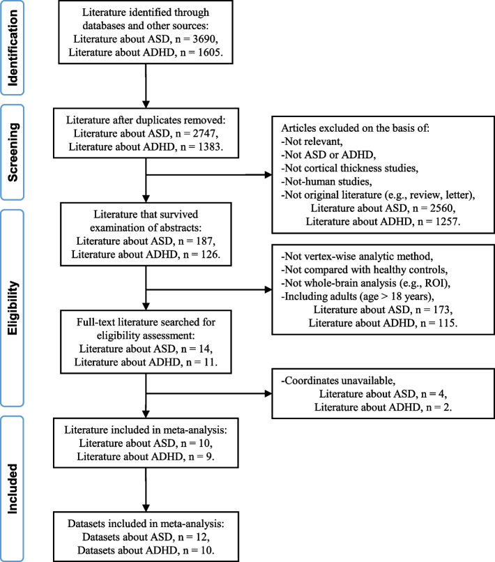
Methods: We searched PubMed, Web of Science, Embase, and Science Direct from inception to 8 December 2023 and included studies of cortical thickness comparing youth (age less than 18) with ASD or ADHD with typically developing controls (TDC). We conducted a comparative meta-analysis of vertex-based studies to identify common and distinct CTh alterations in ASD and ADHD.
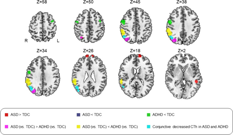
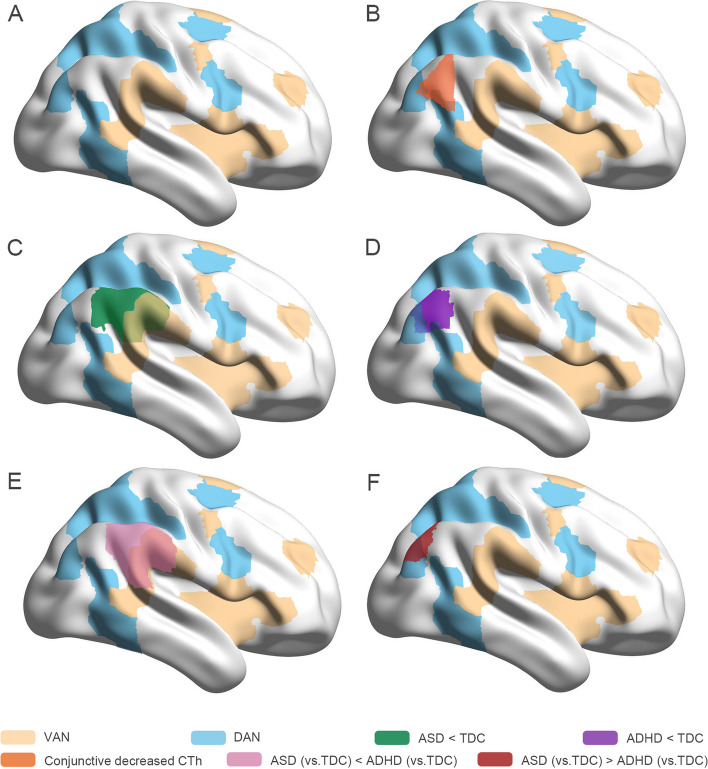
Results: Twelve ASD datasets involving 458 individuals with ASD and 10 ADHD datasets involving 383 individuals with ADHD were included in the analysis. Compared to TDC, ASD showed increased CTh in bilateral superior frontal gyrus, left middle temporal gyrus, and right superior parietal lobule (SPL) and decreased CTh in right temporoparietal junction (TPJ). ADHD showed decreased CTh in bilateral precentral gyri, right postcentral gyrus, and right TPJ relative to TDC. Conjunction analysis showed both disorders shared reduced TPJ CTh located in default mode network (DMN). Comparative analyses indicated ASD had greater CTh in right SPL and TPJ located in dorsal attention network and thinner CTh in right TPJ located in ventral attention network than ADHD.
Conclusions: These results suggest shared thinner TPJ located in DMN is an overlapping neurobiological feature of ASD and ADHD. This alteration together with SPL alterations might be related to altered biological motion processing in ASD, while abnormalities in sensorimotor systems may contribute to behavioral control problems in ADHD. The disorder-specific thinner TPJ located in disparate attention networks provides novel insight into distinct symptoms of attentional deficits associated with the two neurodevelopmental disorders.
Trial registration: PROSPERO CRD42022370620. Registered on November 9, 2022.
Keywords: Attention-deficit/hyperactivity disorder; Autism spectrum disorder; Cortical thickness; Magnetic resonance imaging; Meta-analysis; Surface-based morphometry.
© 2024. The Author(s).
Conflict of interest statement
MPD receives research support from national institutes of health (NIH), PCORI, Acadia, Allergan, Janssen, Johnson and Johnson, Lundbeck, Otsuka, Pfizer, and Sunovion. She is also a consultant, on the advisory board, or has received honoraria for speaking for Alkermes, Allergan, Assurex, CMEology, Janssen, Johnson and Johnson, Lundbeck, Myriad, Neuronetics, Otsuka, Pfizer, Sunovion, and Supernus. RKM has received research support from Martek Biosciences Inc, Royal DSM Nutritional Products, LLC, Inflammation Research Foundation, Ortho-McNeil Janssen, AstraZeneca, Eli Lilly, NARSAD, and NIH, and previously served on the scientific advisory board of the Inflammation Research Foundation. Other authors declare no potential conflicts of interest with regard to this manuscript.
Figures
References
-
- Association AP . Diagnostic and statistical manual of mental disorders: DSM-5. 5. Washington, D.C.: American Psychiatric Association; 2013.
-
- Krakowski AD, Cost KT, Anagnostou E, Lai MC, Crosbie J, Schachar R, et al. Inattention and hyperactive/impulsive component scores do not differentiate between autism spectrum disorder and attention-deficit/hyperactivity disorder in a clinical sample. Mol Autism. 2020;11(1):28. doi: 10.1186/s13229-020-00338-1. - DOI - PMC - PubMed
-
- Luo L, You W, DelBello MP, Gong Q, Li F. Recent advances in psychoradiology. Phys Med Biol. 2022;67(23):23TR01. - PubMed
Publication types
MeSH terms
Grants and funding
LinkOut - more resources
Full Text Sources
Medical
