

doi: 10.1002/ctm2.1603.
Increased oxidized low-density lipoprotein in mice exposed to a high-fat diet impaired spermatogenesis by inhibiting testosterone synthesis via the Klk1bs/Eid3 pathway
Affiliations
- PMID: 38433441
- PMCID: PMC10909978
- DOI: 10.1002/ctm2.1603
Item in Clipboard
Increased oxidized low-density lipoprotein in mice exposed to a high-fat diet impaired spermatogenesis by inhibiting testosterone synthesis via the Klk1bs/Eid3 pathway
Clin Transl Med.
2024 Mar.
No abstract available
Conflict of interest statement
The authors declare no conflict of interest.
Figures
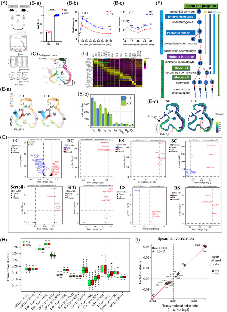
Single‐cell RNA (scRNA) sequencing of testis cells in the ND and HFD groups. (A) Outline of the workflow. (B‐a) Weight of mice in the HFD and ND groups. (B‐b) Blood glucose during the GTT (n = 5 per group). (B‐c) Blood glucose during the ITT (n = 5 per group). (C) UMAPs of types of testis cells. (D) The top 10 marker genes per cell cluster in the scaled expression matrix. The column and row labels represent the cell clusters identified according to the average log‐transform fold change value, spermatogonia (SPG), early spermatocytes (ESC), late spermatocytes (LSC‐1, LSC‐2, LSC‐3, LSC‐4 and LSC‐5), round spermatocytes (RS‐1 and RS‐2), elongated spermatids (ES‐1, ES‐2 and ES‐3), condensed spermatozoa (CS), Leydig cells (LC), Sertoli cells and dendritic (DC) cells. (E‐a) UMAPs of types of testis cells in the ND and HFD groups. (E‐b) Numbers of cell types in the ND and HFD groups. (E‐c) Distribution of cell density for each type of cell. (F) Effects of different ways of blocking the testosterone signaling pathway on spermatogenesis. (G) Volcanic maps of the differential genes in each cell type. (H) Boxplot delineating transcriptional noise in the obese and control groups according to cell type with the number of cells indicated. (I) Scatterplot depicting the log2 ratio of transcriptional noise calculated for the obese and control samples. GTT, glucose tolerance test; HFD, high‐fat diet; ITT, insulin tolerance test; ND, normal diet; UMAP, Uniform Manifold Approximation and Projection.
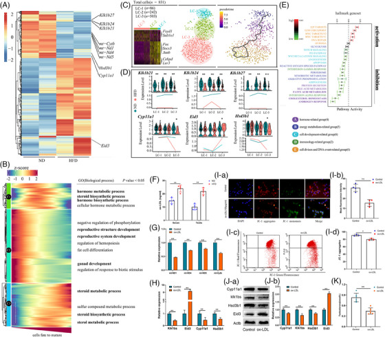
Disruption of Leydig cell function by oxidized low‐density lipoprotein. (A) Heat maps of differential genes in Leydig cells in the HFD and ND groups. (B) The pseudo‐time heatmap classified into three clusters by the hierarchical method. GO analysis was performed for each cluster. (C) Heatmap displaying significant marker genes in each cluster. UMAP of Leydig cells classified into three clusters (i.e., LC‐1, Cebpd+ cluster; LC‐2, Fos+ cluster; LC‐3 and Sult1e1+ cluster). Development of Leydig cells predicted by RNA velocity. (D) Violin plots of Klk1b21, Klk1b24, Klk1b27, Cyp11a1, Eid3 and Hsd3b1 in the HFD and ND groups. (E) Boxplot profiling pathway activity in Leydig cells in the obese and control groups. (F) Serum and testicular ox‐LDL levels were measured by enzyme‐linked immunosorbent assay (n = 4 per group). (G) mRNA expression of mt‐ND1, mt‐ND4, mt‐ND5 and mt‐Cytb was examined by RT‐qPCR (n = 3 per group). (H) mRNA expression of Klk1bs, Eid3, Cyp11a1 and Hsd3b1 was examined by RT‐qPCR (n = 3 per group). (I‐a, I‐b) MMP in Leydig cells was assessed using JC‐1 staining (n = 4 per group). (I‐c, I‐d) MMP was assayed by flow cytometry (n = 3 per group). (J‐a, J‐b) Protein expression of Klk1bs, Eid3, Cyp11a1 and Hsd3b1 was detected by Western blotting (n = 3 per group). (K) Testosterone levels in cell culture medium were measured by enzyme‐linked immunosorbent assay (n = 6 per group). GO, Gene Ontology; HFD, high‐fat diet; ND, normal diet; UMAP, Uniform Manifold Approximation and Projection; HFD, high‐fat diet; MMP, mitochondrial membrane potential; ND, normal diet; ox‐LDL, oxidized low‐density lipoprotein; RT‐qPCR, real‐time quantitative polymerase chain reaction.
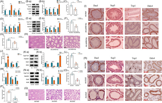
Klk1b21/24//27 and Eid3 are important genes regulating testosterone synthesis in Leydig cells. (A) mRNA expression of Klk1bs, Eid3, Cyp11a1 and Hsd3b1 was examined by RT‐qPCR (n = 3 per group). (B) Protein expression of Klk1bs, Eid3, Cyp11a1 and Hsd3b1 was detected by Western blotting (n = 3 per group). (C) Testosterone levels in cell culture medium were measured by enzyme‐linked immunosorbent assay (n = 6 per group). (D) mRNA expression of Klk1bs, Eid3, Cyp11a1 and Hsd3b1 was examined by RT‐qPCR (n = 3 per group). (E) Protein expression of Klk1bs, Eid3, Cyp11a1 and Hsd3b1 was detected by Western blotting (n = 3 per group). (F) Serum testosterone levels were detected by enzyme‐linked immunosorbent assay (n = 4 per group). (G) Sperm from each cauda epididymis in Ad.Null and Ad.shKlk1bs mice were counted in a hemocytometer under a light microscope (n = 8 for each group). (H) Hematoxylin and eosin staining of histological sections of testes in Ad.Null and Ad.shKlk1bs mice (n = 3 per group; scale bar, 200 μm). (I) Immunohistochemical staining of histological sections of testes in Ad.Null and Ad.shKlk1bs mice (n = 3 per group), Dazl, spermatogonium; Scp3, spermatocyte; Tnp1, sperm cell; Ddx4, germ cell. (J) mRNA expression of Eid3, Cyp11a1 and Hsd3b1 was examined by RT‐qPCR (n = 3 per group). (K) Protein expression of Eid3, Cyp11a1 and Hsd3b1 was detected by Western blotting (n = 3 per group). (L) Testosterone levels in cell culture medium were measured by enzyme‐linked immunosorbent assay (n = 6 per group). (M) mRNA expression of Eid3, Cyp11a1 and Hsd3b1 was examined by RT‐qPCR (n = 3 per group). (N) Protein expression of Eid3, Cyp11a1 and Hsd3b1 was detected by Western blotting (n = 3 per group). (O) Serum testosterone levels were detected by enzyme‐linked immunosorbent assay (n = 4 per group). (P) Sperm from each cauda epididymis in Ad.Null and Ad.Eid3 mice were counted in a hemocytometer under a light microscope (n = 8 for each group). (Q) Hematoxylin and eosin staining of histological sections of testes in Ad.Null and Ad.Eid3 mice (n = 3 per group; scale bar, 200 μm). (R) Immunohistochemical staining of histological sections of testes in Ad.Null and Ad.Eid3 mice (n = 3 per group), Dazl, spermatogonium; Scp3, spermatocyte; Tnp1, sperm cell; Ddx4, germ cell.
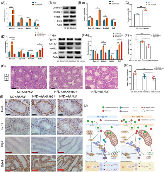
Supplementation with Klk1b21 can reverse reproductive damage caused by HFD. (A) mRNA expression of Klk1b21, Eid3, Cyp11a1 and Hsd3b1 was examined by RT‐qPCR (n = 3 per group). (B) Protein expression of Klk1b21, Eid3, Cyp11a1 and Hsd3b1 was detected by Western blotting (n = 3 per group). (C) Testosterone levels in cell culture medium were measured by enzyme‐linked immunosorbent assay (n = 4 per group). (D) mRNA expression of Klk1b21, Eid3, Cyp11a1 and Hsd3b1 was examined by RT‐qPCR (n = 3 per group). (E) Protein expression of Klk1bs, Eid3, Cyp11a1 and Hsd3b1 was detected by Western blotting (n = 3 per group). (F) Serum testosterone levels were detected by enzyme‐linked immunosorbent assay (n = 4 per group). (G) Hematoxylin and eosin staining of histological sections of testes in ND + Ad.Null, HFD + Ad.Klk1b21 and HFD + Ad.Null mice (n = 3 per group; scale bar, 200 μm). (H) Sperm from each cauda epididymis in ND + Ad.Null, HFD + Ad.Klk1b21 and HFD + Ad.Null mice were counted in a hemocytometer under a light microscope (n = 8 in each group). (I) Immunohistochemical staining of histological sections of testes in ND + Ad.Null, HFD + Ad.Klk1b21 and HFD + Ad.Null mice (n = 3 per group). Dazl, spermatogonium; Scp3, spermatocyte; Tnp1, sperm cell; Ddx4, germ cell. (J) A high‐fat diet increased the ox‐LDL level in Leydig cells, which resulted in a decreased ability to synthesize testosterone in Leydig cells, and the decreased testosterone concentration in the testis caused spermatogenic arrest.
References
-
- Matsui H, Takahashi T. Mouse testicular Leydig cells express Klk21, a tissue kallikrein that cleaves fibronectin and IGF‐binding protein‐3. Endocrinology. 2001;142:4918‐4929. - PubMed
-
- Matsui H, Moriyama A, Takahashi T. Cloning and characterization of mouse klk27, a novel tissue kallikrein expressed in testicular Leydig cells and exhibiting chymotrypsin‐like specificity. Eur J Biochem. 2000;267:6858‐6865. - PubMed
Publication types
MeSH terms
Substances
Grants and funding
- 32172726/National Natural Science Foundation of China
- 32272872/National Natural Science Foundation of China
- 20210202103NC/Key Research and Development Program of Jilin Province
- 20210202048NC/Key Research and Development Program of Jilin Province
- 20190301008NY/Key Research and Development Program of Jilin Province
LinkOut - more resources
Full Text Sources