

Tetrahedral framework nucleic acids-based delivery of MicroRNA-22 inhibits pathological neovascularization and vaso-obliteration by regulating the Wnt pathway
- PMID: 38433462
- PMCID: PMC11216936
- DOI: 10.1111/cpr.13623
Tetrahedral framework nucleic acids-based delivery of MicroRNA-22 inhibits pathological neovascularization and vaso-obliteration by regulating the Wnt pathway
Abstract
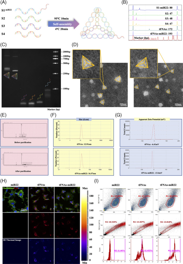
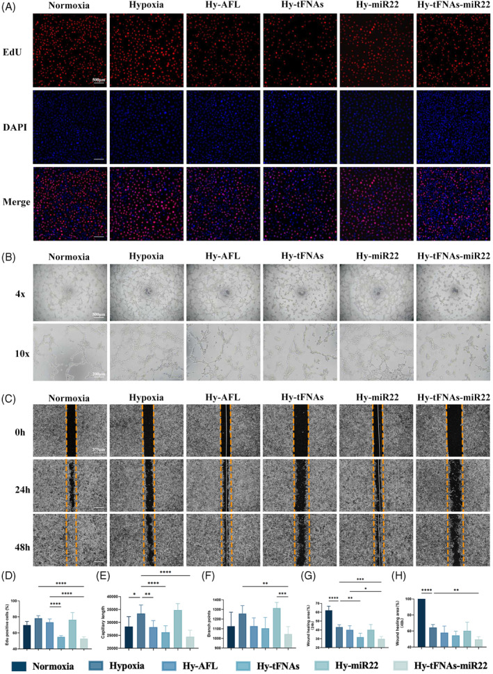
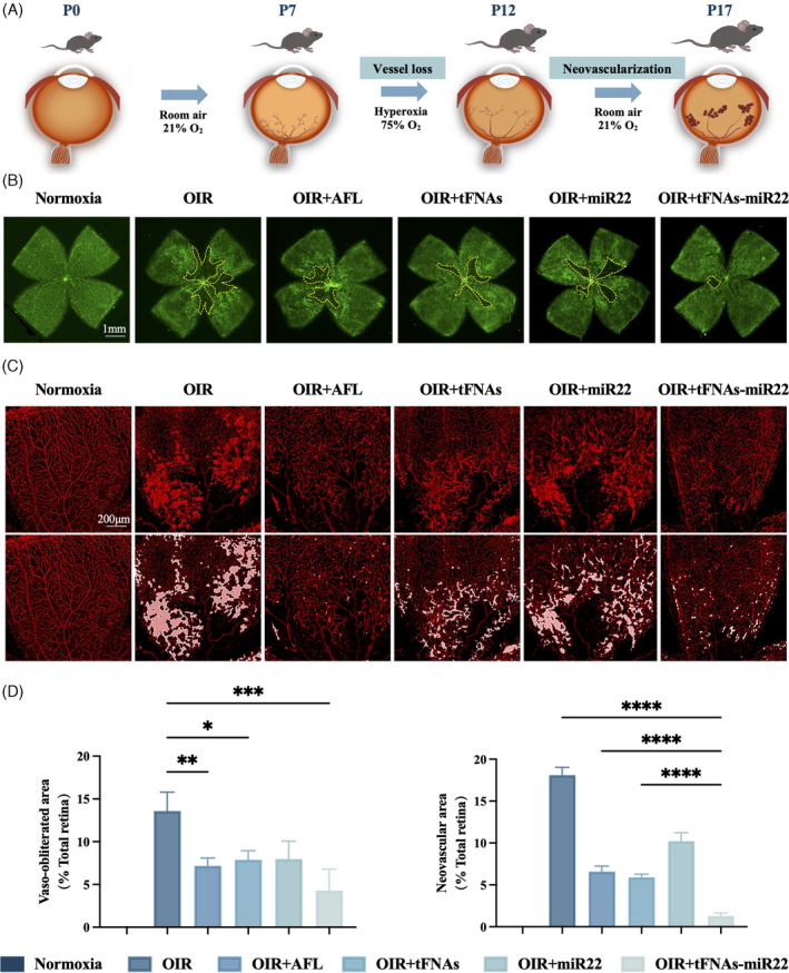
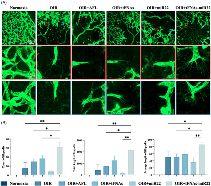
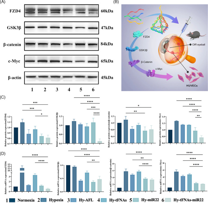
The objective of this study was to investigate the effects and molecular mechanisms of tetrahedral framework nucleic acids-microRNA22 (tFNAs-miR22) on inhibiting pathological retinal neovascularization (RNV) and restoring physiological retinal vessels. A novel DNA nanocomplex (tFNAs-miR22) was synthesised by modifying microRNA-22 (miR22) through attachment onto tetrahedral frame nucleic acids (tFNAs), which possess diverse biological functions. Cell proliferation, wound healing, and tube formation were employed for in vitro assays to investigate the angiogenic function of cells. Oxygen-induced retinopathy (OIR) model was utilised to examine the effects of reducing pathological neovascularization (RNV) and inhibiting vascular occlusion in vivo. In vitro, tFNAs-miR22 demonstrated the ability to penetrate endothelial cells and effectively suppress cell proliferation, tube formation, and migration in a hypoxic environment. In vivo, tFNAs-miR22 exhibited promising results in reducing RNV and promoting the restoration of normal retinal blood vessels in OIR model through modulation of the Wnt pathway. This study provided a theoretical basis for the further understanding of RNV, and highlighted the innovative and potential of tFNAs-miR22 as a therapeutic option for ischemic retinal diseases.
© 2024 The Authors. Cell Proliferation published by Beijing Institute for Stem Cell and Regenerative Medicine and John Wiley & Sons Ltd.
Conflict of interest statement
The authors declare no conflict of interest.
Figures
Similar articles
-
Targeted siRNA Delivery Against RUNX1 Via tFNA: Inhibiting Retinal Neovascularization and Restoring Vessels Through Dll4/Notch1 Signaling.Invest Ophthalmol Vis Sci. 2025 Mar 3;66(3):39. doi: 10.1167/iovs.66.3.39. Invest Ophthalmol Vis Sci. 2025. PMID: 40105819 Free PMC article.
-
Tetrahedral framework nucleic acids inhibit pathological neovascularization and vaso-obliteration in ischaemic retinopathy via PI3K/AKT/mTOR signalling pathway.Cell Prolif. 2023 Jul;56(7):e13407. doi: 10.1111/cpr.13407. Epub 2023 Jan 24. Cell Prolif. 2023. PMID: 36694349 Free PMC article.
-
Therapeutic effects of tetrahedral framework nucleic acids and tFNAs-miR22 on retinal ischemia/reperfusion injury.Cell Prolif. 2024 Nov;57(11):e13695. doi: 10.1111/cpr.13695. Epub 2024 Jul 31. Cell Prolif. 2024. PMID: 39086110 Free PMC article.
-
Tetrahedral framework nucleic acids for improving wound healing.J Nanobiotechnology. 2024 Mar 16;22(1):113. doi: 10.1186/s12951-024-02365-z. J Nanobiotechnology. 2024. PMID: 38491372 Free PMC article. Review.
-
Current Understanding and Translational Prospects of Tetrahedral Framework Nucleic Acids.JACS Au. 2025 Feb 10;5(2):486-520. doi: 10.1021/jacsau.4c01170. eCollection 2025 Feb 24. JACS Au. 2025. PMID: 40017737 Free PMC article. Review.
Cited by
-
Targeted siRNA Delivery Against RUNX1 Via tFNA: Inhibiting Retinal Neovascularization and Restoring Vessels Through Dll4/Notch1 Signaling.Invest Ophthalmol Vis Sci. 2025 Mar 3;66(3):39. doi: 10.1167/iovs.66.3.39. Invest Ophthalmol Vis Sci. 2025. PMID: 40105819 Free PMC article.
References
MeSH terms
Substances
Grants and funding
- 303010303058/the Construction Project of High-Level Hospitals in Guangdong Province
- 303020107/the Construction Project of High-Level Hospitals in Guangdong Province
- 2023A1515010430/Basic and Applied Basic Research Foundation of Guangdong Province
- 82271092/National Natural Science Foundation of China
LinkOut - more resources
Full Text Sources
