

doi: 10.3340/jkns.2024.0027.
Epub 2024 Mar 4.
Management of Pediatric Intracranial Arteriovenous Malformations
Affiliations
- PMID: 38433517
- PMCID: PMC11079567
- DOI: 10.3340/jkns.2024.0027
Item in Clipboard
Management of Pediatric Intracranial Arteriovenous Malformations
J Korean Neurosurg Soc.
2024 May.
Abstract
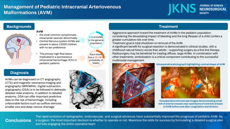
Pediatric intracranial arteriovenous malformations (AVMs) are challenging lesions managed by pediatric neurosurgeons. The high risk of hemorrhage and neurologic injury is compounded by the unique anatomy of each malformation that requires individualizing treatment options. This article reviews the current status of pediatric AVM epidemiology, pathophysiology and clinical care, with a specific focus on the rationale and methodology of surgical resection.
Keywords: Arteriovenous malformations; Pediatrics; Perioperative care; Stroke.
Conflict of interest statement
No potential conflict of interest relevant to this article was reported.
Figures
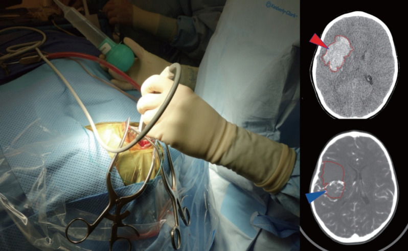
Use of an intraoperative ultrasound to help localize clot (red arrowhead on CT) and AVM nidus (blue arrowhead on CTA) in order to guide controlled decompression of the clot through a small dural opening in advance of a wider exposure. Using the Doppler flow settings can be particularly helpful to visualize AVM, especially when paired with CT/CTA studies. CT : computed tomography, AVM : arteriovenous malformation, CTA : computed tomography angiography.
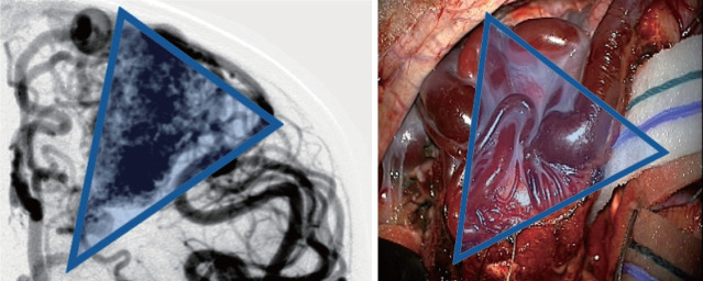
Angiographic correlation with intraoperative photograph highlighting conical shape of arteriovenous malformation, including both surface findings and surgical technique of working around the “cone” of the nidus, walling the margins off with cottonoids.
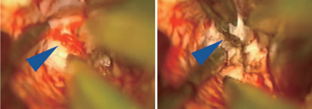
Intraoperative microscope images demonstrating small tuft of arterial vessels near ependyma of ventricle at base of arteriovenous malformation (left image, blue arrowhead) with subsequent cauterization and division of these feeders (right image). In addition, this image also highlights the utility of having an assistant working under the microscope to improve visualization and minimize prolonged retraction on adjacent brain.
Similar articles
-
International multicenter cohort study of pediatric brain arteriovenous malformations. Part 2: Outcomes after stereotactic radiosurgery.J Neurosurg Pediatr. 2017 Feb;19(2):136-148. doi: 10.3171/2016.9.PEDS16284. Epub 2016 Dec 2. J Neurosurg Pediatr. 2017. PMID: 27911249
-
International multicenter cohort study of pediatric brain arteriovenous malformations. Part 1: Predictors of hemorrhagic presentation.J Neurosurg Pediatr. 2017 Feb;19(2):127-135. doi: 10.3171/2016.9.PEDS16283. Epub 2016 Dec 2. J Neurosurg Pediatr. 2017. PMID: 27911248
-
Safety and outcome of combined endovascular and surgical management of low grade cerebral arteriovenous malformations in children compared to surgery alone.Eur J Radiol. 2019 Jul;116:8-13. doi: 10.1016/j.ejrad.2019.02.016. Epub 2019 Feb 18. Eur J Radiol. 2019. PMID: 31153578
-
Prevalence and characteristics of brain arteriovenous malformations in hereditary hemorrhagic telangiectasia: a systematic review and meta-analysis.J Neurosurg. 2017 Aug;127(2):302-310. doi: 10.3171/2016.7.JNS16847. Epub 2016 Oct 21. J Neurosurg. 2017. PMID: 27767404
-
Brain arteriovenous malformations: A review of natural history, pathobiology, and interventions.Neurology. 2020 Nov 17;95(20):917-927. doi: 10.1212/WNL.0000000000010968. Epub 2020 Oct 1. Neurology. 2020. PMID: 33004601 Review.
Cited by
-
One-stop hybrid operation versus microsurgery for treating brain arteriovenous malformation in children-a retrospective case series.Transl Pediatr. 2024 Jul 31;13(7):1051-1060. doi: 10.21037/tp-24-68. Epub 2024 Jul 18. Transl Pediatr. 2024. PMID: 39144421 Free PMC article.
References
-
- Abla AA, Rutledge WC, Seymour ZA, Guo D, Kim H, Gupta N, et al. A treatment paradigm for high-grade brain arteriovenous malformations: volume-staged radiosurgical downgrading followed by microsurgical resection. J Neurosurg. 2015;122:419–432. - PubMed
-
- Aboukaïs R, Vinchon M, Quidet M, Bourgeois P, Leclerc X, Lejeune JP. Reappearance of arteriovenous malformations after complete resection of ruptured arteriovenous malformations: true recurrence or falsenegative early postoperative imaging result? J Neurosurg. 2017;126:1088–1093. - PubMed
-
- Al-Jarallah A, Al-Rifai MT, Riela AR, Roach ES. Nontraumatic brain hemorrhage in children: etiology and presentation. J Child Neurol. 2000;15:284–289. - PubMed
-
- Altschuler E, Lunsford LD, Kondziolka D, Wu A, Maitz AH, Sclabassi R, et al. Radiobiologic models for radiosurgery. Neurosurg Clin N Am. 1992;3:61–77. - PubMed
LinkOut - more resources
Full Text Sources
